. 2004 Nov;168(3):1205-18.
doi: 10.1534/genetics.104.028035.

The amino terminus of the Saccharomyces cerevisiae DNA helicase Rrm3p modulates protein function altering replication and checkpoint activity

Affiliations

The amino terminus of the Saccharomyces cerevisiae DNA helicase Rrm3p modulates protein function altering replication and checkpoint activity

Jessica B Bessler et al. Genetics. 2004 Nov.

Abstract

The Pif1 family of DNA helicases is conserved from yeast to humans. Although the helicase domains of family members are well conserved, the amino termini of these proteins are not. The Saccharomyces cerevisiae genome encodes two Pif1 family members, Rrm3p and Pif1p, that have very different functions. To determine if the amino terminus of Rrm3p contributes to its role in promoting fork progression at >1000 discrete chromosomal sites, we constructed a deletion series that lacked portions of the 249-amino-acid amino terminus. The phenotypes of cells expressing alleles that lacked all or most of the amino terminus were indistinguishable from those of rrm3Delta cells. Rrm3p deletion derivatives that lacked smaller portions of the amino terminus were also defective, but the extent of replication pausing at tRNA genes, telomeres, and ribosomal DNA (rDNA) was not as great as in rrm3Delta cells. Deleting only 62 amino acids from the middle of the amino terminus affected only rDNA replication, suggesting that the amino terminus can confer locus-specific effects. Cells expressing a fusion protein consisting of the Rrm3p amino terminus and the Pif1p helicase domain displayed defects similar to rrm3Delta cells. These data demonstrate that the amino terminus of Rrm3p is essential for Rrm3p function. However, the helicase domain of Rrm3p also contributes to its functional specificity.


Figures

F<sc>igure</sc> 1.—
Figure 1.—
Rrm3p is a member of the Pif1 family of DNA helicases; alignment of Rrm3p with Pif1p is shown. Dashes indicate gaps in the alignment. Conserved residues are in gray; numbers refer to Rrm3p amino acids. The positions of the seven helicase motifs are indicated with black boxes and roman numerals. The dashed box outlines the proposed site of PCNA interaction (Warbrick 2000; Schmidt et al. 2002). RRM3 is predicted to encode both nuclear and mitochondrial forms of the protein (M. K. Mateyak and V. A. Zakian, unpublished results); the arrow indicates the predicted cleavage site for removing the mitochondrial signal sequence. The putative NLSs are underlined.
F<sc>igure</sc> 2.—
Figure 2.—
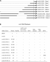
Structure and summary of phenotypes of rrm3 deletion alleles. (A) Amino-terminal deletions of Rrm3p. Deletion alleles are named according to the amino acids removed. The amino terminus of Rrm3p is 249 amino acids long. Full-length Rrm3p is 723 amino acids. The dashed vertical line indicates the end of the amino terminus and the beginning of the helicase domain. Thick lines are the regions of the protein that are present and thin lines indicate the regions deleted. (B) The nine alleles are divided into three classes based upon the phenotypes described in this article, which are summarized here. Plus indicates a wild-type phenotype; minus indicates the null phenotype; +/− indicates an intermediate phenotype; RFB indicates a novel perturbation at the RFB. The protein column indicates a rough estimate of protein levels, with the wild-type level defined as one.
F<sc>igure</sc> 3.—
Figure 3.—
The Rrm3p amino terminus negatively regulates protein abundance. For each strain, proteins were extracted from equal numbers of cells, diluted as indicated, separated by SDS-PAGE, and analyzed by Western blotting using a monoclonal anti-Myc antibody (BD Biosciences Clonetech). The same membranes were also probed with anti-actin antibody to establish that similar amounts of protein were loaded for each of the strains expressing rrm3 deletion alleles. Positions of molecular weight markers are indicated. For each allele, the expected position of full-length protein is marked with an asterisk. In each strain, the YCplac111 plasmid alone or carrying an allele expressed from the RRM3 promoter was introduced into an rrm3Δ strain. (A) A 1:10 dilution of protein extract was loaded for all lanes except the lanes with extracts from RRM3 (lane 1) and rrm3Δ (lane 2) cells; i.e., 10 times more RRM3 and rrm3Δ extract was loaded compared to the amount from each of the deletion alleles. (B) 1:30 dilutions of mutant protein extracts. (C) 1:100 dilutions of mutant protein extracts. Protein abundance was estimated from these gels. Note the band visible for rrm3Δ2-218 is a doublet. Western exposure times were varied according to the dilution level of the extracts from the rrm3 deletion alleles.
F<sc>igure</sc> 4.—
Figure 4.—
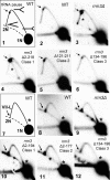
Class 1 and class 2 rrm3 deletion alleles have impaired replication at tRNAA and telomere VII-L. (1) Schematic of 2D gel analysis of the 5.3-kbp BglII fragment that contains tRNAA in RRM3 DNA. The arrow indicates the position of tRNAA on the arc of forked replication intermediates. (7) Schematic of 2D gel analysis of the 3.8-kbp ClaI fragment that contains the modified telomere VII-L in a wild-type strain: the asterisk indicates a pause in subtelomeric DNA, and the arrow indicates the pause within the ∼300-bp telomere. In 1 and 7, the 1N spot, which consists of nonreplicating DNA, falls on the arc of linear molecules (indicated with a dashed line). Almost fully replicated DNA fragments reenter the arc of linear molecules when their mass is nearly 2N. Although replication was examined in each deletion allele, only one example is shown from each class. DNA from the indicated strains was digested with BglII (tRNA gels) or ClaI (telomere gels), separated by 2D gels, and analyzed by Southern blotting using a portion of HIS2 (for tRNAA) or URA3 (telomere VII-L). (2 and 8) Wild type; (3 and 9) rrm3Δ; (4 and 10) class 1 mutants; (5 and 11) class 2 mutants; and (6 and 12) class 3 mutant.
F<sc>igure</sc> 5.—
Figure 5.—
Class 1 and class 2 rrm3 deletion alleles have impaired replication at rDNA. (1) Schematic of 2D gel analysis of the 4.5-kbp BglII rDNA fragment in RRM3 DNA. The positions of forks arrested (RFB; marked by bracket) and converged (X) at the RFB are indicated. Arrows mark the positions of replication forks at inactive origins of DNA replication (ARS), the end of the 35S transcript (35S), and the 5S rRNA gene (5S). (7) Schematic of 2D gel analysis of the 5.0-kbp StuI rDNA fragment in RRM3 DNA; symbols are the same as in 1. In addition, HJ indicates putative Holliday junctions, BR indicates broken replication intermediates, and BU indicates bubble-shaped replication intermediates that arise from the ∼20% of repeats that contain an active origin. Although rDNA replication was examined in each deletion allele, only one example is shown from each class. DNA from the indicated strains was digested with BglII (2–6) or StuI (8–13), separated by 2D gels, and analyzed by Southern blotting using a portion of rDNA as a probe. (2 and 8) Wild type; (3 and 9) rrm3Δ; (4) class 1 mutant; (5) class 2 mutant; and (6 and 10) class 3 mutant. To determine if the RFB phenotype of rrm3Δ134-196 cells was Fob1p dependent, StuI-digested DNA from fob1Δ (11), rrm3Δ fob1Δ (12), and rrm3Δ134-196 fob1Δ (13) was analyzed. The thin arrow in 12 indicates the presence of a new pause in rrm3Δ fob1Δ cells (Torres et al. 2004a).
F<sc>igure</sc> 6.—
Figure 6.—
Fob1p binding to the RFB is not diminished in rrm3Δ134-196 cells. (A) Schematic of a single repeat of S. cerevisiae rDNA. The DNA regions PCR amplified in the ChIP assays are indicated below. (B) Chromatin immunoprecipitation using anti-Myc monoclonal antibody was carried out in wild-type and rrm3Δ134-196 cells expressing Fob1-13Myc and in wild-type cells lacking an Myc-tagged protein (labeled no tag). Twofold serial dilutions of rrm3Δ input DNA were amplified to establish the linearity of PCR reaction. A 363-bp fragment that spans the complete RFB (RFB) and a 407-bp sequence from within the 35S RNA gene (35S) were PCR amplified. PCR products were resolved on a 2.8% ethidium bromide/agarose gel and quantified by densitometric analysis.
F<sc>igure</sc> 7.—
Figure 7.—
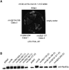
Class 1 alleles, unlike class 2 and 3 alleles, hyperphosphorylate Rad53p and require checkpoint and repair proteins for viability. (A) A mec1Δ sml1Δ derivative of each rrm3 deletion allele was constructed. Each strain also contained the RRM3 URA3 plasmid pIA20. Cells were streaked on plates containing 5-FOA, which kills URA3 cells, and grown for 3 days at 23°. Only cells that retain viability upon loss of the URA3 plasmid-borne RRM3 allele can grow on 5-FOA plates. One example of each deletion class is shown. (B) Cell extracts were prepared from equal numbers of log phase cultures of the indicated strains, separated on acrylamide gels, and analyzed by Western blotting using a Rad53p polyclonal antibody JDI47 kindly supplied by J. Diffley.
F<sc>igure</sc> 8.—
Figure 8.—
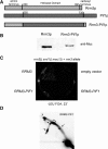
The helicase domain contributes to the functional specificity of Rrm3p. (A) Schematic of the hybrid protein. The top line shows Rrm3p divided into its three regions. The second line shows Pif1p. In both cases, the numbers indicate the first and last amino acid of the respective helicase domain. Rrm3p is 723 amino acids long and Pif1p is 859 amino acids. The third line is the hybrid protein. Hatched regions are from Rrm3p and solid regions are from Pif1p. (B) Protein levels of wild-type protein vs. the Rrm3-Pif1 fusion protein. The Western blot was probed with anti-Myc monoclonal antibody. (C) Viability assay using an rrm3Δ mec1Δ sml1Δ strain. Strains were grown at 23° for 3 days on YC-LEU 5-FOA media. Two independent transformants of Rrm3-Pif1p are shown. (D) A 2D gel of BglII-digested DNA from rrm3Δ cells expressing Rrm3-Pif1p. X indicates the converged forks and the bracket marks the position of the RFB. Arrows point to the position of the 5S rRNA genes, the 35S rRNA termination site, and the ARS on the arc of simple Y's.

References

    1. Bae, S. H., J. A. Kim, E. Choi, K. H. Lee, H. Y. Kang et al., 2001. Tripartite structure of Saccharomyces cerevisiae Dna2 helicase/endonuclease. Nucleic Acids Res. 29: 3069–3079. - PMC - PubMed
    1. Bennett, R. J., M. F. Noirot-Gros and J. C. Wang, 2000. Interaction between yeast sgs1 helicase and DNA topoisomerase III. J. Biol. Chem. 275: 26898–26905. - PubMed
    1. Bessler, J. B., J. Z. Torres and V. A. Zakian, 2001. The Pif1p subfamily of helicases: region specific DNA helicases. Trends Cell Biol. 11: 60–65. - PubMed
    1. Boeke, J. D., J. Trueheart, G. Natsoulis and G. R. Fink, 1987. 5-Fluoroorotic acid as a selective agent in yeast molecular genetics. Methods Enzymol. 154: 164–175. - PubMed
    1. Brewer, B. J., and W. L. Fangman, 1987. The localization of replication origins on ARS plasmids in S. cerevisiae. Cell 51: 463–471. - PubMed

Publication types

MeSH terms