. 2005 Jan;6(1):31-41.
doi: 10.1038/ni1150. Epub 2004 Dec 5.

Locus 'decontraction' and centromeric recruitment contribute to allelic exclusion of the immunoglobulin heavy-chain gene

Affiliations

Locus 'decontraction' and centromeric recruitment contribute to allelic exclusion of the immunoglobulin heavy-chain gene

Esther Roldán et al. Nat Immunol. 2005 Jan.

Abstract

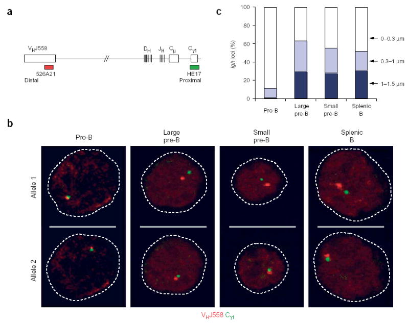
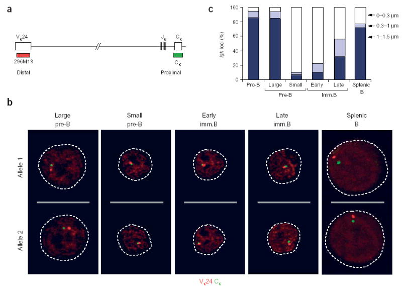
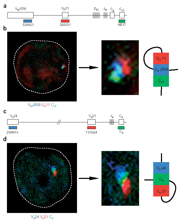
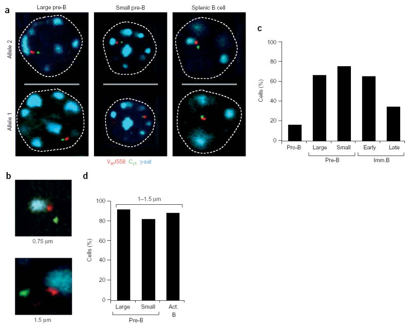
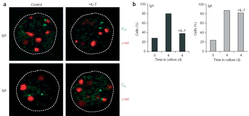
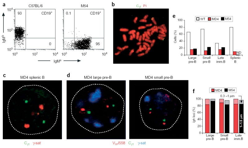
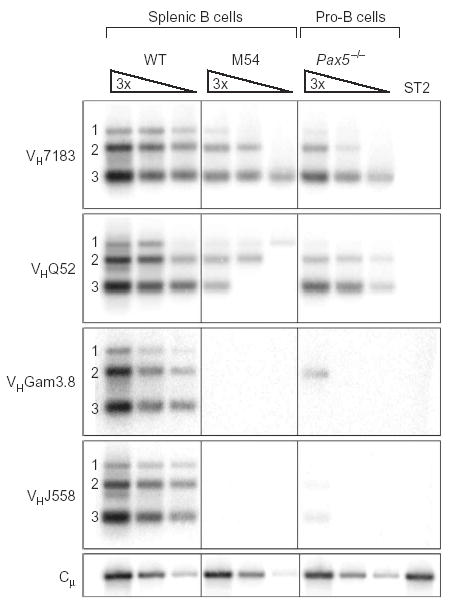
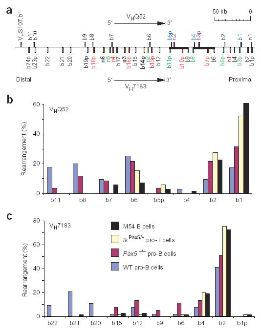
Allelic exclusion of immunoglobulin genes ensures the expression of a single antibody molecule in B cells through mostly unknown mechanisms. Large-scale contraction of the immunoglobulin heavy-chain (Igh) locus facilitates rearrangements between Igh variable (V(H)) and diversity gene segments in pro-B cells. Here we show that these long-range interactions are mediated by 'looping' of individual Igh subdomains. The Igk locus also underwent contraction by looping in small pre-B and immature B cells, demonstrating that immunoglobulin loci are in a contracted state in rearranging cells. Successful Igh recombination induced the rapid reversal of locus contraction in response to pre-B cell receptor signaling, which physically separated the distal V(H) genes from the proximal Igh domain, thus preventing further rearrangements. In the absence of locus contraction, only the four most proximal V(H) genes escaped allelic exclusion in immature mu-transgenic B lymphocytes. Pre-B cell receptor signaling also led to rapid repositioning of one Igh allele to repressive centromeric domains in response to downregulation of interleukin 7 signaling. These data link both locus 'decontraction' and centromeric recruitment to the establishment of allelic exclusion at the Igh locus.


Conflict of interest statement

COMPETING INTERESTS STATEMENT

The authors declare that they have no competing financial interests.

Figures

Figure 1
Figure 1
Decontraction of the Igh locus in large pre–B cells. (a) Igh locus (not drawn to scale), indicating the positions of bacterial artificial chromosome (BAC) 526A21 (ref. 12) and plasmid HE17 (ref. 24), which were used to generate the VHJ558 (red) and Cγ1 (green) probes, respectively. (b) Representative confocal sections through the nuclei of B lymphocytes at various developmental stages (above images) in which three-dimensional DNA FISH analysis of the Igh locus was done with VHJ558 (red) and Cγ1 (green) probes. The two Igh alleles of each cell are presented on separate optical sections. Broken lines outline the contours of the nuclei. Pre–B cells were identified as large or small cells under the microscope. (c) Separation of VHJ558 and Cγ1 gene segments. The distance (in μm) separating the VHJ558 and Cγ1 segments was evaluated statistically for B lymphocytes of various developmental stages (horizontal axis). Actual numbers and sample sizes are in Supplementary Table 1 online.
Figure 2
Figure 2
Contraction and decontraction of the Igk locus. (a) Igk locus (not drawn to scale), indicating the positions of BAC 296M13 (ref. 50) and plasmid IgkC, which were used to generate the Vκ24 (red) and Cκ (green) probes, respectively. (b) Representative confocal sections through B cells at various stages of development (above images) in which three-dimensional DNA FISH analysis of the Igk locus was done with Vκ24 (red) and Cκ (green) probes. The two Igk alleles of each cell are presented in separate optical sections. Flow cytometry sorting of the developmental stages is in Supplementary Fig. 1 online. (c) Separation of Vκ24 and Cκ gene segments. The distance (in μm) separating the Vκ24 and Cκ segments was evaluated statistically for cells of various developmental stages (horizontal axis). Actual numbers and sample sizes are in Supplementary Table 1 online. Imm.B, immature B cell.
Figure 3
Figure 3
Contraction by looping of the Igh and Igk loci. (a) Igh locus, indicating the position of the third VH11 probe (BAC 282021). (b) Confocal section through the nucleus of a Rag2/ pro–B cell, which was analyzed by three-dimensional DNA FISH with VHJ558 (blue), VH11 (red) and Cγ1 (green) probes. Middle, enlargement of the Igh allele shown at left; right, looping configuration. (c) Igk locus, indicating the position of the third Vκ21 probe (BAC 113G24; ref. 51). (d) Confocal image of an early immature B cell showing the relative positions of the Vκ24 (blue), Vκ21 (red) and Cκ (green) probe signals. Middle, higher magnification of the Igk allele shown at left; right, looping configuration. There was similar looping of Igk and Igh loci in 20 and 24 cells, respectively. Additional alleles with Igk and Igh looping are in Supplementary Fig. 2 online.
Figure 4
Figure 4
Monoallelic centromeric recruitment of the Igh locus during B cell development. (a) Centromeric location of one Igh allele in bone marrow pre–B cells. Large and small pre–B cells and activated splenic B cells were analyzed by three-color three-dimensional DNA FISH with VHJ558 (red), Cγ1 (green) and γ-satellite (γ-sat; blue) probes. The relative positions of the three signals are shown in confocal sections through the nuclei of these cells. (b) Association of the distal VHJ558 gene domain with centromeric clusters. Enlargements show the orientation and decontraction of the Igh locus at γ-satellite clusters. Below images, distance between the VHJ558 and Cγ1 probe signals. (c) Monoallelic recruitment of the Igh locus to centromeres. Data represent the percentage of cells showing association of one Igh allele with centromeric heterochromatin in various B cell developmental stages, sorted as indicated in Supplementary Fig. 1 online. Actual numbers and sample sizes are in Supplementary Table 2 online. (d) Preferential location of widely separated Igh alleles at the centromeres. The cells showing monoallelic centromeric recruitment were subdivided into a population of cells containing an Igh allele with wide separation (1–1.5 μm) of the VHJ558 and Cγ1 genes. Data represent the percentage of centromeric recruitment of the widely separated Igh allele in this cell population for large and small pre–B cells and activated (Act.) splenic B cells.
Figure 5
Figure 5
IL-7 signaling prevents centromeric recruitment of the Igh locus in splenic B cells. (a) Two-color three-dimensional DNA FISH analysis of activated splenic B cells with (+IL-7) or without (Control) IL-7, showing the proximity of the Cγ1 (Igh) and Cκ (Igk) signals (green) with γ-satellite clusters (red). Both Igh or Igk alleles are on the same optical section. (b) Statistical analysis of cells with centromeric association of one Igh or Igk allele in splenic B cells before and after in vitro activation with anti-CD40 in the presence (+IL-7) or absence of IL-7. Actual numbers and sample sizes are in Supplementary Table 4 online.
Figure 6
Figure 6
Absence of centromeric recruitment and locus contraction of endogenous Igh alleles in μ-transgenic B cells. (a) Flow cytometry of IgM expression on CD19+ B cells of C57BL/6 and M54 transgenic mice. The M54 transgene of the IgMa haplotype was derived from the BALB/c hybridoma 17.2.25 (ref. 34) and was crossed into the C57BL/6 mouse strain of the IgMb haplotype. Lymph node B cells from a 3-week-old M54 mouse express transgenic IgMa but no endogenous IgMb, as published,. Numbers in dot plots indicate the percentages of cells in quadrants. The same degree of allelic exclusion was noted for MD4 transgenic B cells (data not shown). (b) Metaphase spread of MD4 transgenic B cells. Specific hybridization of the Cγ1 probe (green) is detected at the telomeres of chromosome 12. Chromosomes were counterstained with propidium iodide (PI; red). (c) Confocal section through the nucleus of an activated splenic MD4 B cell. The Cγ1 regions (green) of both endogenous Igh alleles are not associated with γ-satellite clusters (red), as visualized by three-dimensional DNA FISH. (d) Confocal sections through the nuclei of large and small pre–B cells of the MD4 transgenic mouse. The Cγ1 (green) and VHJ558 (red) signals are widely separated and are not associated with γ-satellite clusters (blue). (e) Statistical analysis of the monoallelic centromeric association of endogenous Igh loci in B lymphocytes of wild-type (WT) and MD4 and M54 transgenic mice. ND, not determined. Actual numbers and sample sizes are in Supplementary Table 2 online. (f) Statistical analysis of the distance separating the distal VHJ558 and proximal Cγ1 regions of the endogenous Igh loci in developing MD4 (red) and M54 (black) transgenic B lymphocytes (Supplementary Table 4 online). Light or dark shading indicates VHJ558-Cγ1 gene separation of 0.3–1 μm or 1–1.5 μm, respectively.
Figure 7
Figure 7
Proximal VH-DJH rearrangements in M54 transgenic B cells. IgMa CD19+ B cells expressing the μa transgene were isolated by flow cytometry sorting from the spleens of M54 transgenic mice (Supplementary Fig. 4 online). PCR was used to determine the frequency of the different VH-DJH rearrangements in these transgenic B cells versus Pax5−/− bone marrow pro–B cells and wild-type B220+ splenocytes. Threefold (3×) serial DNA dilutions were analyzed by PCR with VH family–specific forward primers and a JH3 reverse primer, which was unable to amplify the VHDJH4-rearranged M54 transgene. Bottom, input DNA was normalized by PCR amplification of an Igh Cμ fragment; far right lane, DNA of stromal ST2 cells (negative control). Numbers along the left margin indicate rearrangements to the JH1, JH2 and JH3 segments. The same result was obtained in two independent experiments.
Figure 8
Figure 8
The most proximal VH genes escape allelic exclusion in M54 transgenic B lymphocytes. (a) VHQ52 and VH7183 gene families of the mouse C57BL/6 strain. The family members were identified by annotation of the Igh sequences of the mouse genome database (http://mendel.imp.univie.ac.at/SEQUENCES/VH/). The VH genes are numbered according to published nomenclature. b, C57BL/6; p, pseudogenes. Our annotation identified previously unknown family members (n). Thick horizontal black bars indicate a large sequence duplication; genes with high sequence similarity are in the same color. The VH7183.b2 gene segment of the C57BL/6 mouse corresponds to the VH81X gene segment of the BALB/c strain. (b,c) Statistical analysis of VH-DJH rearrangements involving different members of the VHQ52 (b) and VH7183 (c) gene families in wild-type and Pax5−/− pro–B cells, IkPax5/+ pro–T cells and M54 transgenic IgMa B cells. VH-DJH4 rearrangements of pro–B and pro–T cells and VH-DJH3 rearrangements of M54 B cells were amplified by PCR, cloned, sequenced and assigned to the various family members. Data represent the percentage of rearrangements involving individual genes for each cell type. Total number of distinct rearrangements analyzed: wild-type pro–B cells, 76 (VHQ52) and 123 (VH7183); Pax5−/− pro–B cells, 61 (VHQ52) and 83 (VH7183); IkPax5/+ pro–T cells, 73 (VHQ52) and 81 (VH7183); splenic M54 B cells, 71 (VHQ52) and 79 (VH7183).

Comment in

  • A move to exclude.
    Sen R. Sen R. Nat Immunol. 2005 Feb;6(2):128-30. doi: 10.1038/ni0205-128. Nat Immunol. 2005. PMID: 15662439 No abstract available.

Similar articles

Cited by

References

    1. Hesslein DG, Schatz DG. Factors and forces controlling V(D)J recombination. Adv Immunol. 2001;78:169–232. - PubMed
    1. Bassing CH, Swat W, Alt FW. The mechanism and regulation of chromosomal V(D)J recombination. Cell. 2002;109:S45–S55. - PubMed
    1. Mostoslavsky R, Alt FW, Rajewsky K. The lingering enigma of the allelic exclusion mechanism. Cell. 2004;118:539–544. - PubMed
    1. Yancopoulos GD, Alt FW. Developmentally controlled and tissue-specific expression of unrearranged VH gene segments. Cell. 1985;40:271–281. - PubMed
    1. Stanhope-Baker P, Hudson KM, Shaffer AL, Constantinescu A, Schlissel MS. Cell type-specific chromatin structure determines the targeting of V(D)J recombinase activity in vitro. Cell. 1996;85:887–897. - PubMed

Publication types

MeSH terms

Substances