. 2005 Feb 18;280(7):5430-4.
doi: 10.1074/jbc.M413513200. Epub 2004 Dec 6.

Vacuolar ATPase regulates zymogen activation in pancreatic acini

Affiliations

Vacuolar ATPase regulates zymogen activation in pancreatic acini

Stephen D Waterford et al. J Biol Chem. .

Abstract

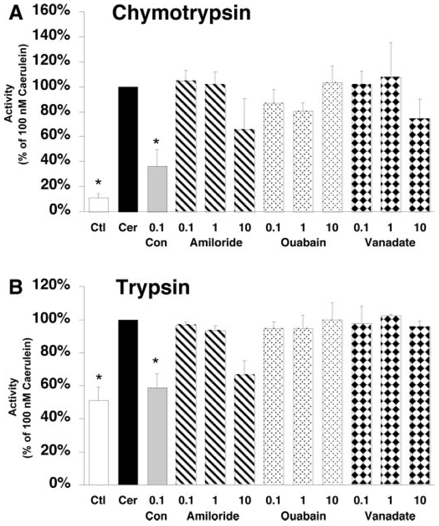
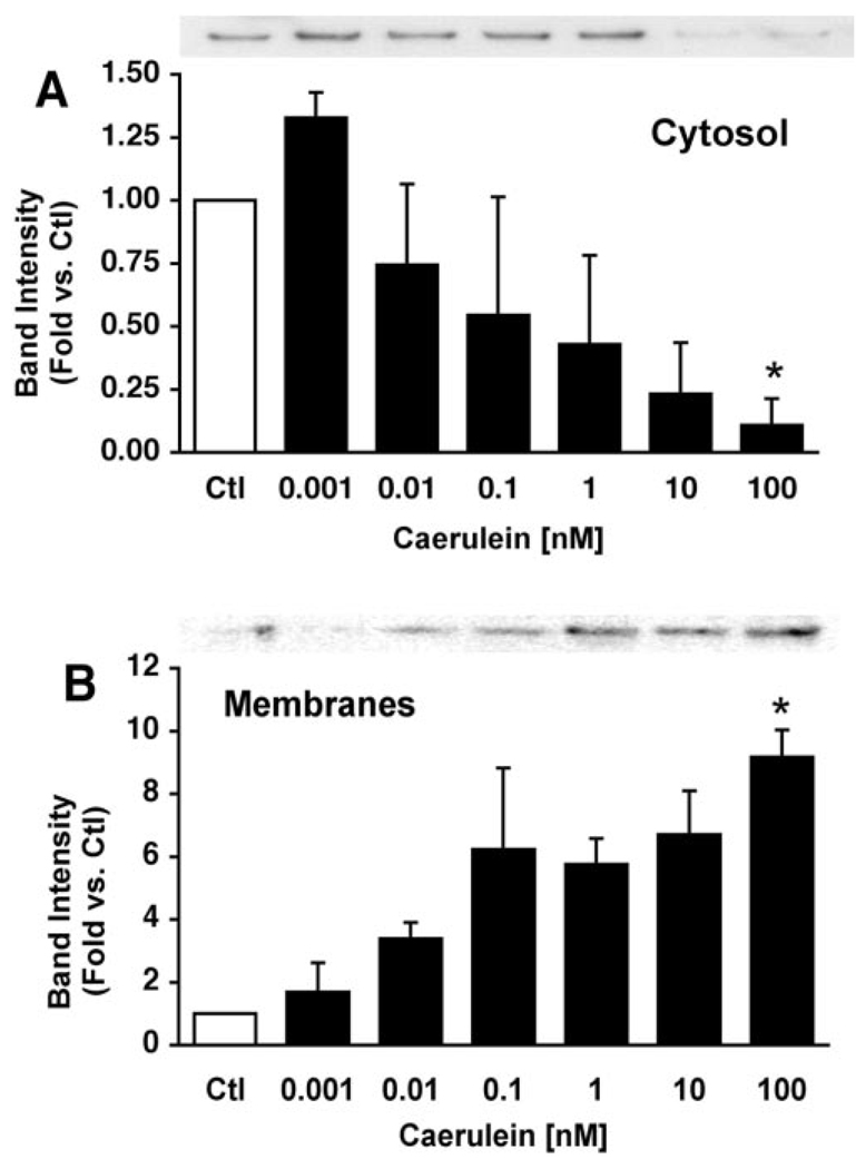
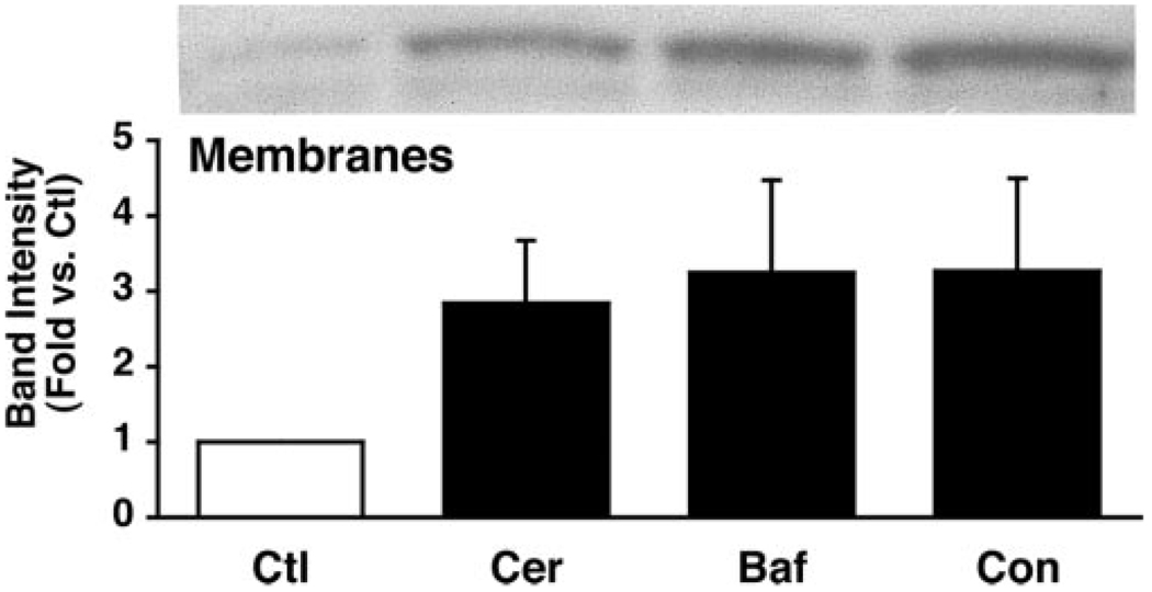
Supramaximal concentrations of cholecystokinin or its analogue caerulein have been shown to stimulate the proteolytic activation of zymogens within the pancreatic acinar cell and initiate acute pancreatitis. Previous studies suggest that a low pH compartment might be required for activation. To test this hypothesis, the effects of agents that modulate intracellular pH on caerulein-induced trypsin and chymotrypsin activation were studied. Pretreatment of pancreatic acini with the proto-ionophore monensin (10 microM) and the weak base chloroquine (40 microM) inhibited activation. Pre-incubation with the vacuolar ATPase (V-ATPase) inhibitors bafilomycin A(1) and concanamycin A also decreased activation in a concentration-dependent manner with 50% inhibition at approximately 50 and 25 nM, respectively. Caerulein stimulation caused a time- and concentration-dependent translocation of soluble V-ATPase V(1) subunits to a membrane fraction, a marker of V-ATPase activation. Carbachol also stimulated translocation at supramaximal concentrations. Elevation of cytosolic Ca(2+) by thapsigargin was sufficient to induce translocation. Thus, stimulation of V-ATPase activity appears to be required for agonist-induced zymogen activation in the pancreatic acinar cell.


Figures

FIG. 1
FIG. 1. Agents that raise intracellular pH decrease zymogen activation
Isolated acini were pretreated with medium alone as control (Ctl), 40 µM chloroquine (Chlor), or 10 µM monensin (Mon) for 30 min and then exposed to 100 µM caerulein (Cer) for 1 h. Black bars indicate caerulein alone; gray bars indicate caerulein plus chloroquine or monensin. Chymotrypsin and trypsin activities were determined by fluorometric assay as described under “Materials and Methods.” Values represent mean ± S.E. *, p < 0.05 versus caerulein alone.
FIG. 2
FIG. 2. V-ATPase inhibitors reduce caerulein-induced chymotrypsinogen and trypsinogen activation
Isolated acini were pretreated for 30 min with the specific V-ATPase inhibitors bafilomycin A1 or concanamycin A at the indicated concentrations and then exposed to 100 nM caerulein (CER) for 1 h. Chymotrypsinogen activation (left) and trypsinogen activation (right) were inhibited by both bafilomycin A1 and concanamycin A, respectively, in a concentration-dependent manner. Black bars indicate caerulein alone; gray bars indicate caerulein plus V-ATPase inhibitor. CTL, control. Values represent mean ± S.E. *, p < 0.05 versus caerulein alone.
FIG. 3
FIG. 3. P-ATPase inhibitors and ion exchange inhibitors do not reduce caerulein-induced zymogen activation
Isolated acini were pretreated with the V-ATPase inhibitor concanamycin A (Con), the Na+-H+ exchange inhibitor amiloride, the Na+-K+ ATPase inhibitor ouabain, or the plasma membrane ATPase inhibitor vanadate (ranging from 0.1 to 10 µM) before stimulation with 100 nM caerulein (Cer). Ctl, control. Chymotrypsin (A) and trypsin (B) activities were quantitated as described under “Materials and Methods.” Values represent mean + S.E. *, p < 0.05 versus caerulein alone.
FIG. 4
FIG. 4. Translocation of the soluble E-subunit to membranes is agonist-dependent
Acini were treated with medium as control (Ctl) or supraphysiologic concentrations of 0.1 µM caerulein (Cer), 1 mM carbachol (Carb) (A), or 10 µM thapsigargin (Thapsi) (B) for 15 min and then homogenized. A membrane fraction was formed from a post-nuclear supernatant, and the E subunit was detected using immunoblot analysis (see “Materials and Methods”). Results are representative of three separate studies.
FIG. 5
FIG. 5. Caerulein stimulation causes a concentration-dependent translocation of soluble V-ATPase subunits to membranes in pancreatic acini
Acini were treated with increasing concentrations of caerulein for 15 min and fractionated to obtain cytosolic (A) and particulate (B) fractions. Proteins were separated by SDS-PAGE and immunoblotted with anti-E subunit antibodies. Representative immunoblots are shown (n = 3). Band intensities were quantified by laser densitometry and expressed as mean ± S.E. *, p < 0.05 versus control (Ctl) for panels A and B.
FIG. 6
FIG. 6. Caerulein stimulation causes a time-dependent translocation of soluble V-ATPase subunits to membranes in pancreatic acini
Acini were treated with 100 nM caerulein for the indicated periods and fractionated to obtain cytosolic (A) and particulate (B) fractions. Proteins were separated by SDS-PAGE and immunoblotted with anti-E subunit antibodies. Representative immunoblots are shown (n = 3). Band intensities were quantified by laser densitometry and expressed as mean ± S.E. *, p < 0.02 versus control (Ctl) for panels A and B.
FIG. 7
FIG. 7. V-ATPase inhibitors do not affect caerulein-induced redistribution of soluble V-ATPase subunits
Acini were pretreated with 100 nM bafilomycin A1 (Baf) or 100 nM concanamycin A (Con) for 15 min and then treated with 100 nM caerulein (Cer) for an additional 15 min. Ctl, control. Following fractionation to obtain cytosolic and particulate fractions, proteins in the membrane fraction were separated by SDS-PAGE and immunoblotted with anti-E subunit antibodies. A representative immunoblot is shown. Band intensities from independent experiments were quantified by laser densitometry and expressed as mean ± S.E.

References

    1. Leach SD, Modlin IM, Scheele GA, Gorelick FS. J. Clin. Investig. 1991;87:362–366. - PMC - PubMed
    1. Saluja A, Saito I, Saluja M, Houlihan MJ, Powers RE, Meldolesi J, Steer M. Am. J. Physiol. 1985;249:G702–G710. - PubMed
    1. Niederau C, Grendell JH. J. Clin. Investig. 1988;81:229–236. - PMC - PubMed
    1. Leach SD, Bilchik AJ, Karapetian O, Gorelick FS, Modlin IM. Pancreas. 1993;8:64–69. - PubMed
    1. Guillaumes S, Blanco I, Villanueva A, Sans MD, Clave P, Chabas A, Farre A, Lluis F. Pancreas. 1997;14:262–266. - PubMed

Publication types

MeSH terms

LinkOut - more resources