. 2004 Dec 6;200(11):1359-70.
doi: 10.1084/jem.20031763.

Apoptotic cells with oxidation-specific epitopes are immunogenic and proinflammatory

Affiliations

Apoptotic cells with oxidation-specific epitopes are immunogenic and proinflammatory

Mi-Kyung Chang et al. J Exp Med. .

Abstract

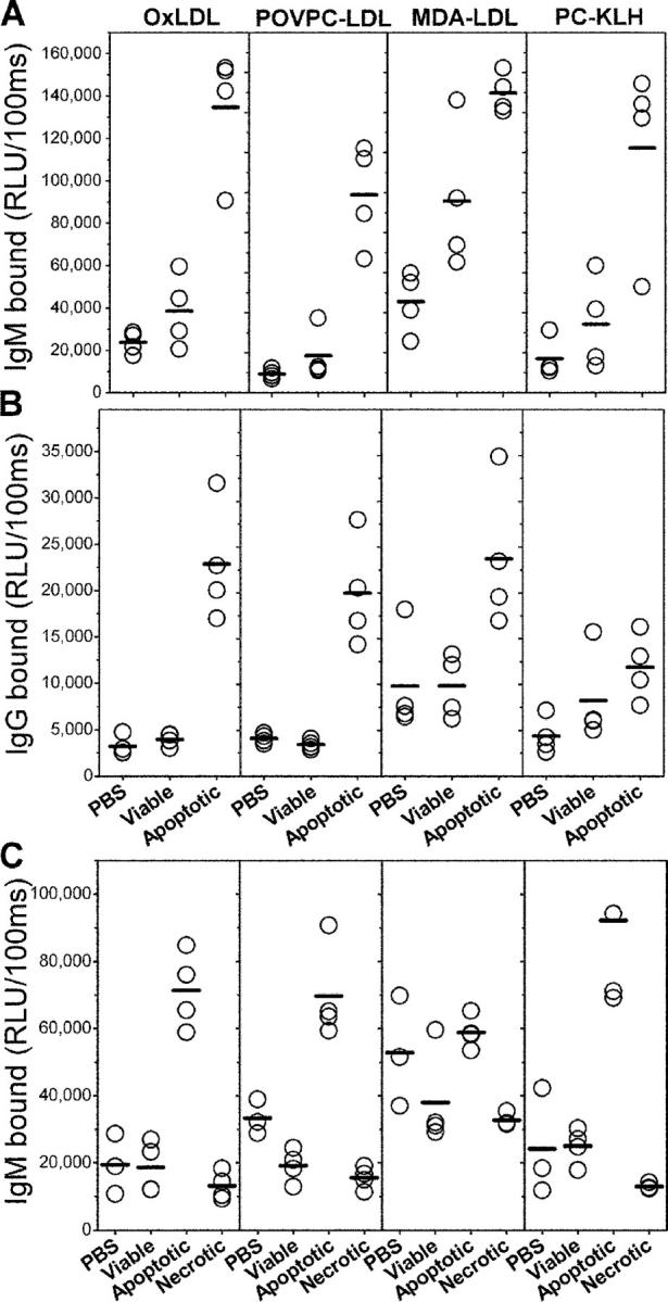
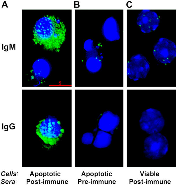
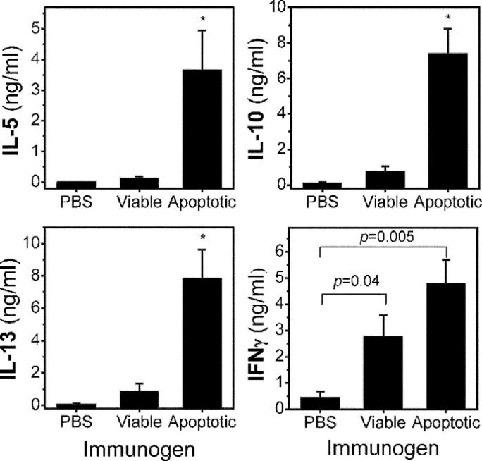
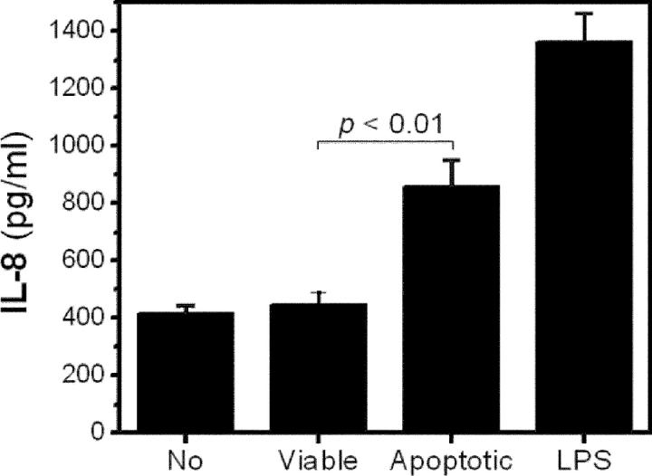
Oxidation of low density lipoprotein (LDL) generates a variety of oxidatively modified lipids and lipid-protein adducts that are immunogenic and proinflammatory, which in turn contribute to atherogenesis. Cells undergoing apoptosis also display oxidized moieties on their surface membranes, as determined by binding of oxidation-specific monoclonal antibodies. In the present paper, we demonstrated by mass spectrometry that in comparison with viable cells, membranes of cells undergoing apoptosis contain increased levels of biologically active oxidized phospholipids (OxPLs). Indeed, immunization of mice with syngeneic apoptotic cells induced high autoantibody titers to various oxidation-specific epitopes of oxidized LDL, including OxPLs containing phosphorylcholine, whereas immunization with viable thymocytes, primary necrotic thymocytes, or phosphate-buffered saline did not. Reciprocally, these antisera specifically bound to apoptotic cells through the recognition of oxidation-specific epitopes. Moreover, splenocyte cultures from mice immunized with apoptotic cells spontaneously released significant levels of T helper cell (Th) 1 and Th2 cytokines, whereas splenocytes from controls yielded only low levels. Finally, we demonstrated that the OxPLs of apoptotic cells activated endothelial cells to induce monocyte adhesion, a proinflammatory response that was abrogated by an antibody specific to oxidized phosphatidylcholine. These results suggest that apoptotic cell death generates oxidatively modified moieties, which can induce autoimmune responses and a local inflammatory response by recruiting monocytes via monocyte-endothelial cell interaction.


Figures

Figure 1.
Figure 1.
LC/MRM analysis of OxPLs from cellular lipid extracts. Quantification of OxPLs obtained from lipid extracts of viable or apoptotic thymocytes induced either by dexamethasone (DEXA) or by PMA treatment was performed using LC/MRM. POVPC, 1-palmitoyl-2-(5-oxovaleroyl)-sn-glycero-3-PC; PGPC, 1-palmitoyl-2-glutaroyl-sn-glycero-3-PC; PEIPC, 1-palmitoyl-2-epoxyisoprostane-sn-glycero-3-PC; lyso-PC, 1-palmitoyl-2-lyso-sn-glycero-3-PC. Amounts of the four OxPLs were determined using 1,2-dimyristoyl-sn-glycero-3-PC as a standard. Data are expressed as micrograms per milligram of the parent lipid 1-palmitoyl-2-arachidonyl-sn-glycero-3-phosphorylcholine (PAPC) ± SD from three separate extractions. *, P < 0.001 compared with viable thymocytes.
Figure 2.
Figure 2.
Binding of immune sera to oxidatively modified epitopes. Chemiluminescent immunoassay for the binding of IgM (A) and IgG (B) from pooled immune sera of NIH/Swiss-Webster mice to indicated antigens. NIH/Swiss-Webster mice were immunized in the presence of adjuvant with the following: PBS, viable, normal thymocytes (Viable), and dexamethasone-treated apoptotic thymocytes (Apoptotic). (C) Binding of IgM from pooled immune sera of C57BL/6 mice immunized in the absence of adjuvant with the following: PBS, viable, normal thymocytes (Viable), dexamethasone treated apoptotic thymocytes (Apoptotic), or suspension of primary necrotic thymocytes generated by repeated freeze-thaw cycles (Necrotic). Sera from NIH/Swiss-Webster and C57BL/6 mice were diluted at 1:400 and 1:250 in PBS with 2% BSA, respectively, and IgM and IgG bindings were measured as described in Materials and Methods. Data are expressed as relative light units (RLU) per 100 ms. Each point is the mean of triplicate determinations.
Figure 3.
Figure 3.
Competition immunoassay for immune sera binding to OxLDL. (A) Competition immunoassay for binding of IgM from a representative serum of NIH/Swiss-Webster mice immunized with apoptotic thymocytes as described in Materials and Methods. The serum was diluted at 1:5,000 in PBS with 1% BSA and was incubated in the absence or presence of indicated concentrations of native LDL, OxLDL, MDA-LDL, or PC-KLH as competitors. Immune complexes were pelleted by centrifugation, and supernatants were tested for binding of IgM to OxLDL using chemiluminescent immunoassay. Competition immunoassay for IgM (B) and IgG (C) from another serum of NIH/Swiss-Webster mice immunized with apoptotic thymocytes was performed to determine the binding affinity of the antisera for OxLDL as described in Materials and Methods. IgM binding was performed with 1:2,000 dilution and IgG binding was performed with 1:300 dilution. Data are expressed as a ratio of the binding of serum in the presence of competitor (B) over the binding in the absence of competitor (Bo). Each point is the mean of triplicate determinations.
Figure 4.
Figure 4.
Immunofluorescence deconvolution microscopy of sera binding to apoptotic cells: Pooled (IgM) or a representative (IgG) preimmune and postimmune sera from NIH/Swiss-Webster mice immunized with apoptotic thymocytes were diluted 1:200 in PBS with 1% BSA and incubated with apoptotic or normal thymocytes. IgM or IgG binding to the cells was detected by fluorescein-conjugated F(ab)2 fragments against mouse IgM or IgG, respectively. (A) Note the marked binding of postimmune sera (green) to apoptotic thymocytes with their characteristic condensed, fragmented nuclei detected by Hoechst staining (blue). (B) Note the negative staining of preimmune sera to apoptotic cells and (C) postimmune sera to normal thymocytes. Bar, 5 μm.
Figure 5.
Figure 5.
Flow cytometry analysis of sera binding to apoptotic thymocytes and competition immunoassay for immune sera binding to apoptotic cells. (A) Apoptosis-induced thymocytes were gated into three populations according to forward scatter and the intensity of PI staining. (R1) Region 1: viable cells or cells at the very early stage of apoptosis; (R2) region 2: cells at intermediate stage of apoptosis with dim PI staining, and (R3) region 3: cells at later stage of apoptosis with shrunken cell size and bright PI staining. (B) Pooled preimmune or postimmune sera from NIH/Swiss-Webster mice immunized with apoptotic thymocytes were diluted at 1:500 in PBS with 1% BSA and tested for IgM binding to apoptotic thymocytes in region 3. (C) Aliquots of diluted, pooled sera were incubated in the absence or presence of OxLDL or MDA-LDL, or both as competitors at indicated concentrations (μg/ml). After the incubations, immune complexes were pelleted by centrifugation and supernatants were tested for IgM binding to apoptotic thymocytes using flow cytometry. Mean fluorescence intensity of binding of immune sera incubated in the absence (No) or presence of competitors to the apoptotic thymocytes in region 3 was measured. Data display the inhibition of binding by competitors as percent of control in the absence of competitors and representative of three independent experiments.
Figure 6.
Figure 6.
Cytokine assay of splenocyte cultures. Splenocytes from each mouse in each group of NIH/Swiss-Webster mice immunized with PBS, viable cells, or apoptotic cells were harvested. Splenocytes were seeded at 5 × 10⁶ cells per well into a 96-well tissue culture plate and were cultured in triplicates in the absence of any added antigens for 72 h. After the incubation, the amount of cytokine released into the supernatants was determined using chemiluminescent immunoassays as described in Materials and Methods. Data reflect mean ± SEM values of each group. *, P < 0.04 compared with groups immunized with either PBS or normal cells.
Figure 7.
Figure 7.
Monocyte adhesion to endothelial cells induced by apoptotic cells. (A) The ability of apoptotic cells to stimulate endothelial cells for monocyte binding was tested. Porcine aortic endothelial cells (PAECs) were incubated for 4 h at 37°C with culture media alone or EO6 antibody in the absence or presence of apoptotic thymocytes (5 × 10⁶ per well) generated by incubation with serum-starved media for 18 h as stimulant. 100 ng/ml LPS was used as positive control. For these experiments, EO6 antibody was obtained from the culture supernatant of EO6 hybridoma that was maintained under LPS-free culture condition. After the incubation, stimulants were washed off with PBS, and PAECs were incubated with THP-1 cells for 30 min at 37°C. Nonadherent THP-1 cells were removed by washing, and the number of adherent THP-1 cells was determined in 10× high-power field per well. The results of four to six separate wells were averaged for each experiment. Data reflect mean ± SEM values of data from three different experiments. (B) Human coronary artery endothelial cells (HCAECs) were tested. In this experiment, viable, normal thymocytes were used as another control.
Figure 8.
Figure 8.
Apoptotic cells stimulate endothelial cells to secrete IL-8. HCAECs were incubated for 4 h at 37°C with culture media alone; viable, normal thymocytes; or apoptotic thymocytes induced by UV irradiation as stimulant. Apoptotic thymocytes themselves did not release IL-8. 200 ng/ml LPS was used as a positive control. After the incubation, IL-8 in the supernatants was determined as described in Materials and Methods. Data represent the mean ± SEM of quadruplicates.

References

    1. Casciola-Rosen, L. 1994. Autoantigen targeted in systemic lupus erythematosus are clustered in two population of surface structure on apoptotic keratinocyte. J. Exp. Med. 179:1317–1330. - PMC - PubMed
    1. Cocca, B.A., A.M. Cline, and M.Z. Radic. 2002. Blebs and apoptotic bodies are B cell autoantigens. J. Immunol. 169:159–166. - PubMed
    1. Price, B.E., J. Rauch, M.A. Shia, M.T. Walsh, W. Lieberthal, H.M. Gilligan, T. O'Laughlin, J.S. Koh, and J.S. Levine. 1996. Anti-phospholipid autoantibodies bind to apoptotic, but not viable, thymocytes in a beta 2-glycoprotein I-dependent manner. J. Immunol. 157:2201–2208. - PubMed
    1. Levine, J.S., J.S. Koh, R. Subang, and J. Rauch. 1999. Apoptotic cells as immunogen and antigen in the antiphospholipid syndrome. Exp. Mol. Pathol. 66:82–98. - PubMed
    1. Mevorach, D., J.L. Zhou, X. Song, and K.B. Elkon. 1998. Systemic exposure to irradiated apoptotic cells induces autoantibody production. J. Exp. Med. 188:387–392. - PMC - PubMed

Publication types