. 2004 Dec 6;167(5):953-60.
doi: 10.1083/jcb.200404149.

Neuronal membrane cholesterol loss enhances amyloid peptide generation

Affiliations

Neuronal membrane cholesterol loss enhances amyloid peptide generation

Jose Abad-Rodriguez et al. J Cell Biol. .

Abstract

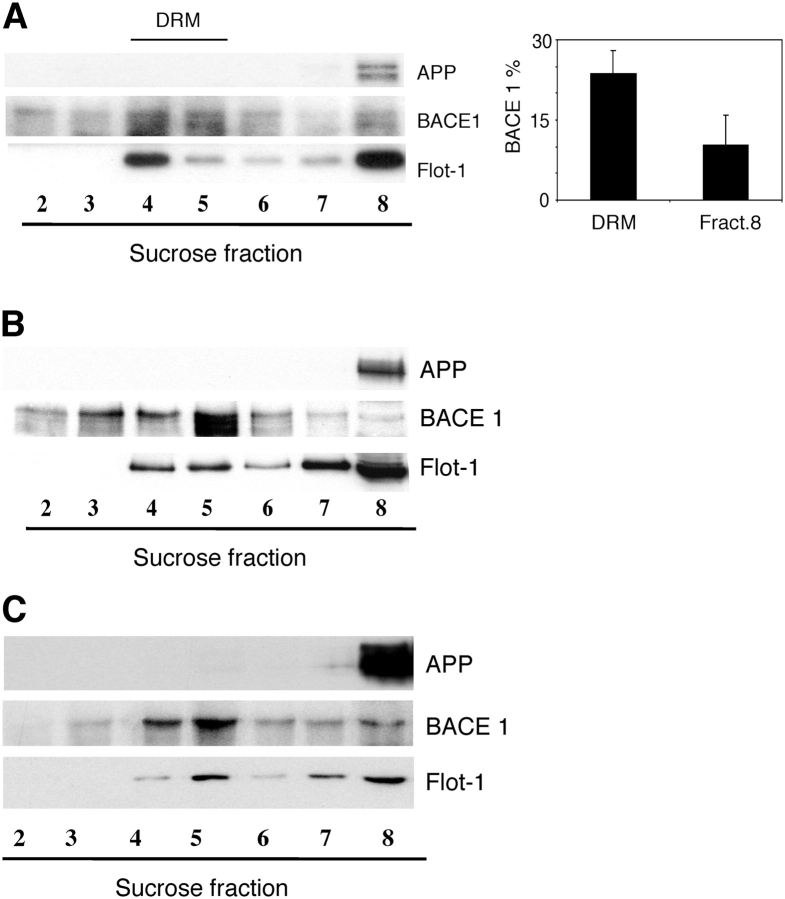
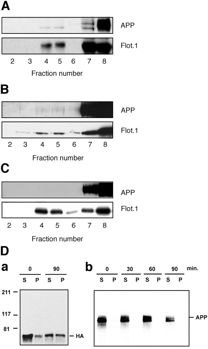
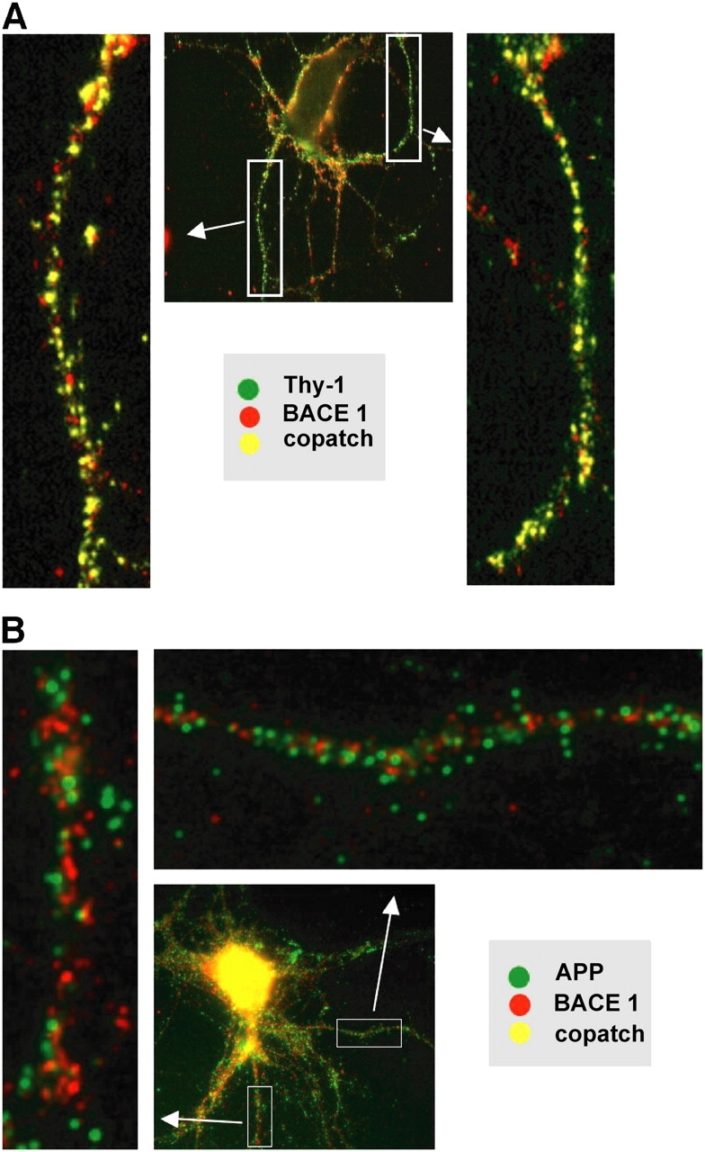
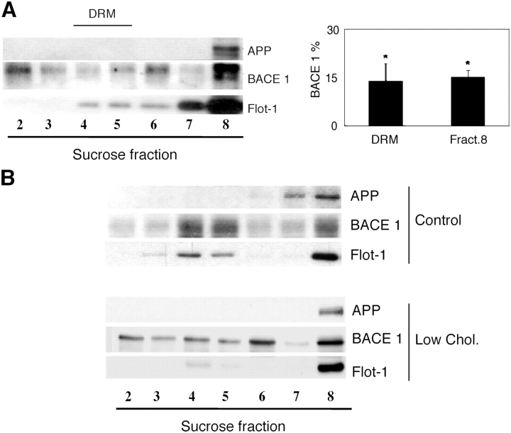
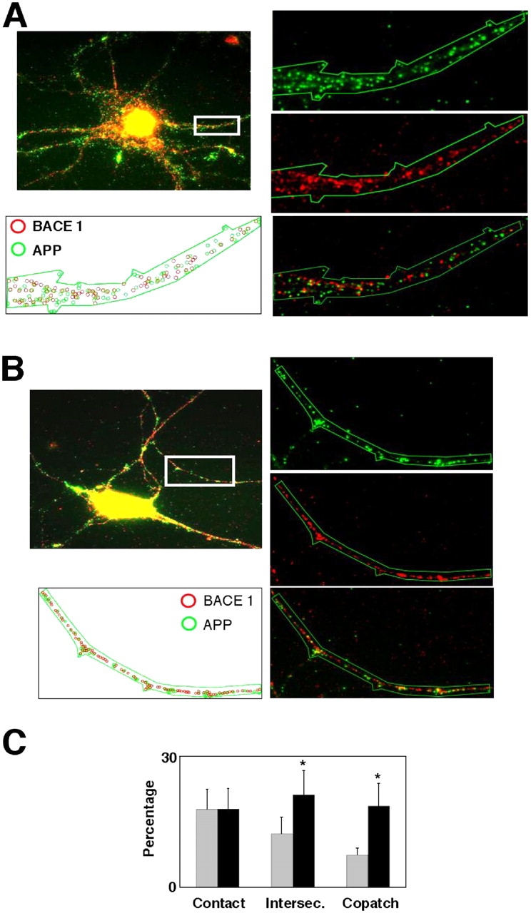
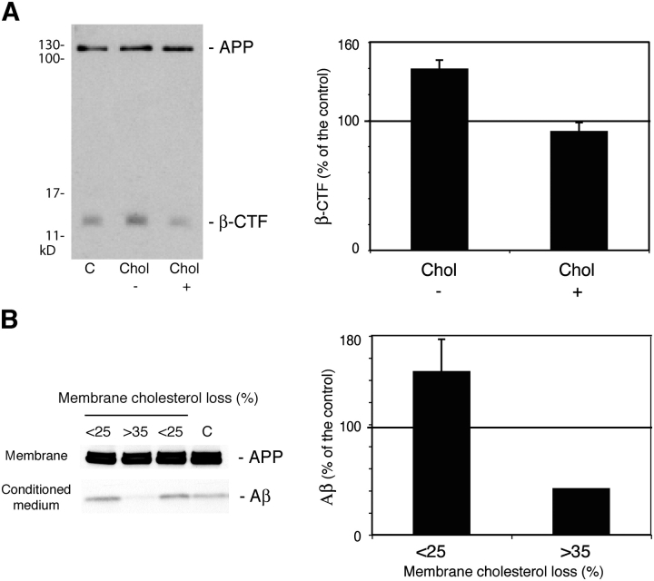
Recent experimental and clinical retrospective studies support the view that reduction of brain cholesterol protects against Alzheimer's disease (AD). However, genetic and pharmacological evidence indicates that low brain cholesterol leads to neurodegeneration. This apparent contradiction prompted us to analyze the role of neuronal cholesterol in amyloid peptide generation in experimental systems that closely resemble physiological and pathological situations. We show that, in the hippocampus of control human and transgenic mice, only a small pool of endogenous APP and its beta-secretase, BACE 1, are found in the same membrane environment. Much higher levels of BACE 1-APP colocalization is found in hippocampal membranes from AD patients or in rodent hippocampal neurons with a moderate reduction of membrane cholesterol. Their increased colocalization is associated with elevated production of amyloid peptide. These results suggest that loss of neuronal membrane cholesterol contributes to excessive amyloidogenesis in AD and pave the way for the identification of the cause of cholesterol loss and for the development of specific therapeutic strategies.


Figures

Figure 1.
Figure 1.
BACE 1 floats in the DRM fraction from control human and mice brain membranes, whereas APP remains in non-DRM heavy fractions. (A) Immunoblots for APP, BACE 1, and flotilin1 in a representative control human hippocampal sample after Lubrol WX extraction and sucrose gradient centrifugation. Note that BACE 1 is enriched in fractions 4 and 5 corresponding to DRMs as indicated by the presence of the DRM marker flotilin 1 (Flot-1). APP, on the contrary, is only detected in heavy fraction 8. The percentages of total BACE 1 in DRMs and fraction 8 are shown in the graph as means and SDs from 10 control human samples. (B) Immunoblots for APP, BACE 1, and flotilin 1 of hippocampal extracts from mice expressing the human APP after Lubrol WX extraction and sucrose gradient centrifugation. As for the human brain, BACE 1 is enriched in fractions 4 and 5 corresponding to DRMs as indicated by the presence of the DRM marker flotilin1 (Flot-1), whereas APP is only detected in heavy fraction 8. (C) Immunoblots for APP, BACE 1, and flotilin 1 of Golgi-endosomal–enriched brain membranes from mice expressing the human APP after Lubrol WX extraction and sucrose gradient centrifugation. Although BACE 1 floats to DRM light fractions similar to flotilin 1, APP remains in the heavy fractions of the gradient.
Figure 2.
Figure 2.
Overexpression of human APP leads to the incorporation of the protein in DRMs from rodent neuroblastoma N2A cells but not in human neuroblastoma SH-SY5Y cells or rat hippocampal neurons in primary culture. Nondifferentiated N2A cells (A), nondifferentiated SH-SY5Y cells (B) and mature primary rat hippocampal neurons (C) were infected with SFV-APP for 8 h. Cell extracts were detergent extracted at 4°C and centrifuged in sucrose gradients. DRMs were obtained in fractions 4 and 5 as indicated by the enrichment of the DRM marker flotilin1. Although a small amount of human APP (<5%) appears in DRMs from overexpressing N2A cells, no significant amount of the protein was found in the DRM fractions from SH-SY5Y or primary neurons even when the levels of APP overexpression are very high (see fractions 7 and 8 of the gradients). The absence of overexpressed APP in detergent insoluble membranes of primary hippocampal neurons was further confirmed along the biosynthetic pathway with a pulse-chase experiment after metabolic labeling (D). Mature neurons were infected either with Fowl plague virus to express the DRM marker HA (D, a) or with SFV-APP (D, b) and extracted with 20 mM CHAPS at 4°C. CHAPS-soluble material (S) and insoluble (P) was resolved in SDS-PAGE (6%) and the images obtained by autoradiography. Although HA CHAPS insolubility increases during the biosynthetic pathway (D, a) and is evident after 90-min chase, when the protein has already reached the plasma membrane, APP remains CHAPS soluble along the biosynthetic pathway and transport to the membrane (D, b).
Figure 3.
Figure 3.
BACE 1 copatches with the DRM marker Thy-1 but is largely segregated from APP in the neuronal plasma membrane. Hippocampal neurons were cultured for 10 d and pairs of membrane proteins were studied by the copatching technique (see Materials and methods). (A) BACE 1 and the DRM marker Thy-1 copatch extensively, indicating that both molecules are located in membrane DRM domains (note yellow dots in the enlarged images). (B) APP and BACE 1, in contrast, appear extensively segregated.
Figure 4.
Figure 4.
BACE 1 is displaced from DRMs and cofractionates with APP in heavier membrane fractions of low membrane cholesterol AD hippocampi and hippocampal neurons in culture. (A) Immunoblots for APP, BACE 1, and flotilin1 in representative low membrane cholesterol AD hippocampal sample after Lubrol WX extraction and sucrose gradient centrifugation. Note that BACE 1 migration is shifted to the heavy APP-containing fraction 8. DRM modification is shown by a similar shift in the flotation characteristics of the DRM marker flotilin 1 (compare with Fig. 1 A). For quantification, the amount of BACE 1 in fractions 4 and 5 of sucrose gradients (DRM fraction) and fraction 8 (heavy APP-containing fraction) was measured by densitometry. The percentage of BACE 1 in DRMs is significantly reduced to 14% (graph, * indicates P < 0.005) compared with 24% in control samples (Fig. 1 A, graph). Conversely in the APP-containing fraction 8 BACE 1 content is increased to 15% compared with 10% in control samples (Fig. 1 A, graph). Data are means and SDs from 10 low cholesterol AD samples. (B) Moderate membrane cholesterol reduction in vitro displaces BACE 1 from DRMs. Hippocampal neurons grown for 5 d in culture were treated (low chol.) or not (control) with low concentrations of mevilonin and MCD for 5 d (see Materials and methods). This treatment induced <30% reduction in membrane cholesterol. Sucrose gradient fractionations after Lubrol WX extraction and Western blotting for APP and BACE 1 show that in control neurons BACE 1 peaks in fractions 4 and 5 (DRMs), whereas in low cholesterol neurons BACE 1 is spread along the gradient, with a relative enrichment in the APP-containing fraction 8. Disruption of DRMs is shown by the almost complete absence of flotilin 1 in fractions 4 and 5 and the relative enrichment in heavy fraction 8 (compare Flot-1 lines in control and low chol. samples).
Figure 5.
Figure 5.
Moderate cholesterol reduction induces coclustering of BACE 1 and APP on the plasma membrane of cultured hippocampal neurons. BACE 1–APP colocalization was studied in 10 d in vitro hippocampal neurons using the copatching technique (see Materials and methods). (A) In control neurons BACE 1 (red clusters) and APP (green clusters) are extensively segregated to different membrane domains. (B) In contrast, low cholesterol neurons (treated as indicated in Materials and methods to lower the membrane cholesterol up to 30%) show a clear enhancement of BACE 1–APP colocalization. (C) For quantification, the degree of intersection among APP and BACE 1 clusters was considered as “copatching” (intersection >80%), “partial copatching” (intersection between 30% and 50%),“contact” (intersection between 0% and 30%), or random (no intersection; not depicted). For control cells 7% of APP clusters copatch and 12% partially copatch with BACE 1 (gray bars). These values are significantly increased to 19% and 21%, respectively (* indicates P < 0.005) in low cholesterol neurons (black bars). Data are means and SDs of three independent experiments.
Figure 6.
Figure 6.
Moderate membrane cholesterol reduction in vitro enhances APP-β-cleavage and Aβ production. (A) 10 d in vitro hippocampal neurons were not treated (C) or treated as indicated in Materials and methods to lower the membrane cholesterol up to 30% (Chol−). Cholesterol was added back to some of the treated cells for 15 min (Chol+) as cholesterol–MCD inclusion complexes. The amount of total APP and APP-β-CTF fragment in the different cell extracts was determined by Western blot. Densitometry of the β-CTF fragment normalized to the amount of total APP revealed a significant 39% increase in low cholesterol neurons with respect to untreated neurons. This effect was reverted by cholesterol replenishment. Thus, a 15-min treatment with the cholesterol inclusion complexes results in the production of similar amount of β-CTF fragment than control neurons (91% of control). Data shown in the graph are means and SDs from three different experiments. (B) Crude membrane pellets and conditioned media of control or low membrane cholesterol CHO-7w cells (stably expressing human APP) were submitted to PAGE-SDS in 10% Bis-Tris NuPage gels. Western blot detection was performed with an anti-APP COOH-terminal antibody to visualize holo-APP and with anti-APP mAb (WO2) to detect the amyloid peptide. Aβ peptide production was increased in cases of moderate low cholesterol an average of 47% over the control (lanes 1 and 3 in the blot and <25% bar in the graph, n = 6). Confirming previous data from other groups, extensive cholesterol loss over 35% leads to a strong decrease in Aβ production (lane 2 in the blot and >35% bar in the graph, n = 2).

Comment in

References

    1. Austen, B., G. Christodoulou, and J.E. Terry. 2002. Relation between cholesterol levels, statins and Alzheimer's disease in the human population. J. Nutr. Health Aging. 6:377–382. - PubMed
    1. Burns, M., and K. Duff. 2002. Cholesterol in Alzheimer's disease and tauopathy. Ann. N. Y. Acad. Sci. 977:367–375. - PubMed
    1. Corder, E.H., A.M. Saunders, W.J. Strittmatter, D.E. Schmechel, P.C. Gaskell, G.W. Small, A.D. Roses, J.L. Haines, and M.A. Pericak-Vance. 1993. Gene dose of apolipoprotein E type 4 allele and the risk of Alzheimer's disease in late onset families. Science. 261:921–923. - PubMed
    1. Cordy, J.M., I. Hussain, C. Dingwall, N.M. Hooper, and A.J. Turner. 2003. Exclusively targeting beta-secretase to lipid rafts by GPI-anchor addition up-regulates beta-site processing of the amyloid precursor protein. Proc. Natl. Acad. Sci. USA. 100:11735–11740. - PMC - PubMed
    1. Cucchiara, B., and S.E. Kasner. 2001. Use of statins in CNS disorders. J. Neurol. Sci. 187:81–89. - PubMed

Publication types

MeSH terms