. 2004 Dec 6;167(5):973-83.
doi: 10.1083/jcb.200409077.

Ameloblastin is a cell adhesion molecule required for maintaining the differentiation state of ameloblasts

Affiliations

Ameloblastin is a cell adhesion molecule required for maintaining the differentiation state of ameloblasts

Satoshi Fukumoto et al. J Cell Biol. .

Abstract

Tooth morphogenesis results from reciprocal interactions between oral epithelium and ectomesenchyme culminating in the formation of mineralized tissues, enamel, and dentin. During this process, epithelial cells differentiate into enamel-secreting ameloblasts. Ameloblastin, an enamel matrix protein, is expressed by differentiating ameloblasts. Here, we report the creation of ameloblastin-null mice, which developed severe enamel hypoplasia. In mutant tooth, the dental epithelium differentiated into enamel-secreting ameloblasts, but the cells were detached from the matrix and subsequently lost cell polarity, resumed proliferation, and formed multicell layers. Expression of Msx2, p27, and p75 were deregulated in mutant ameloblasts, the phenotypes of which were reversed to undifferentiated epithelium. We found that recombinant ameloblastin adhered specifically to ameloblasts and inhibited cell proliferation. The mutant mice developed an odontogenic tumor of dental epithelium origin. Thus, ameloblastin is a cell adhesion molecule essential for amelogenesis, and it plays a role in maintaining the differentiation state of secretory stage ameloblasts by binding to ameloblasts and inhibiting proliferation.


Figures

Figure 1.
Figure 1.
Generation of ameloblastin-null mice. (A) Structure of the Ambn gene, targeting vector, and targeted allele after homologous recombination. The location of the fragment used as a probe in Southern blotting is shown, as well as the sizes of the XbaI fragment detected for wild-type and targeted alleles. Exons are depicted as closed boxes. The PGK-neo and PGK-TK cassettes are depicted as open and oblique boxes. (B) Southern blot analysis of 6-wk-old mouse genomic DNA. Genomic DNA isolated from tails was digested with XbaI and hybridized with the probe containing exons 8–10. The wild-type and mutant alleles were detected as 4.5- and 2.3-kb fragments, respectively. (C) RT-PCR analysis of Ambn mRNA expression in wild-type, heterozygous, and homozygous mice. 2 μg of total RNA isolated from P3 mouse molars were reverse transcribed and amplified with primers for Ambn and GAPDH. (D) Western blot analysis of ameloblastin in wild-type, heterozygous, and homozygous mice. Two P3 maxillary first molars were dissected and lysed with 100 μl of lysis buffer, and 20 μl were separated by SDS-PAGE and immunoblotted with polyclonal anti-ameloblastin antibodies. (E) Incisors of 8-wk-old heterozygous and homozygous mice.
Figure 2.
Figure 2.
Defects in enamel formation of ameloblastin-null mice. (A) Faxitron analysis of the craniofacial region of 8-wk-old heterozygote and homozygote mice. Boxed areas were enlarged in B, 1–3. (B) High magnification of incisor tips (1), bottom incisors and alveolar bone space (2), and bottom molars (3). Round incisor tips, wider incisor-alveolar bone space, and a flat occlusal plane are observed in mutant mice. Arrows indicate incisor tips, incisor-alveolar bone space, and occlusal planes. inc, incisor; alv, alveolar bone. (C) CT-scan analysis of top first molar of heterozygous and homozygous mice. E, enamel; D, dentin; P, pulp. (D) SEM analysis of incisor. E, enamel; D, dentin; p, pulp, dE, defective enamel.
Figure 3.
Figure 3.
Histology of molars and incisors and characterization of multilayered cells. (A) Hematoxylin-eosin staining of top first molars of E18, P1, P3, and P7 heterozygous (top) and homozygous (bottom) mice. Loss of cell polarity and detachment from the matrix were observed in P3. P7 molar ameloblast cells accumulate and fail to form columnar shaped single cell layers. (B) Incisors in 6-wk-old heterozygous (1) and homozygous (2) mice. High magnifications of the presecretory and secretory stages (3) and the early maturation stage (4) of mutant incisors. Ambn / ameloblasts form multiple layers containing abnormal calcified structures (arrows). Connective tissue (CT) was migrated into the detached space between ameloblasts and the matrix surface. Am, ameloblasts; D, dentin: EO, enamel organ; Od, odontoblasts; PS, presecretory stage; PD, predentin; P, pulp; E, enamel layer; arrows, calcified structures, dE; defective enamel. (C) Immunostaining of P7 normal (top) and mutant molars (bottom) with anti-amelogenin, enamelin, and tuftelin antibodies and Cy-3-conjugated secondary antibody. DAPI staining was used for nuclear localization. Dashed lines show the border between ameloblasts and the stratum intermedium interface (top) and margin of multilayered cells (bottom). Arrows indicate abnormal matrix accumulating enamel matrix proteins. (D) PCNA immunostaining of ameloblasts (right) and nuclear staining with Hoechst dye (left). am, ameloblast; si, stratum intermedium; en, enamel; sr, stellate reticulum. Bars, 25 μm.
Figure 4.
Figure 4.
Expression of Msx2, p27, and p75 in molar and incisor ameloblasts. P3 and P7 molars and 6-wk-old incisors were fixed and decalcified. Paraffin sections were stained with DAPI for nuclear localization and immunostained with antibodies for Msx2, p27, and p75. (A) P3 and P7 molars of Ambn+/− and (EB) Ambn−/− mice. Msx2 was expressed in ameloblasts of both P3 and P7 Ambn−/− tooth, whereas it was expressed in P3 but not P7 Ambn+/− ameloblasts. p27 was expressed in P7 Ambn+/− ameloblasts but not P7 Ambn−/− ameloblasts, but p75 was expressed in P7 Ambn−/− ameloblasts but not in P7 Ambn+/− ameloblasts. sr, stellate reticulum; si, stratum intermedium; ab, ameloblast.
Figure 5.
Figure 5.
Expression of tooth markers in mutant mice. (A) mRNA expression in P3 mandibular first molars by RT-PCR. (B) Immunostaining of tooth marker proteins, ameloblastin, amelogenin, tuftelin, enamelin, and dentin sialoprotein (DSP) in P3 molars. There is no ameloblastin staining in mutant molars. (C) Silver staining of total lysates from P3 mandibular molars from two different heterozygous and mutant mice separated by SDS-PAGE. Arrows indicate the amelogenin protein and their cleaved derivatives. (D) Western blot analysis of amelogenin in P3 mandibular first molars.
Figure 6.
Figure 6.
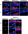
Immunostaining of basement membrane and integrin in incisors. 6-wk-old mouse maxillas were fixed and decalcified. Paraffin sections of incisors of heterozygous and mutant mice were immunostained with antibodies to amelogenin, collagen IV, laminin 1, integrin α6, and laminin γ2 and visualized with Cy-3-conjugated secondary antibody. Polyclonal antibodies to laminin 1 react with α1, β1, and γ1. Laminin α1 is not present and the tooth basement membrane contains laminin 10 (α5, β1, and γ1). am, ameloblast; si, stratum intermedium; od, odontoblast; en, enamel; de, dentin.
Figure 7.
Figure 7.
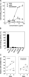
Ameloblastin binds dental epithelium and inhibits proliferation of Ambn / ameloblasts. (A) Adhesion of dental epithelial cells isolated from P3 molars to dishes coated with various amounts of recombinant ameloblastin. pEF6 is a control protein containing only the His-tagged portion purified from mock-transfected cells (pEF6, square). (B) Cell type–specific adhesion to recombinant ameloblastin. Various types of cells were plated on dishes coated with ameloblastin (10 μg/plate), and bound cells were counted. DE, dental epithelial cells. (C) Inhibition of ameloblast proliferation by maeloblastin. Dental epithelial cells from normal and mutant P7 molars were incubated on plates without (left) or with recombinant ameloblastin (right; 10 μg/plate). Plating cell numbers at day 0 are set as 100%. Proliferation of cells from P7 mutant mice are significantly inhibited by ameloblastin. Data are expressed as mean of triplicate results.
Figure 8.
Figure 8.
Tumor formation in mutant mice. (A) Tumor appearance in 36-wk-old homozygous ameloblastin-null mouse. Tumors were observed only on the right side of the maxillar of mutant mice. Right panel shows micro X-ray radiographs by Faxitron. Arrow and arrowheads indicate tumor. (B) Frequency of tumor formation with age. The earliest tumor was observed at 26 wk old. Only one heterozygous mouse developed a tumor. (C) Hematoxylin-eosin staining of tumor lesion of 36-wk-old mutant moue shown in A. Dotted lines indicate tumor lesions and the boxed area is enlarged at the bottom panel. (D) Immunostaining of tumor lesions. Top panel shows immunostaining of focal area and bottom panel shows surrounding cells. The focal area is stained with antibodies to enamel matrix proteins except ameloblastin, indicating accumulation of enamel matrix proteins. Cells surrounding focal areas are immunopositive for enamel matrix proteins, suggesting ameloblast origin. AMBN, ameloblastin; AMEL, amelogenin; ENAM, enamelin; int α6, integrin α6. (E) RT-PCR analysis of RNA from tumor lesions. RNA samples for lanes +/− and −/− were from dental epithelium of P7 molar of homozygous and heterozygous mice, respectively. RNA of lanes T1 and T2 was from tumor lesions of 39- and 51-wk-old homozygous mice. RNA of lane T3 was from a 51-wk-old heterozygous mouse. Note: heterozygous mouse did not express ameloblastin mRNA in the tumor but expressed mRNA for other enamel matrix proteins similar to the homozygous mice.

References

    1. Abiko, Y., M. Murata, Y. Ito, T. Taira, M. Nishimura, M. Arisue, T. Inoue, M. Shimono, Y. Kuboki, and T. Kaku. 2001. Immunohistochemical localization of amelogenin in human odontogenic tumors, using a polyclonal antibody against bovine amelogenin. Med. Electron Microsc. 34:185–189. - PubMed
    1. Adams, J.C., and F.M. Watt. 1993. Regulation of development and differentiation by the extracellular matrix. Development. 117:1183–1198. - PubMed
    1. Bloch-Zupan, A., T. Leveillard, P. Gorry, J.L. Fausser, and J.V. Ruch. 1998. Expression of p21(WAF1/CIP1) during mouse odontogenesis. Eur. J. Oral Sci. 106(Suppl 1):104–111. - PubMed
    1. Bosshardt, D.D., and A. Nanci. 1998. Immunolocalization of epithelial and mesenchymal matrix constituents in association with inner enamel epithelial cells. J. Histochem. Cytochem. 46:135–142. - PubMed
    1. Bouwman, P., H. Gollner, H.P. Elsasser, G. Eckhoff, A. Karis, F. Grosveld, S. Philipsen, and G. Suske. 2000. Transcription factor Sp3 is essential for post-natal survival and late tooth development. EMBO J. 19:655–661. - PMC - PubMed

Publication types

MeSH terms

Grants and funding