. 2004 Dec 14;101(50):17345-50.
doi: 10.1073/pnas.0408153101. Epub 2004 Dec 6.

In silico study of amyloid beta-protein folding and oligomerization

Affiliations

In silico study of amyloid beta-protein folding and oligomerization

B Urbanc et al. Proc Natl Acad Sci U S A. .

Abstract

Experimental findings suggest that oligomeric forms of the amyloid beta protein (Abeta) play a critical role in Alzheimer's disease. Thus, elucidating their structure and the mechanisms of their formation is critical for developing therapeutic agents. We use discrete molecular dynamics simulations and a four-bead protein model to study oligomerization of two predominant alloforms, Abeta40 and Abeta42, at the atomic level. The four-bead model incorporates backbone hydrogen-bond interactions and amino acid-specific interactions mediated through hydrophobic and hydrophilic elements of the side chains. During the simulations we observe monomer folding and aggregation of monomers into oligomers of variable sizes. Abeta40 forms significantly more dimers than Abeta42, whereas pentamers are significantly more abundant in Abeta42 relative to Abeta40. Structure analysis reveals a turn centered at Gly-37-Gly-38 that is present in a folded Abeta42 monomer but not in a folded Abeta40 monomer and is associated with the first contacts that form during monomer folding. Our results suggest that this turn plays an important role in Abeta42 pentamer formation. Abeta pentamers have a globular structure comprising hydrophobic residues within the pentamer's core and hydrophilic N-terminal residues at the surface of the pentamer. The N termini of Abeta40 pentamers are more spatially restricted than Abeta42 pentamers. Abeta40 pentamers form a beta-strand structure involving Ala-2-Phe-4, which is absent in Abeta42 pentamers. These structural differences imply a different degree of hydrophobic core exposure between pentamers of the two alloforms, with the hydrophobic core of the Abeta42 pentamer being more exposed and thus more prone to form larger oligomers.


Figures

Fig. 1.
Fig. 1.
Oligomer size distributions for Aβ40 and Aβ42, showing the occurrence probability [%] of monomers and oligomers. Standard errors of average probabilities are indicated by vertical bars. (Inset) The potential energy per peptide of Aβ40, E40 (black curve), and Aβ42, E42 (red curve), each averaged over eight trajectories. Potential energies of individual trajectories are shown as black (Aβ40) and red (Aβ42) dots. The difference between the average potential energies (E42E40) is depicted by a green curve.
Fig. 2.
Fig. 2.
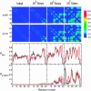
Time evolution of contacts and secondary structure elements during monomer folding from high-temperature, zero-potential-energy initial conformations. Columns correspond to states at the start of the simulation (initial conformations) and after 103, 104, and 105 simulation steps. (Upper) The contact maps for Aβ40 and Aβ42 are averages of >150 monomer conformations each. (Asp-1, Asp-1) is at the upper-left corner of the contact maps and (Val-40, Val-40) for Aβ40 or (Ala-42, Ala-42) for Aβ42 is at the lower-right corner. The strength of the contact is color-coded following the rainbow scheme: from blue (no contact), through green, yellow, and orange, to red (strongest contact), as shown on the bar with the scale on the right. (Lower) Time evolution of the turn propensity Pturn and the β-strand propensity Pβ-strand is presented. The black curves correspond to Aβ40, and the red ones correspond to Aβ42. Error bars indicate the SEM values.
Fig. 3.
Fig. 3.
Intramolecular contacts in pentamers. (a and b) Average contact maps of intramolecular contacts within a pentamer conformation. The averages are calculated by using individual contact maps of 11 Aβ40 and 34 Aβ42 pentamers using three fixed simulation steps (9 million, 9.5 million, and 10 million simulation steps). a and c correspond to Aβ40, and b and d correspond to Aβ42. (Asp-1, Asp-1) is at the upper-left corner of the contact maps and (Val-40, Val-40) for Aβ40 or (Ala-42, Ala-42) for Aβ42 is at the lower-right corner. The strength of the contacts is color-coded as described for Fig. 2. The centers of the TRA and TRB regions are marked by black and red squares, respectively. (c and d) Diagrams of the most important intramolecular contacts within a pentamer calculated by using the contact map data from a and b. Solid lines represent stronger contacts, and dashed lines represent weaker contacts. Color code: magenta, contacts inside the MHR; green, contacts between the MHR and the CHC or the CTR; blue, contacts between the CHC and the CTR; red, contacts inside the CTR.
Fig. 4.
Fig. 4.
Structural features of pentamers. Typical Aβ40 (a) and Aβ42 (b) pentamers. The secondary structure of pentamers is shown as a silver tube (random coil-like structure), light-blue tube (turn), and yellow ribbon (β-strand). Red spheres in both a and b represent the N-terminal Asp-1. (a) The C-terminal amino acids Val-39 and Val-40 are shown in purple. (b) The C-terminal amino acid Ile-41 is shown in green, and Ala-42 is shown in blue. (c) Distribution of intramolecular distances between Asp-1 and Val-40 within Aβ40 (black) and Aβ42 (red) pentamers. The distributions are significantly different (P < 10–4, χ2 test). (d) The average distances from the center of mass of Aβ40 and Aβ42 pentamers per residue. The error bars represent SEM values. (d Inset) The atom density (number of atoms per volume unit) in dependence on the radial distance from the center of mass for pentamers of Aβ40 (black) and Aβ42 (red).

References

    1. Walsh, D. M., Lomakin, A., Benedek, G. B., Condron, M. M. & Teplow, B. D. (1997) J. Biol. Chem. 272, 22364–22372. - PubMed
    1. Hartley, D. M., Walsh, D. M., Ye, C. P., Diehl, T., Vasquez, S., Vassilev, P. M., Teplow, D. B. & Selkoe, D. J. (1999) J. Neurosci. 19, 8876–8884. - PMC - PubMed
    1. Harper, J. D., Lieber, C. M. & Lansbury, P. T., Jr. (1997) Chem. Biol. 4, 951–959. - PubMed
    1. Lambert, M.P., Barlow, A.K., Chromy, B. A., Edwards, C., Freed, R., Liosatos, M., Morgan, T. E., Rozovsky, I., Trommer, B., Viola, K. L., et al. (1998) Proc. Natl. Acad. Sci. USA 95, 6448–6453. - PMC - PubMed
    1. Hsia, A. Y., Masliah, E., McConlogue, L., Yu, G. Q., Tatsuno, G., Hu, K., Kholodenko, D., Malenka, R. C., Nicoll, R. A. & Mucke, L. (1999) Proc. Natl. Acad. Sci. USA 96, 3228–3233. - PMC - PubMed

Publication types

Substances