

Amoebal coculture of "Mycobacterium massiliense" sp. nov. from the sputum of a patient with hemoptoic pneumonia
- PMID: 15583272
- PMCID: PMC535245
- DOI: 10.1128/JCM.42.12.5493-5501.2004
Amoebal coculture of "Mycobacterium massiliense" sp. nov. from the sputum of a patient with hemoptoic pneumonia
Expression of concern in
-
Expression of Concern for Adékambi et al., 'Amoebal Coculture of "Mycobacterium massiliense" sp. nov. from the Sputum of a Patient with Hemoptoic Pneumonia'.J Clin Microbiol. 2023 Nov 21;61(11):e0085423. doi: 10.1128/jcm.00854-23. Epub 2023 Oct 31. J Clin Microbiol. 2023. PMID: 37905796 Free PMC article. No abstract available.
Abstract
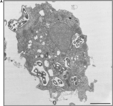
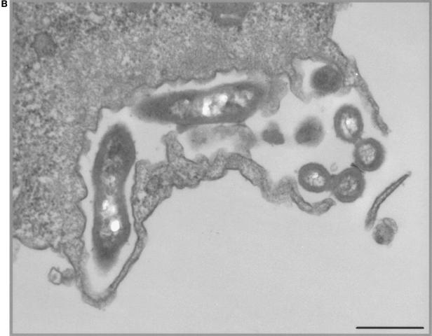
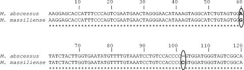
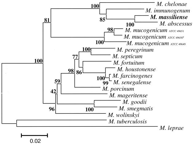
A nonphotochromogenic, rapidly growing Mycobacterium strain was isolated in pure culture from the sputum and the bronchoalveolar fluid of a patient with hemoptoic pneumonia by using axenic media and an amoebal coculture system. Both isolates grew in less than 7 days at 24 to 37 degrees C with an optimal growth temperature of 30 degrees C. The isolates exhibited biochemical and antimicrobial susceptibility profiles overlapping those of Mycobacterium abscessus, Mycobacterium chelonae, and Mycobacterium immunogenum, indicating that they belonged to M. chelonae-M. abscessus group. They differed from M. abscessus in beta-galactosidase, beta-N-acetyl-beta-glucosaminidase, and beta-glucuronidase activities and by the lack of nitrate reductase and indole production activities, as well as in their in vitro susceptibilities to minocycline and doxycycline. These isolates and M. abscessus differed from M. chelonae and M. immunogenum by exhibiting gelatinase and tryptophane desaminase activities. Their 16S rRNA genes had complete sequence identity with that of M. abscessus and >99.6% similarity with those of M. chelonae and M. immunogenum. Further molecular investigations showed that partial hsp65 and sodA gene sequences differed from that of M. abscessus by five and three positions over 441 bp, respectively. Partial rpoB and recA gene sequence analyses showed 96 and 98% similarities with M. abscessus, respectively. Similarly, 16S-23S rRNA internal transcribed spacer sequence of the isolates differed from that of M. abscessus by a A-->G substitution at position 60 and a C insertion at position 102. Phenotypic and genotypic features of these two isolates indicated that they were representative of a new mycobacterial species within the M. chelonae-M. abscessus group. Phylogenetic analysis suggested that these isolates were perhaps recently derived from M. abscessus. We propose the name of "Mycobacterium massiliense" for this new species. The type strain has been deposited in the Collection Institut Pasteur as CIP 108297(T) and in Culture Collection of the University of Goteborg, Goteborg, Sweden, as CCUG 48898(T).
Figures
References
-
- Adékambi, T., and M. Drancourt. Dissection of phylogenic relationships among nineteen rapidly growing Mycobacterium species by 16S rRNA, hsp65, sodA, recA, and rpoB gene sequencing. Int. J. Syst. Evol. Microbiol., in press. - PubMed
Publication types
MeSH terms
Substances
Associated data
- Actions
- Actions
- Actions
- Actions
- Actions
- Actions
LinkOut - more resources
Full Text Sources
Other Literature Sources
Medical
Molecular Biology Databases
Research Materials
