Comparative Study
. 2005 Apr;14(3):277-86.
doi: 10.1007/s00586-004-0782-5. Epub 2004 Oct 2.

Morphological changes of the ligamentum flavum as a cause of nerve root compression

Affiliations
Comparative Study

Morphological changes of the ligamentum flavum as a cause of nerve root compression

Teruaki Okuda et al. Eur Spine J. 2005 Apr.

Abstract

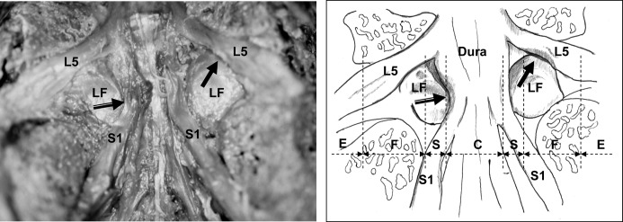
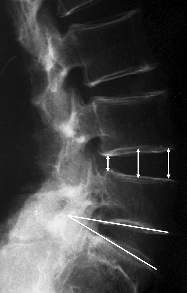
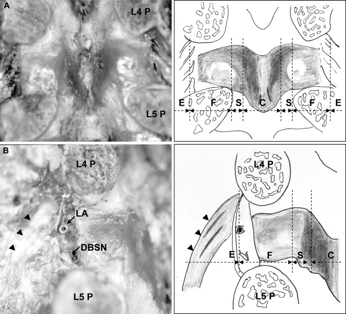
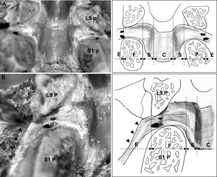
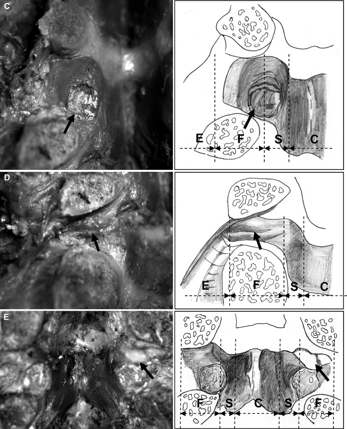
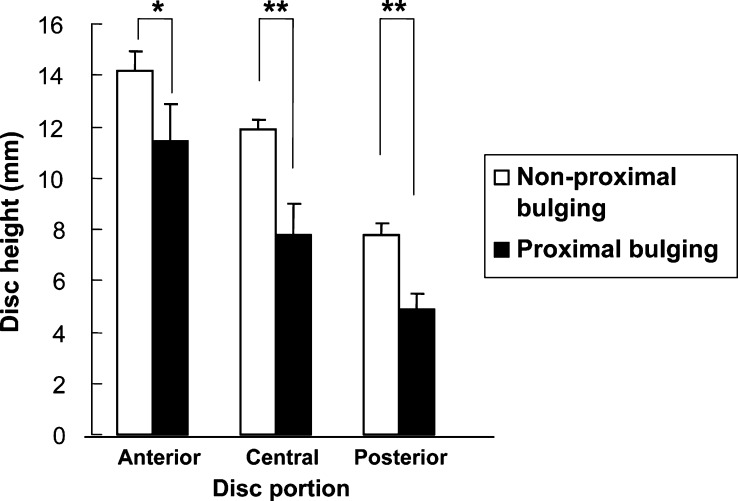
The ligamentum flavum is considered to be one of the important causes of radiculopathy in lumbar degenerative disease. Although there have been several reports anatomically examining the positional relationship between the ligamentum flavum and nerve root, there are few reports on ventral observation. The purpose of this study is to clarify the shape of the ligamentum flavum seen ventrally, and to obtain anatomic findings related to nerve root compression. The subjects were 18 adult embalmed cadavers, with an average age of 78 years at the time of death. The ventral shapes of the ligamentum flavum were observed. The relationships between the morphological change of the ligamentum flavum and nerve root compression or radiographic findings were statistically evaluated. Among the shapes of the ligamentum flavum, bulging of the ligament was most frequently observed. Proximal bulging indicates the type with the cranial portion bulging from the subarticular zone to the foraminal zone of the ligamentum flavum. In this type associated with a decrease in disc height, nerve root compression was frequently observed. Thus, we could more realistically grasp the relationship between bulging morphology of the ligamentum flavum and nerve root compression.


Figures

Fig. 1
Fig. 1
Ventral observation at the L5–S1 level showed nerve root compression by the ligamentum flavum (LF) in the subarticular zone (black arrow with white line) and foraminal zone (solid black arrow). The dotted lines of the schema show the zone classification [13, 15]. (Dura dura mater, L5 L5 nerve root, S1 S1 nerve root, E extraforaminal zone, F foraminal zone, S subarticular zone, C central canal)
Fig. 2
Fig. 2
As radiographical measurements, the angle made by the endplate in each interbody and disc height were measured (anterior, central, posterior)
Fig. 3
Fig. 3
The ligamentum flavum at the L4–L5 level is shown. A Inside the spinal canal, both the cranial and caudal borders of the ligament dilated from the center to the lateral side in a V shape. In the foraminal zone, the proximal border of the ligament attached almost horizontally and was in contact with the lateral inferior portion of the superior pedicle. The distal border of the ligament attached gradually toward the cranial side, while advancing toward the lateral side. B From the intraforaminal zone to the extraforaminal zone, the fiber bundle, starting from the superior pedicle, ran as a fan toward the transverse process, forming the transverse ligament (arrowheads). Between the fiber bundle and the ligamentum flavum, the bifurcation of the lumbar artery and vein ran in the proximal side and the dorsal branch of the spinal nerve root in the distal side. The dotted lines of the schema show the zone classification. (P pedicle, LA lumbar artery, DBSN dorsal branch of the spinal nerve root, E extraforaminal zone, F foraminal zone, S subarticular zone, C central canal)
Fig. 4
Fig. 4
The ligamentum flavum at the L5–S1 level is shown. A In the foraminal zone, the cranial border of the ligament attached almost horizontally and the lateral portion was in contact with almost the center of the L5 pedicle. The distal border of the ligament attached gradually to the cranial side, while advancing toward the lateral side, similarly to other levels. B At the intraforaminal zone, the bifurcation of the lumbar artery and vein and the dorsal branch of the spinal nerve root passed. Also, the ligament component extending from the ligamentum flavum attached to the lateral surface of the S1 pedicle, composing the lateral side wall of the neural tube at the extraforaminal zone (arrowheads). The dotted lines of the schema show the zone classification. (P pedicle, E extraforaminal zone, F foraminal zone, S subarticular zone, C central canal)
Fig. 5
Fig. 5
With the morphological changes of the ligamentum flavum, A in the whole bulging type, the whole ligament bulged from the central canal to the intraforaminal zone centering on the subarticular zone (20 sites). B With the proximal bulging type, the cranial portion from the subarticular zone to the foraminal zone bulged like a boomerang (22 sites); C Erosion due to contact with disc (15 sites); D Fold (six sites); E Bone exposure of the tip of the superior articular process (three sites) was observed. The dotted lines of the schema show the zone classification. ( E extraforaminal zone, F foraminal zone, S subarticular zone, C central canal)
Fig. 6
Fig. 6
In the proximal bulging type, disc height was significantly decreased in the anterior, central and posterior portions. Data are mean ± SEM. (*p<0.05, **p<0.01)

References

    1. Abdullah J Neurosurg. 1974;41:229. - PubMed
    1. Avrahami Spine. 1990;15:21. - PubMed
    1. Boden J Bone Joint Surg Am. 1990;72:403. - PubMed
    1. Brown J Bone Joint Surg. 1938;20:325.
    1. Dunlop J Bone Joint Surg Br. 1984;66:706. - PubMed

Publication types

LinkOut - more resources