. 2004 Dec 7;32(21):6425-36.
doi: 10.1093/nar/gkh979. Print 2004.

Basic helix-loop-helix transcription factor Tcfl5 interacts with the Calmegin gene promoter in mouse spermatogenesis

Affiliations

Basic helix-loop-helix transcription factor Tcfl5 interacts with the Calmegin gene promoter in mouse spermatogenesis

Michel Siep et al. Nucleic Acids Res. .

Abstract

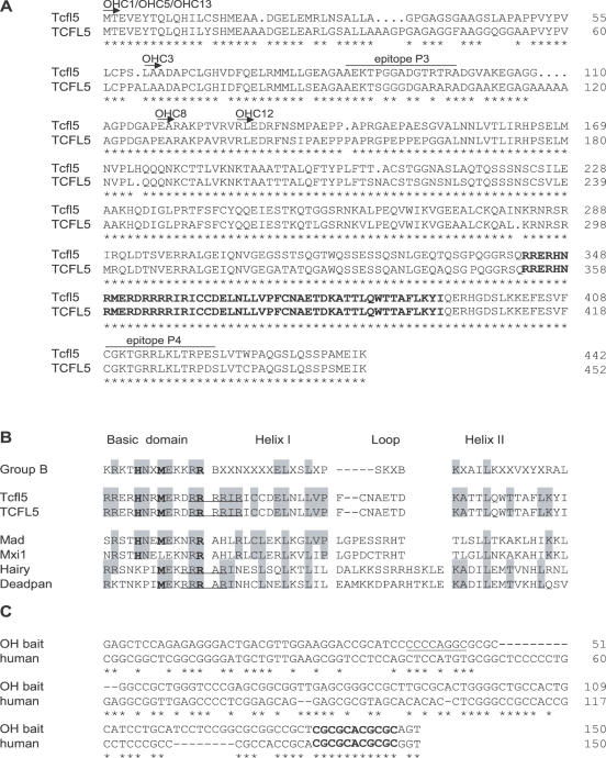
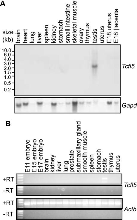
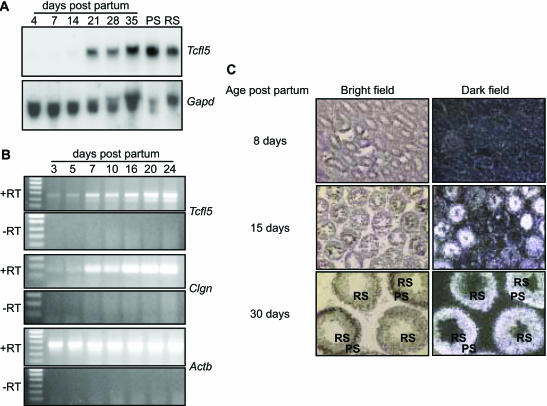
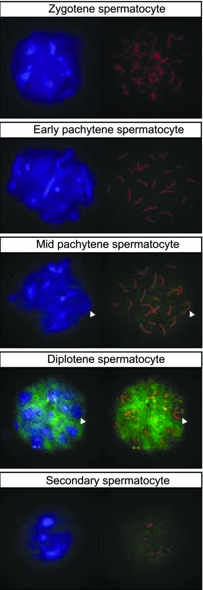
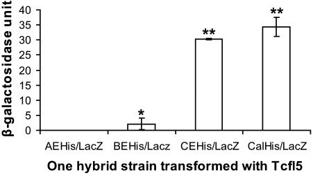
In mouse spermatogenesis, differentiating germ line cells initiate expression of specific genes at subsequent developmental steps. The Calmegin (Clgn) gene is first expressed in meiotic prophase, in primary spermatocytes, and encodes a protein that acts as a chaperone. To identify testis-specific transcription factors that control expression of the Clgn gene in spermatogenesis, we performed a yeast one-hybrid screening with a Clgn promoter sequence as bait DNA. This screening resulted in the identification of mouse Tcfl5 as a candidate Clgn promoter-binding protein. Tcfl5 is a member of the basic helix-loop-helix (bHLH) family of transcription factors, and mouse Tcfl5 shows 83% amino acid sequence identity with human TCFL5. Gel-shift and yeast one-hybrid experiments showed that Tcfl5 interacts with a non-canonical CACGCG site that is present in the Clgn promoter. By using northern blot, RT-PCR and in situ hybridization, mouse Tcfl5 mRNA was detected only in testis, with the highest expression level in primary spermatocytes and round spermatids. The highest level of Tcfl5 protein was found in primary spermatocytes at the diplotene stage of meiotic prophase, where the protein colocalizes with transcriptionally active chromatin.


Figures

Figure 1
Figure 1
Alignment of bHLH proteins Tcfl5 and TCFL5. (A) Alignment of mouse Tcfl5 (accession no. AY234363) and human TCFL5 (accession no. AB012124). Arrows and clone names indicate the part of the protein that is encoded by the six positive one-hybrid clones. Both proteins show high-sequence conservation (83% identity), the bHLH domain (in boldface) at the C-terminus is 100% conserved. Asterisks indicate identical amino acid residues. The epitopes that were selected to generate antibodies against Tcfl5 are indicated (P3 and P4). (B) Alignment of Tcfl5, TCFL5 and other bHLH protein family members. Group B depicts the consensus bHLH amino acid sequence for this bHLH protein family (38). Amino acid residues at positions 5, 8 and 13 (in boldface) determine the family of bHLH proteins. Tcfl5 and TCFL5 follow the group B consensus at these positions. Mad and Mxi1 are the closest family members of Tcfl5 and TCFL5. Hairy and deadpan show similarity in arginine (R) content (underlined) with Tcfl5. (C) Alignment of the one-hybrid bait sequence and the putative human Calmegin promoter. The AP-2 site that is present in the mouse calmegin promoter is underlined and the 11 bp conserved element containing the non-canonical E-box CACGCG is indicated in boldface. Asterisks indicate identical nucleotides.
Figure 2
Figure 2
Testis-specific expression of Tcfl5 mRNA. (A) Northern-blot analysis of poly(A)+ mRNA from 15 mouse tissues shows a specific signal in testis, while no signal was detected in any of the other tissues. Loading of the different lanes was checked by hybridization with a mouse glyceraldehyde-3-phosphate dehydrogenase (Gapd) cDNA probe. (B) RT–PCR analysis on total RNA panel from 15 tissues. The 455 bp Tcfl5 PCR product is only detected in testis. Primers specific for the ubiquitously expressed beta actin gene (Actb) were used to show that equal amounts of material were used. The 448 bp Actb PCR product is not detected in the control experiment without reverse transcriptase (−RT), ruling out contamination with genomic DNA.
Figure 3
Figure 3
Expression of Tcfl5 mRNA during spermatogenesis. (A) Northern-blot analysis of total RNA isolated from testis of mice of different ages (4-, 7-, 14-, 21-, 28- and 35-day-old mice) and purified spermatocytes (PS) and spermatids (RS). Only a faint signal is detected in day 4, 7 and 14 testis and the signal increases significantly between day 14 and day 21. Spermatocytes show a slightly higher expression level of Tcfl5 mRNA than spermatids. Loading of the different lanes was checked by hybridization with a Gapd cDNA. (B) Using RT–PCR, testicular RNAs from mice aged 3–24 days were analysed for Tcfl5 and Clgn expression. Both, the 455 bp Tcfl5 and the 166 bp Clgn PCR product intensified in later stages of spermatogenesis. Primers specific for the ubiquitously expressed Actb gene were used to show that equal amounts of material were used. The 448 bp Actb PCR product is not detected in the control experiment without reverse transcriptase (−RT), ruling out contamination with genomic DNA. (C) In situ hybridization on postnatal mouse testes from 8-, 15- and 30-day-old mice with an antisense Tcfl5 RNA probe shows that Tcfl5 mRNA expression starts in the spermatocytes. Little Tcfl5 mRNA signal is detected in the 8-day-old mouse testis. In the 15-day-old mouse testis, many tubule cross-sections show Tcfl5 mRNA signal, while other tubules show no signal. The cells that show the Tcfl5 mRNA signal are spermatocytes. Expression of Tcfl5 mRNA continues in spermatids as is shown in the 30-day-old mouse testis, where both spermatocytes and spermatids show the Tcfl5 mRNA signal. PS, pachytene spermatocytes; RS, round spermatids. With the sense probe no signal is visible, indicating the specificity of the antisense probe (data not shown).
Figure 4
Figure 4
Tcfl5 protein expression in mouse testis. (A) Left panel: western-blot analysis using P3-αTcfl5 antibody shows that a specific 55 kDa protein is recognized in total testis and spermatocyte protein extracts. PS, pachytene spermatocytes; RS, round spermatids; and Te, total testis. Right panel: competition experiment with immunizing peptide P3. Western blots of spermatocyte protein extracts were incubated with P3-αTcfl5 in the absence (−) or presence (+) of excess amount of peptide P3. (B) Immunohistochemical staining of Tcfl5 protein on testis sections. The upper panel shows the results obtained with P3-αTcfl5. The middle panel represents the results of the control competition experiment using the immunizing peptide P3. Tcfl5 is detected mainly in mid-to-late pachytene spermatocytes (ps), and this signal is lost in the competition experiment. The lower panel shows larger magnifications of P3-αTcfl5 immunostaining (left) and the control competition experiment (right). rs, round spermatids; es, elongating spermatids; m, metaphase spermatocytes; and roman numerals indicate the stage of the tubule cross-section. Size bar indicates 100 μm in the upper and middle panel, and 20 μm in the lower panel.
Figure 5
Figure 5
Tcfl5 protein expression in nuclei of late spermatocytes. Using immunocytochemistry on spread nuclei of spermatocytes Tcfl5 protein is detected from mid-pachytene spermatocytes to secondary spermatocytes. Specificity of the antibody for Tcfl5 was verified by peptide competition assay (data not shown). Blue: DAPI DNA staining; green: P3-αTcfl5; red: αSycp3; and arrowheads indicate the XY body.
Figure 6
Figure 6
Gel-mobility shift assay of Tcfl5 binding to bHLH E-box oligomers. (A) Reactions in lanes 1, 3 and 5 contain the in vitro translation product of the vector pGBKT7 (control), and the reactions in lanes 2, 4 and 6 contain the in vitro translated Tcfl5 protein. The Tcfl5 protein forms a complex with both the group B bHLH canonical E-box oligomer (BE) and the hairy-related bHLH non-canonical E-box oligomer (CE), whereas no retarded band is obtained with the group A bHLH canonical E-box oligomer (AE). (B) The retarded band obtained with Tcfl5 for oligomers BE and CE is supershifted by the addition of P4-αTcfl5, in lanes 3 and 6. Reactions in lanes 1 and 4 contain the in vitro translation product of the vector pGBKT7 (control).
Figure 7
Figure 7
One-hybrid analysis of Tcfl5 binding site. Graphical representation of quantified one-hybrid interactions of Tcfl5 and the constructed promoter reporters. An aliquot of 1 U β-galactosidase is the amount that hydrolyses 1 μmol of ONPG to o-nitrophenol and d-galactose per minute per cell. The values represent the mean average of three independent measurements. Asteriks indicate a significant difference with reporter AEHis/LacZ (P < 0.01, Tuckey test). Double asteriks indicate that reporters CEHis/LacZ and CalHis/LacZ also are significantly different from BEHis/LacZ (P < 0.01, Tuckey test).

Similar articles

Cited by

References

    1. Schultz N., Hamra,F.K. and Garbers,D.L. (2003) A multitude of genes expressed solely in meiotic or postmeiotic spermatogenic cells offers a myriad of contraceptive targets. Proc. Natl Acad. Sci. USA, 100, 12201–12206. - PMC - PubMed
    1. Baarends W.A. and Grootegoed,J.A. (1999) Molecular biology of male gametogenesis. In Fauser,B.C.J.M. (ed.), Molecular Biology in Reproductive Medicine. The Parthenon Publishing Group, NY, pp. 271–295.
    1. Eddy E.M. (2002) Male germ cell gene expression. Recent Prog. Horm. Res., 57, 103–128. - PubMed
    1. Wang P.J. (2004) X chromosomes, retrogenes and their role in male reproduction. Trends Endocrinol. Metab., 15, 79–83. - PubMed
    1. Oakberg E.F. (1956) Duration of spermatogenesis in the mouse and timing of stages of the cycle of the seminiferous epithelium. Am. J. Anat., 99, 507–516. - PubMed

Publication types

MeSH terms

Associated data