

Selective growth, in vitro and in vivo, of individual T cell clones from tumor-infiltrating lymphocytes obtained from patients with melanoma
- PMID: 15585890
- PMCID: PMC2174603
- DOI: 10.4049/jimmunol.173.12.7622
Selective growth, in vitro and in vivo, of individual T cell clones from tumor-infiltrating lymphocytes obtained from patients with melanoma
Abstract
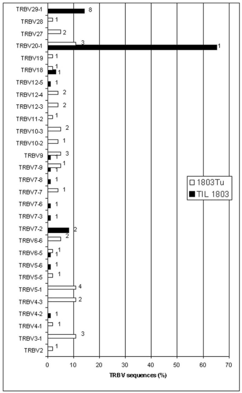
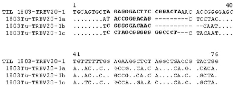
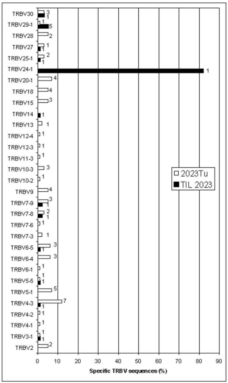
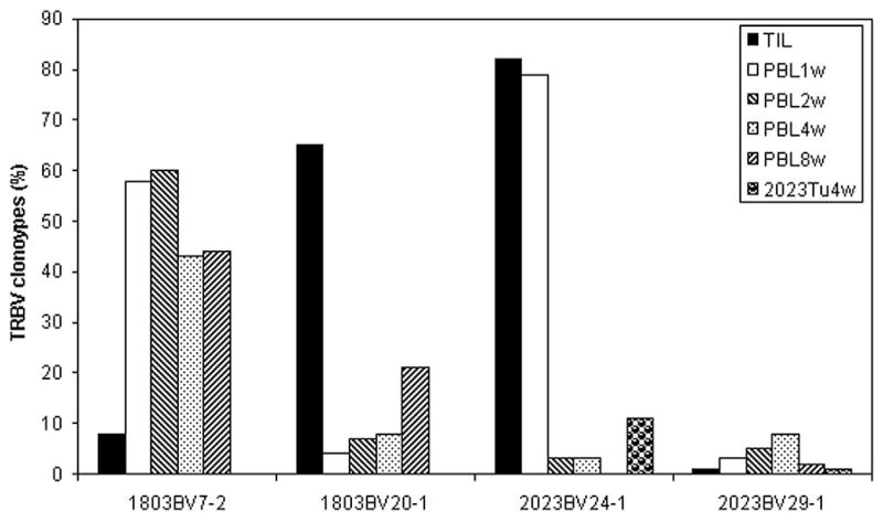
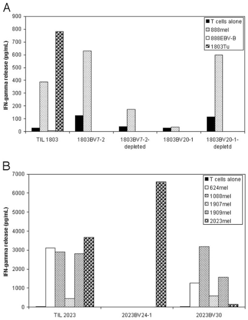
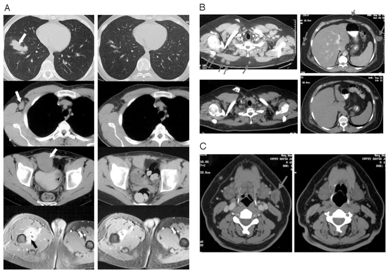
In recent clinical trials in patients with metastatic melanoma, adoptive transfer of tumor-reactive lymphocytes mediated the regression of metastatic tumor deposits. To better understand the role of individual T cell clones in mediating tumor regression, a 5' RACE technique was used to determine the distribution of TCR beta-chain V region sequences expressed in the transferred cells as well as in tumor samples and circulating lymphocytes from melanoma patients following adoptive cell transfer. We found that dominant T cell clones were present in the in vitro-expanded and transferred tumor-infiltrating lymphocyte samples and certain T cell clones including the dominant T cell clones persisted at relatively high levels in the peripheral blood of the patients that demonstrated clinical responses to adoptive immunotherapy. However, these dominant clones were either undetected or present at a very low level in the resected tumor samples used for tumor-infiltrating lymphocyte generation. These data demonstrated that there was selective growth and survival, both in vitro and in vivo, of individual T cell clones from a relatively small number of T cells in the original tumor samples. These results suggest that the persistent T cell clones played an active role in mediating tumor regression and that 5' RACE analysis may provide an important tool for the analysis of the role of individual T cell clones in mediating tumor regression. A similar analysis may also be useful for monitoring autoimmune responses.
Figures
Similar articles
-
Persistence of multiple tumor-specific T-cell clones is associated with complete tumor regression in a melanoma patient receiving adoptive cell transfer therapy.J Immunother. 2005 Jan-Feb;28(1):53-62. doi: 10.1097/00002371-200501000-00007. J Immunother. 2005. PMID: 15614045 Free PMC article.
-
T cell receptor (TCR) structure of autologous melanoma-reactive cytotoxic T lymphocyte (CTL) clones: tumor-infiltrating lymphocytes overexpress in vivo the TCR beta chain sequence used by an HLA-A2-restricted and melanocyte-lineage-specific CTL clone.J Exp Med. 1993 Oct 1;178(4):1231-46. doi: 10.1084/jem.178.4.1231. J Exp Med. 1993. PMID: 8376931 Free PMC article.
-
Cutting edge: persistence of transferred lymphocyte clonotypes correlates with cancer regression in patients receiving cell transfer therapy.J Immunol. 2004 Dec 15;173(12):7125-30. doi: 10.4049/jimmunol.173.12.7125. J Immunol. 2004. PMID: 15585832 Free PMC article. Clinical Trial.
-
Antitumor cytotoxic T-lymphocyte response in human lung carcinoma: identification of a tumor-associated antigen.Immunol Rev. 2002 Oct;188:114-21. doi: 10.1034/j.1600-065x.2002.18810.x. Immunol Rev. 2002. PMID: 12445285 Review.
-
Adoptive T-cell therapy using autologous tumor-infiltrating lymphocytes for metastatic melanoma: current status and future outlook.Cancer J. 2012 Mar-Apr;18(2):160-75. doi: 10.1097/PPO.0b013e31824d4465. Cancer J. 2012. PMID: 22453018 Free PMC article. Review.
Cited by
-
Th cells promote CTL survival and memory via acquired pMHC-I and endogenous IL-2 and CD40L signaling and by modulating apoptosis-controlling pathways.PLoS One. 2013 Jun 13;8(6):e64787. doi: 10.1371/journal.pone.0064787. Print 2013. PLoS One. 2013. PMID: 23785406 Free PMC article.
-
Adoptive transfer of autologous, HER2-specific, cytotoxic T lymphocytes for the treatment of HER2-overexpressing breast cancer.Cancer Immunol Immunother. 2008 Feb;57(2):271-80. doi: 10.1007/s00262-007-0355-7. Epub 2007 Jul 24. Cancer Immunol Immunother. 2008. PMID: 17646988 Free PMC article. Clinical Trial.
-
Advances in the development of cancer immunotherapies.Trends Immunol. 2013 Feb;34(2):90-8. doi: 10.1016/j.it.2012.08.004. Epub 2012 Sep 30. Trends Immunol. 2013. PMID: 23031830 Free PMC article. Review.
-
Toward T Cell-Mediated Control or Elimination of HIV Reservoirs: Lessons From Cancer Immunology.Front Immunol. 2019 Sep 10;10:2109. doi: 10.3389/fimmu.2019.02109. eCollection 2019. Front Immunol. 2019. PMID: 31552045 Free PMC article. Review.
-
Biased usage of T cell receptor β-chain variable region genes of Wilms' tumor gene (WT1)-specific CD8+ T cells in patients with solid tumors and healthy donors.Cancer Sci. 2012 Mar;103(3):408-14. doi: 10.1111/j.1349-7006.2011.02163.x. Epub 2012 Jan 17. Cancer Sci. 2012. PMID: 22126448 Free PMC article.
References
-
- Rosenberg SA, Packard BS, Aebersold PM, Solomon D, Topalian SL, Toy ST, Simon P, Lotze MT, Yang JC, Seipp CA, et al. Use of tumor infiltrating lymphocytes and interleukin-2 in the immunotherapy of patients with metastatic melanoma: preliminary report. N Engl J Med. 1988;319:1676. - PubMed
-
- Rosenberg SA, Yannelli JR, Yang JC, Topalian SL, Schwartzentruber DJ, Weber JS, Parkinson DR, Seipp CA, Einhorn JH, White DE. Treatment of patients with metastatic melanoma with autologous tumor-infiltrating lymphocytes and interleukin 2. J Natl Cancer Inst. 1994;86:1159. - PubMed
-
- Dudley ME, Wunderlich J, Nishimura MI, Yu D, Yang JC, Topalian SL, Schwartzentruber DJ, Hwu P, Marincola FM, Sherry R, et al. Adoptive transfer of cloned melanoma-reactive T lymphocytes for the treatment of patients with metastatic melanoma. J Immunother. 2001;24:363. - PubMed
Publication types
MeSH terms
Grants and funding
LinkOut - more resources
Full Text Sources
Other Literature Sources
Medical
