. 2005 May 1;387(Pt 3):747-54.
doi: 10.1042/BJ20041536.

Orientation and mode of lipid-binding interaction of human apolipoprotein E C-terminal domain

Affiliations

Orientation and mode of lipid-binding interaction of human apolipoprotein E C-terminal domain

Vincent Raussens et al. Biochem J. .

Abstract

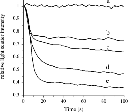
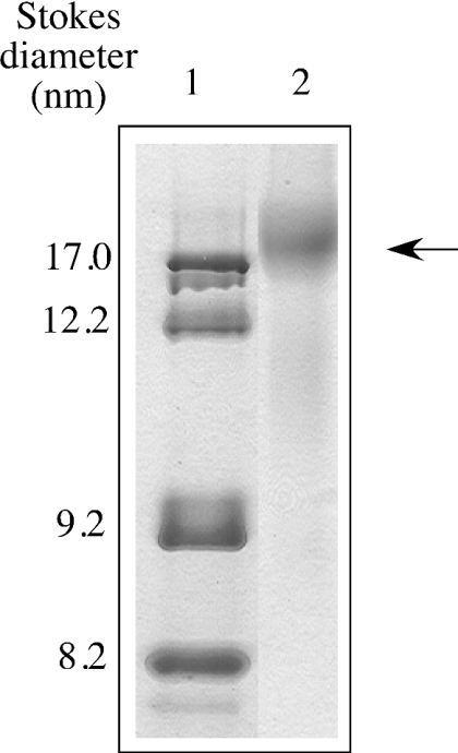
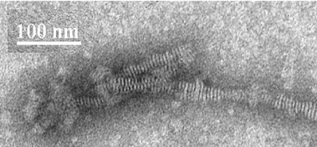
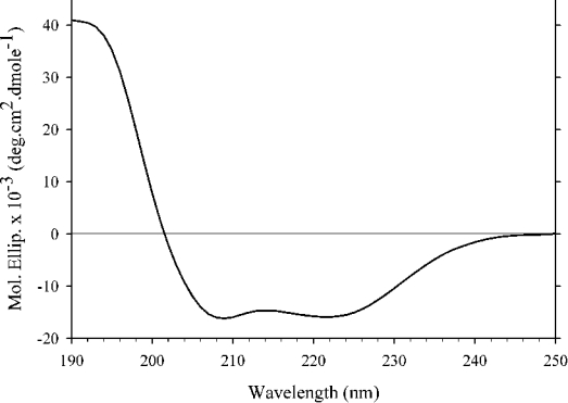
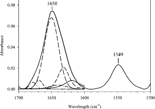
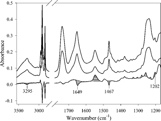
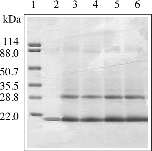
ApoE (apolipoprotein E) is an anti-atherogenic lipid transport protein that plays an integral role in lipoprotein metabolism and cholesterol homoeostasis. Lipid association educes critical functional features of apoE, mediating reduction in plasma and cellular cholesterol levels. The 10-kDa CT (C-terminal) domain of apoE facilitates helix-helix interactions in lipid-free state to promote apoE self-association and helix-lipid interactions during binding with lipoproteins, although the mode of lipid-binding interaction is not well understood. We investigated the mode of lipid-binding interaction and orientation of apoE CT domain on reconstituted lipoproteins. Isolated recombinant human apoE CT domain (residues 201-299) possesses a strong ability to interact with phospholipid vesicles, yielding lipoprotein particles with an apparent molecular mass of approximately 600 kDa, while retaining the overall alpha-helical content. Electron microscopy and non-denaturing PAGE analysis of DMPC (dimyristoylphosphatidylcholine)--apoE CT domain lipoprotein complexes revealed discoidal complexes with a diameter of approx. 17 nm. Cross-linking apoE CT domain on discoidal particles yielded dimeric species as the major product. Attenuated total reflectance Fourier transform IR spectroscopy of phospholipid-apoE CT domain complexes reveals that the helical axis is oriented perpendicular to fatty acyl chains of the phospholipid. Fluorescence quenching analysis of DMPC-apoE CT domain discoidal complexes by spin-labelled stearic acid indicated a relatively superficial location of the native tryptophan residues with respect to the plane of the phospholipid bilayer. Taken together, we propose that apoE CT domain interacts with phospholipid vesicles, forming a long extended helix that circumscribes the discoidal bilayer lipoprotein complex.


Figures

Figure 1
Figure 1. Lipid-binding ability of apoE CT domain
DMPG vesicles (200 μg) were pre-incubated in 20 mM Tris/HCl, pH 7.2, and 150 mM NaCl at 32 °C followed by addition of varying concentrations of apoE CT domain: a, no added protein; b–e, 10, 20, 50 and 100 μg of apoE CT domain respectively. Transformation of vesicular structures to discoidal particles was followed by right-angle light scattering in a spectrofluorimeter, with the excitation and emission wavelengths set at 560 nm, and the slit width at 3 nm.
Figure 2
Figure 2. Native PAGE analysis of DMPC–apoE CT domain complexes
Native PAGE analysis was carried out on a 4–20% polyacrylamide continuous gradient gel. Electrophoresis was performed in Tris-glycine buffer, pH 8.4, for 20 h at 150 V, and stained with Amido Black. Lane 1, protein standards; lane 2, DMPC–apoE CT domain complexes (arrow). The particle sizes were calculated from a calibration curve using the following standards and their corresponding Stokes diameters: thyroglobulin, 17 nm; ferritin, 12.2 nm; catalase, 9.2 nm; lactate dehydrogenase, 8.2 nm.
Figure 3
Figure 3. Electron micrographs of DMPC–apoE CT domain complexes
DMPC–apoE CT domain complexes prepared and isolated as described in the Experimental section. The reconstituted lipoprotein particles were stained with 2% phosphotungstate for visualization. Scale bar, 100 nm.
Figure 4
Figure 4. Far-UV CD spectrum of DMPC–apoE CT domain complex
Far-UV CD spectrum of DMPC–apoE CT domain complex was recorded in 25 mM sodium phosphate, pH 7.0, from 250 to 190 nm at 24 °C at 0.5 nm intervals, and averaged over 20000 points.
Figure 5
Figure 5. IR spectrum of DMPC–apoE CT domain discoidal complexes in the 1700–1500 cm−1 region (solid curve)
Secondary structure of DMPC–apoE CT domain complex was obtained by analysis of IR spectrum between 1700 and 1600 cm−1 by curve fitting. The different curves used to fit the amide I peak are displayed as broken lines.
Figure 6
Figure 6. IR spectra of DMPC–apoE CT domain complexes
The top spectrum was obtained with parallel polarized light and the middle spectrum with perpendicular polarized light. The bottom spectrum is the dichroic spectrum obtained by subtracting the middle spectrum (perpendicular polarization) from the top spectrum (parallel polarization). The optical density amplitude of the bottom spectrum has been increased three times with respect to the other spectra. Striped peaks are the most important for the orientation determination (see text).
Figure 7
Figure 7. SDS/PAGE of DMPC–apoE CT domain discoidal complexes cross-linked with DSS
DMPC–apoE CT domain complexes (0.05 mg of protein) were incubated with increasing concentration of DSS for 30 min at room temperature. The reaction was stopped by addition of 1 M Tris/HCl, pH 7.4, and SDS sample treatment buffer, followed by electrophoresis in a 4–20% acrylamide gradient gel. Lane 1, molecular mass markers with the indicated masses in kDa; lane 2, DMPC–apoE CT domain; lanes 3–6, DMPC–apoE CT domain treated with 1-, 10-, 20- and 50-fold molar excess DSS respectively.

References

    1. Shimano H., Yamada N., Katsuki M., Yamamoto K., Gotoda T., Harada K., Shimada M., Yazaki T. Plasma lipoprotein metabolism in transgenic mice overexpressing apolipoprotein E: accelerated clearance of lipoproteins containing apolipoprotein B. J. Clin. Invest. 1992;90:2084–2091. - PMC - PubMed
    1. Zhang S. H., Reddick R. L., Piedrahita J. A., Maeda N. Spontaneous hypercholesterolemia and arterial lesions in mice lacking apolipoprotein E. Science. 1992;258:468–471. - PubMed
    1. Schaefer E. J., Gregg R. E., Ghiselli G., Forte T. M., Ordoyas J. M., Zech L. A., Brewer N. B., Jr Familial apolipoprotein E deficiency. J. Clin. Invest. 1986;78:1206–1219. - PMC - PubMed
    1. Brown M. S., Goldstein J. L. A receptor-mediated pathway for cholesterol homeostasis. Science. 1986;232:34–47. - PubMed
    1. Fielding C. J., Fielding P. E. Molecular physiology of reverse cholesterol transport. J. Lipid Res. 1995;36:211–228. - PubMed

Publication types