

Tissue engineering, stem cells, cloning, and parthenogenesis: new paradigms for therapy
- PMID: 15588286
- PMCID: PMC539246
- DOI: 10.1186/1743-1050-1-3
Tissue engineering, stem cells, cloning, and parthenogenesis: new paradigms for therapy
Abstract
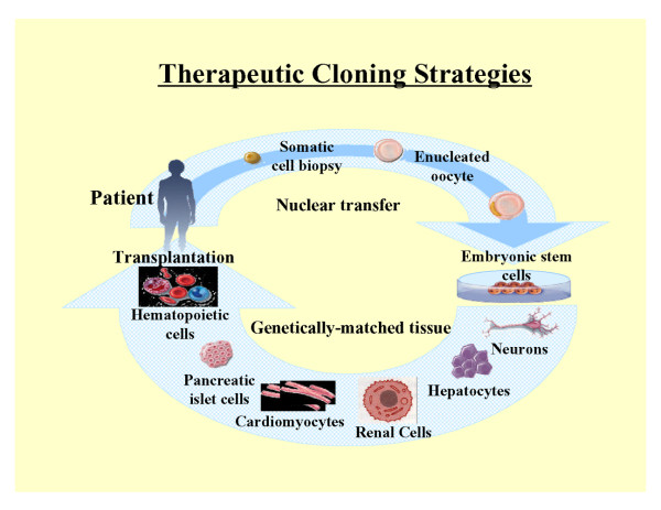
: BACKGROUND: Patients suffering from diseased and injured organs may be treated with transplanted organs. However, there is a severe shortage of donor organs which is worsening yearly due to the aging population. Scientists in the field of tissue engineering apply the principles of cell transplantation, materials science, and bioengineering to construct biological substitutes that will restore and maintain normal function in diseased and injured tissues. Both therapeutic cloning (nucleus from a donor cell is transferred into an enucleated oocyte), and parthenogenesis (oocyte is activated and stimulated to divide), permit extraction of pluripotent embryonic stem cells, and offer a potentially limitless source of cells for tissue engineering applications. The stem cell field is also advancing rapidly, opening new options for therapy. The present article reviews recent progress in tissue engineering and describes applications of these new technologies that may offer novel therapies for patients with end-stage organ failure.
Figures
Similar articles
-
Tissue engineering and regenerative medicine: concepts for clinical application.Rejuvenation Res. 2004 Spring;7(1):15-31. doi: 10.1089/154916804323105053. Rejuvenation Res. 2004. PMID: 15256042 Review.
-
Engineering tissues, organs and cells.J Tissue Eng Regen Med. 2007 Mar-Apr;1(2):83-96. doi: 10.1002/term.18. J Tissue Eng Regen Med. 2007. PMID: 18038397 Review.
-
Therapeutic cloning applications for organ transplantation.Transpl Immunol. 2004 Apr;12(3-4):193-201. doi: 10.1016/j.trim.2003.12.006. Transpl Immunol. 2004. PMID: 15157913 Review.
-
Tissue engineering applications of therapeutic cloning.Annu Rev Biomed Eng. 2004;6:27-40. doi: 10.1146/annurev.bioeng.6.012204.115707. Annu Rev Biomed Eng. 2004. PMID: 15255761 Review.
-
Therapeutic cloning and tissue engineering.Curr Top Dev Biol. 2004;60:1-15. doi: 10.1016/S0070-2153(04)60001-9. Curr Top Dev Biol. 2004. PMID: 15094294 Review.
Cited by
-
Healing of Artificially Created Gap Non-union Using Autologous Cultured Osteoblasts Impregnated Over Three-Dimensional Biodegradable Scaffold: An Experimental Study (Rabbit).Indian J Orthop. 2021 Apr 19;55(Suppl 2):460-465. doi: 10.1007/s43465-020-00288-z. eCollection 2021 Jul. Indian J Orthop. 2021. PMID: 34306561 Free PMC article.
-
An improved transplantation strategy for mouse mesenchymal stem cells in an acute myocardial infarction model.PLoS One. 2011;6(6):e21005. doi: 10.1371/journal.pone.0021005. Epub 2011 Jun 17. PLoS One. 2011. PMID: 21698117 Free PMC article.
-
Extracellular Vesicles in Musculoskeletal Regeneration: Modulating the Therapy of the Future.Cells. 2021 Dec 24;11(1):43. doi: 10.3390/cells11010043. Cells. 2021. PMID: 35011605 Free PMC article. Review.
-
Nanomedicine: techniques, potentials, and ethical implications.J Biomed Biotechnol. 2006;2006(5):51516. doi: 10.1155/JBB/2006/51516. J Biomed Biotechnol. 2006. PMID: 17489016 Free PMC article.
-
Electrofusion Stimulation Is an Independent Factor of Chromosome Abnormality in Mice Oocytes Reconstructed via Spindle Transfer.Front Endocrinol (Lausanne). 2021 Jul 28;12:705837. doi: 10.3389/fendo.2021.705837. eCollection 2021. Front Endocrinol (Lausanne). 2021. PMID: 34413830 Free PMC article.
References
-
- Cilento BG, Freeman MR, Schneck FX, Retik AB, Atala A. Phenotypic and cytogenetic characterization of human bladder urothelia expanded in vitro. J Urol. 1994;152:665–670. - PubMed
-
- Liebert M, Hubbel A, Chung M, Wedemeyer G, Lomax MI, Hegeman A, Yuan TY, Brozovich M, Wheelock MJ, Grossman HB. Expression of mal is associated with urothelial differentiation in vitro: identification by differential display reverse-transcriptase polymerase chain reaction. Differentiation. 1997;61:177–185. doi: 10.1046/j.1432-0436.1997.6130177.x. - DOI - PubMed
-
- Puthenveettil JA, Burger MS, Reznikoff CA. Replicative senescence in human uroepithelial cells. Adv Exp Med Biol. 1999;462:83–91. - PubMed
-
- Freeman MR, Yoo JJ, Raab G, Soker S, Adam RM, Schneck FX, Renshaw AA, Klagsbrun M, Atala A. Heparin-binding EGF-like growth factor is an autocrine growth factor for human urothelial cells and is synthesized by epithelial and smooth muscle cells in the human bladder. J Clin Invest. 1997;99:1028–1036. - PMC - PubMed
LinkOut - more resources
Full Text Sources