. 2004 Dec 8;1(1):24.
doi: 10.1186/1742-2094-1-24.

Passive immunotherapy against Abeta in aged APP-transgenic mice reverses cognitive deficits and depletes parenchymal amyloid deposits in spite of increased vascular amyloid and microhemorrhage

Affiliations

Passive immunotherapy against Abeta in aged APP-transgenic mice reverses cognitive deficits and depletes parenchymal amyloid deposits in spite of increased vascular amyloid and microhemorrhage

Donna M Wilcock et al. J Neuroinflammation. .

Abstract

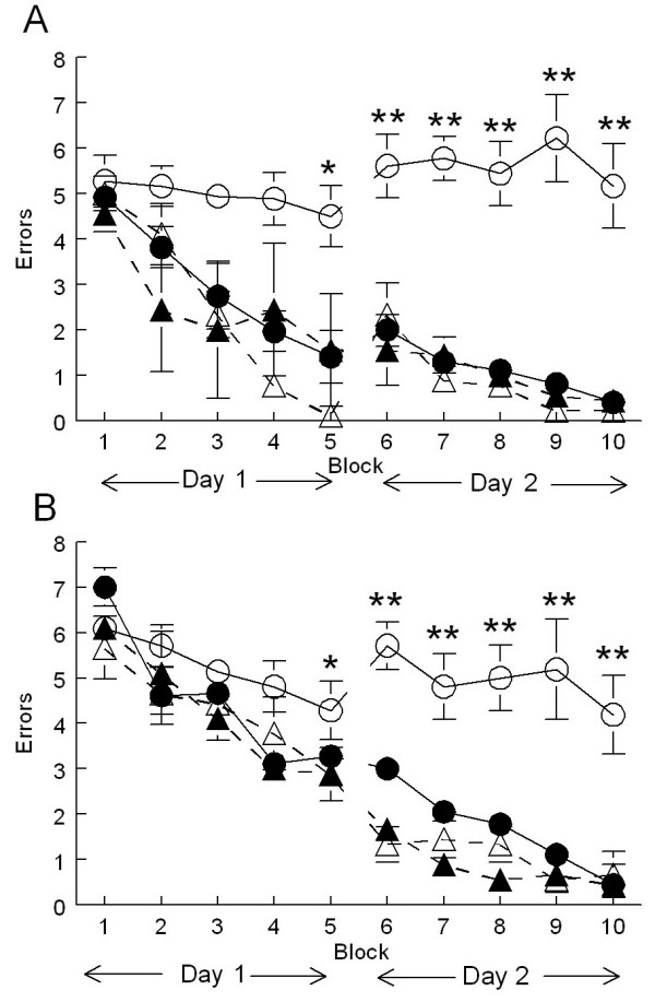
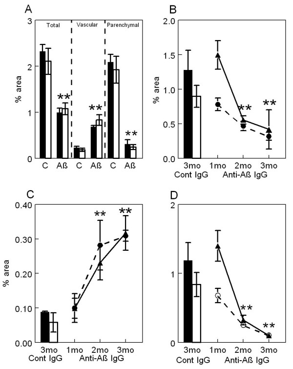
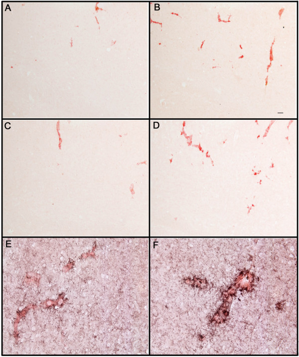
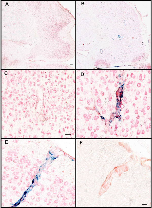
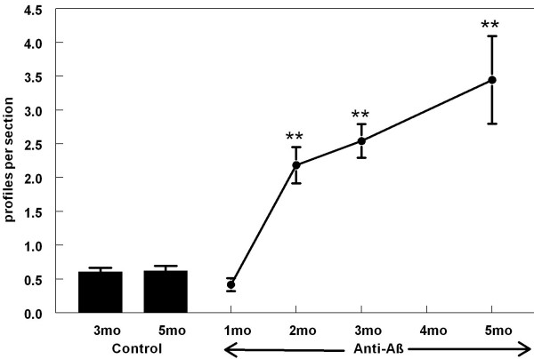
BACKGROUND: Anti-Abeta immunotherapy in transgenic mice reduces both diffuse and compact amyloid deposits, improves memory function and clears early-stage phospho-tau aggregates. As most Alzheimer disease cases occur well past midlife, the current study examined adoptive transfer of anti-Abeta antibodies to 19- and 23-month old APP-transgenic mice. METHODS: We investigated the effects of weekly anti-Abeta antibody treatment on radial-arm water-maze performance, parenchymal and vascular amyloid loads, and the presence of microhemorrhage in the brain. 19-month-old mice were treated for 1, 2 or 3 months while 23-month-old mice were treated for 5 months. Only the 23-month-old mice were subject to radial-arm water-maze testing. RESULTS: After 3 months of weekly injections, this passive immunization protocol completely reversed learning and memory deficits in these mice, a benefit that was undiminished after 5 months of treatment. Dramatic reductions of diffuse Abeta immunostaining and parenchymal Congophilic amyloid deposits were observed after five months, indicating that even well-established amyloid deposits are susceptible to immunotherapy. However, cerebral amyloid angiopathy increased substantially with immunotherapy, and some deposits were associated with microhemorrhage. Reanalysis of results collected from an earlier time-course study demonstrated that these increases in vascular deposits were dependent on the duration of immunotherapy. CONCLUSIONS: The cognitive benefits of passive immunotherapy persist in spite of the presence of vascular amyloid and small hemorrhages. These data suggest that clinical trials evaluating such treatments will require precautions to minimize potential adverse events associated with microhemorrhage.


Figures

Figure 1
Figure 1
Spatial learning deficits in APP-transgenic mice were reversed following 3 and 5 months of immunization. Mice were tested in a two-day version of the radial-arm water maze. Solid lines represent APP-transgenic mice while dashed lines represent nontransgenic mice. Open symbols indicate anti-AMN, control-antibody treatment (○: APP-transgenic, control antibody; △: nontransgenic, control antibody) while closed symbols indicate anti-Aβ antibody treatment (●: APP-transgenic, Aβ antibody; ▲: nontransgenic, Aβ antibody). Panel A shows mean number of errors made over the two-day trial period following 3 months of immunization. Each data point is the average of 3 trials. Panel B shows the mean number of errors made over the 2-day trial period following 5 months of immunization. For both graphs * indicates p < 0.05, ** indicates p < 0.001 when the APP-transgenic mice receiving control antibody are compared with the remaining groups.
Figure 2
Figure 2
Passive immunization with anti-Aβ antibodies decreases total and parenchymal amyloid loads while increasing vascular amyloid in frontal cortex and hippocampus of APP-transgenic mice. Panel A shows total amyloid load measured with Congo red, vascular amyloid load and parenchymal amyloid load from APP-transgenic mice administered control IgG (C) or anti-Aβ IgG (Aβ) for a period of 5 months. Panels B-D show total amyloid load (Panel B), vascular amyloid load (Panel C) and parenchymal amyloid load (Panel D) from APP-transgenic mice administered control IgG for 3 months (Cont IgG) or anti-Aβ IgG for a period of 1, 2, or 3 months (Anti-Aβ IgG). For all panels, the solid bar and solid line represent values from the frontal cortex, while the open bar and dashed line represent values from the hippocampus. ** p < 0.01.
Figure 3
Figure 3
Increased Congo red staining of blood vessels following anti-Aβ antibody administration is associated with activated microglia. Panels A and B are from the frontal cortex of 22-month-old APP-transgenic mice immunized for 3 months with either control antibody (3A) or anti-Aβ antibody (3B). Panels C and D are from the frontal cortex of 28-month-old APP-transgenic mice immunized for 5 months with either control antibody (3C) or anti-Aβ antibody (3D). Panels E and F show a high-magnification image of CD45 immunohistochemistry (black) counterstained with Congo red (red) from 28-month-old APP-transgenic mice immunized for 5 months with either control antibody (Panel E) or anti-Aβ antibody (Panel F). Panels A-D, magnification = 100X. Scale bar in Panel B = 50 μ for panels A-D. Panels E-F, magnification = 200X. Scale bar in Panel E = 25 μm for panels E-F.
Figure 4
Figure 4
Microhemorrhage associated with CAA following systemic administration of anti-Aβ antibodies. Panels A and B are low magnification images of the frontal cortex of APP-transgenic mice receiving either control antibodies (Panel A) or anti-Aβ antibodies (Panel B) for a period of 5 months. Panels C and D show representative images of amyloid containing vessels stained for Prussian blue (blue), counterstained with neutral red (red), from APP-transgenic mice receiving either control antibodies (Panel C) or anti-Aβ antibodies (Panel D) for a period of 3 months. Panel E shows a blood vessel in the frontal cortex stained for Prussian blue (blue), counterstained with neutral red, from an APP transgenic mouse administered anti-Aβ antibodies for 5 months. Panel F shows the same blood vessel on an adjacent section stained for Congo red, indicating that the blood vessel does in fact contain amyloid. Scale bar panel A = 120 μm for panels A-B. Scale bar panel C = 25 μm for panels C-D. Scale bar in panel F = 25 μm for panels E-F.
Figure 5
Figure 5
Number of Prussian blue-positive profiles increases with duration of anti-Aβ antibody exposure. The graph shows quantification of the average number of Prussian blue-positive profiles per section from mice administered control IgG for 3 or 5 months (Cont) or anti-Aβ IgG for 1, 2, 3 or 5 months (anti-Aβ). ** p < 0.01.

References

    1. Iwatsubo T, Odaka A, Suzuki N, Mizusawa H, Nukina N, Ihara Y. Visualization of A beta 42(43) and A beta 40 in senile plaques with end-specific A beta monoclonals: evidence that an initially deposited species is A beta 42(43) Neuron. 1994;13:45–53. doi: 10.1016/0896-6273(94)90458-8. - DOI - PubMed
    1. Gordon MN, Holcomb LA, Jantzen PT, DiCarlo G, Wilcock D, Boyett KW, Connor K, Melachrino J, O'Callaghan JP, Morgan D. Time course of the development of Alzheimer-like pathology in the doubly transgenic PS1+APP mouse. Exp Neurol. 2002;173:183–195. doi: 10.1006/exnr.2001.7754. - DOI - PubMed
    1. Solomon B, Koppel R, Hanan E, Katzav T. Monoclonal antibodies inhibit in vitro fibrillar aggregation of the Alzheimer beta-amyloid peptide. Proc Natl Acad Sci USA. 1996;93:452–5. doi: 10.1073/pnas.93.1.452. - DOI - PMC - PubMed
    1. Schenk D, Barbour R, Dunn W, Gordon G, Grajeda H, Guido T, Hu K, Huang J, Johnson-Wood K, Khan K, Kholodenko D, Lee M, Liao Z, Lieberburg I, Motter R, Mutter L, Soriano F, Shopp G, Vasquez N, Vandevert C, Walker S, Wogulis M, Yednock T, Games D, Seubert P. Immunization with amyloid-beta attenuates Alzheimer-disease-like pathology in the PDAPP mouse. Nature. 1999;400:173–177. doi: 10.1038/22124. - DOI - PubMed
    1. Orgogozo JM, Gilman S, Dartigues JF, Laurent B, Puel M, Kirby LC, Jouanny P, Dubois B, Eisner L, Flitman S, Michel BF, Boada M, Frank A, Hock C. Subacute meningoencephalitis in a subset of patients with AD after Abeta42 immunization. Neurology. 2003;61:46–54. - PubMed