

Morphological brain differences between adult stutterers and non-stutterers
- PMID: 15588309
- PMCID: PMC539354
- DOI: 10.1186/1471-2377-4-23
Morphological brain differences between adult stutterers and non-stutterers
Abstract
Background: The neurophysiological and neuroanatomical foundations of persistent developmental stuttering (PDS) are still a matter of dispute. A main argument is that stutterers show atypical anatomical asymmetries of speech-relevant brain areas, which possibly affect speech fluency. The major aim of this study was to determine whether adults with PDS have anomalous anatomy in cortical speech-language areas.
Methods: Adults with PDS (n = 10) and controls (n = 10) matched for age, sex, hand preference, and education were studied using high-resolution MRI scans. Using a new variant of the voxel-based morphometry technique (augmented VBM) the brains of stutterers and non-stutterers were compared with respect to white matter (WM) and grey matter (GM) differences.
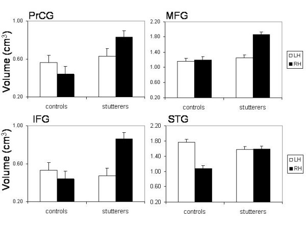
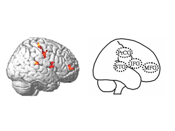
Results: We found increased WM volumes in a right-hemispheric network comprising the superior temporal gyrus (including the planum temporale), the inferior frontal gyrus (including the pars triangularis), the precentral gyrus in the vicinity of the face and mouth representation, and the anterior middle frontal gyrus. In addition, we detected a leftward WM asymmetry in the auditory cortex in non-stutterers, while stutterers showed symmetric WM volumes.
Conclusions: These results provide strong evidence that adults with PDS have anomalous anatomy not only in perisylvian speech and language areas but also in prefrontal and sensorimotor areas. Whether this atypical asymmetry of WM is the cause or the consequence of stuttering is still an unanswered question.
Figures
References
Publication types
MeSH terms
LinkOut - more resources
Full Text Sources
Medical
Miscellaneous
