. 2004 Dec 10:5:57.
doi: 10.1186/1471-2202-5-57.

Membrane trafficking and mitochondrial abnormalities precede subunit c deposition in a cerebellar cell model of juvenile neuronal ceroid lipofuscinosis

Affiliations

Membrane trafficking and mitochondrial abnormalities precede subunit c deposition in a cerebellar cell model of juvenile neuronal ceroid lipofuscinosis

Elisa Fossale et al. BMC Neurosci. .

Abstract

Background: JNCL is a recessively inherited, childhood-onset neurodegenerative disease most-commonly caused by a approximately 1 kb CLN3 mutation. The resulting loss of battenin activity leads to deposition of mitochondrial ATP synthase, subunit c and a specific loss of CNS neurons. We previously generated Cln3Deltaex7/8 knock-in mice, which replicate the common JNCL mutation, express mutant battenin and display JNCL-like pathology.

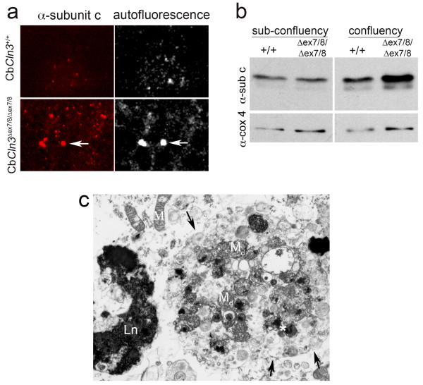
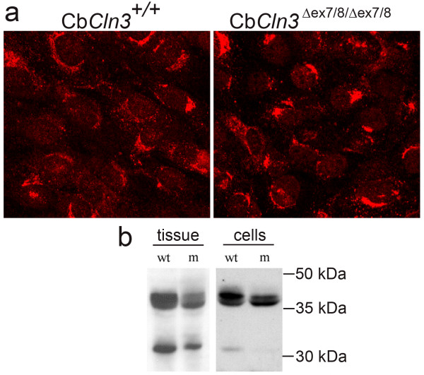
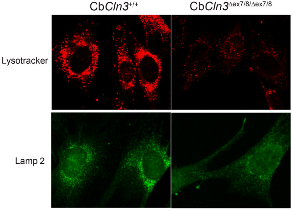
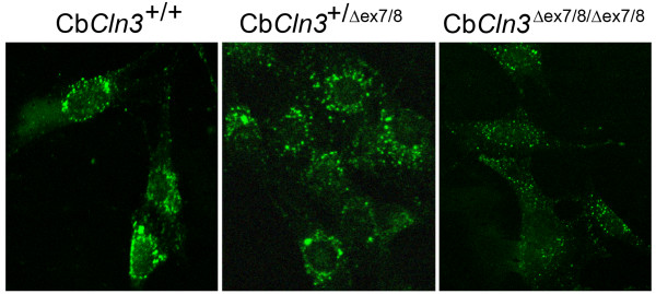
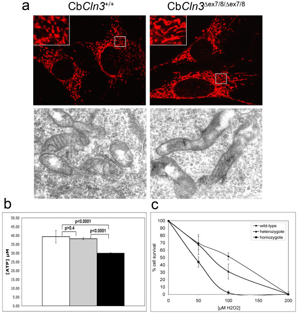
Results: To elucidate the consequences of the common JNCL mutation in neuronal cells, we used P4 knock-in mouse cerebella to establish conditionally immortalized CbCln3 wild-type, heterozygous, and homozygous neuronal precursor cell lines, which can be differentiated into MAP-2 and NeuN-positive, neuron-like cells. Homozygous CbCln3Deltaex7/8 precursor cells express low levels of mutant battenin and, when aged at confluency, accumulate ATPase subunit c. Recessive phenotypes are also observed at sub-confluent growth; cathepsin D transport and processing are altered, although enzyme activity is not significantly affected, lysosomal size and distribution are altered, and endocytosis is reduced. In addition, mitochondria are abnormally elongated, cellular ATP levels are decreased, and survival following oxidative stress is reduced.

Conclusions: These findings reveal that battenin is required for intracellular membrane trafficking and mitochondrial function. Moreover, these deficiencies are likely to be early events in the JNCL disease process and may particularly impact neuronal survival.


Figures

Figure 1
Figure 1
Neuronal marker expression in CbCln3+/+ cells Characterization of CbCln3+/+ cells by immunofluorescence with marker antibodies is shown. CbCln3+/+ precursors exhibit nestin expression (a) but not GFAP expression (b), consistent with a neuronal precursor identity. Upon stimulation with a differentiation cocktail (see Methods), CbCln3+/+ cells achieved neuron-like morphology, with rounded cell bodies and extension of processes, and MAP2 (c) and NeuN (d) expression was increased. CbCln3+/+ cells are negative for the Purkinje neuron marker calbindin (e). CbCln3+/Δex7/8 and CbCln3Δex7/8/Δex7/8 cell lines exhibited identical marker immunofluorescence results. a, b) 20 × magnification; c, d, e) 40 × magnification.
Figure 2
Figure 2
RT-PCR of Cln3 mRNA in wild-type and homozygous CbCln3Δex7/8 cells Cln3 Exon1-forward, Exon 15-reverse RT-PCR products are shown, from total wild-type (+/+) or homozygous mutant (Δex7/8/Δex7/8) brain and cell line RNA. Brain and cell line RT-PCR reaction products had identical band patterns on ethidium-bromide stained agarose gels. Wild-type RT-PCR product was a single ~1.6 kb band and mutant products were ~1.6, ~1.5, ~1.4, ~1.35, and ~1.3 kb, representing multiple mutant splice variants.
Figure 3
Figure 3
Battenin and lysosomal and endosomal marker co-staining in wild-type and homozygous CbCln3Δex7/8 cerebellar precursor cells Batp1 immunostaining of wild-type (CbCln3+/+) and homozygous mutant (CbCln3Δex7/8/Δex7/8) cerebellar precursor cells is shown, with co-staining for lysosomes (Lamp 1), early endosomes (EEA1), and late endosomes (Rab7). Significant overlap of Batp1 signal (red) with EEA1 (green, middle panels) and Rab7 (green, bottom panels) can be seen as yellow when the two channels are merged (Merge). The degree of Batp1 overlap is greatest with Rab7. Only limited overlap between Batp1 (red) and Lamp 1 (green, top panels) can be seen. Batp1 signal in homozygous CbCln3Δex7/8 cells is significantly reduced, but significant overlap with EEA1 and Rab7, and very little Lamp 1 overlap, can be seen as yellow in the respective merged panels. Notably, Lamp 1 and EEA1 localization appear altered, and Rab7 staining was frequently less intense in homozygous CbCln3Δex7/8 cells. Wild-type and homozygous CbCln3Δex7/8 confocal images were captured with identical exposure settings. 60 × magnification.
Figure 4
Figure 4
Subunit c accumulation in homozygous CbCln3Δex7/8 cerebellar precursor cells a. Subunit c immunostaining and autofluorescence of 7-day confluency-aged wild-type and homozygous CbCln3Δex7/8 cells is shown. Wild-type cultures (CbCln3+/+) exhibited limited subunit c immunostaining and autofluorescence. However, CbCln3Δex7/8/Δex7/8 cells contained numerous subunit c puncta. Autofluorescence (7 days AF) was also significantly elevated (right panels), although limited overlap with subunit c puncta was observed (arrows). 40 × magnification. b. Immunoblot analysis of subunit c protein at sub-confluency or 7-day confluency incubation is shown. Total protein extracts from sub-confluency wild-type (+/+) and homozygous mutant (Δex7/8/Δex7/8) cultures contained approximately equal levels of subunit c protein (α-sub c). 7-day confluency extract from homozygous CbCln3Δex7/8 cells (Δex7/8/Δex7/8) had elevated levels of subunit c protein (~1.5X), relative to wild-type extract (+/+). Protein levels were normalized to cytochrome c oxidase subunit IV (α-cox4). c. TEM analysis of inclusions in 7-day confluency-aged homozygous CbCln3Δex7/8 cells is shown. A large autophagosome contained by double membrane (arrows) is filled with degenerating mitochondria (Md), electron dense cores (left and right of *) and other smaller vesicular structures. A large electron-dense inclusion, with a lipofuscin (Ln) appearance, is also present. M, mitochondria. 10,000 × magnification.
Figure 5
Figure 5
Cathepsin D localization and processing in wild-type and homozygous CbCln3Δex7/8 cells a. Immunostaining of wild-type and homozygous CbCln3Δex7/8 precursor cells with anti-cathepsin D antibody, recognizing unprocessed and processed forms of cathepsin D protein is shown. CbCln3+/+ cells (left panel) exhibited a perinuclear and cytoplasmic punctate signal. Cathepsin D signal in homozygous CbCln3Δex7/8 cells (right panel) was more often perinuclear, with less cytoplasmic punctate signal, compared to wild-type CbCln3+/+ cells. 40 × magnification. b. α-Cathepsin D-probed immunoblots of total wild-type versus homozygous Cln3Δex7/8 knock-in tissue or CbCln3Δex7/8 cellular extracts are shown. The ~45 kDa cathepsin D band, representing precursor, was the predominant band in wild-type (wt) tissue and cellular extracts, with lower levels of mature enzyme (single chain, ~43 kDa, and heavy chain, ~31 kDa). Conversely, homozygous Cln3Δex7/8 and CbCln3Δex7/8 mutant (m) extracts exhibited reduced levels of precursor and heavy chain of the double-chain form of the enzyme, with elevated levels of single-chain mature enzyme.
Figure 6
Figure 6
Lysotracker and Lamp 2 labeling of wild-type and homozygous CbCln3Δex7/8 lysosomes Lysosomal labeling of wild-type and homozygous CbCln3Δex7/8 precursor cells with lysotracker and Lamp 2 antibody is shown. Lysotracker dye (top panels) labeled large, perinuclear-clustered lysosomes and scattered lysosomes in the periphery of wild-type cells (CbCln3+/+). Lysotracker stain was dramatically reduced in homozygous mutant cells (CbCln3Δex7/8/Δex7/8), with smaller labeled vesicles and less apparent perinuclear clustering. Lamp 2 (bottom panels) immunostaining also showed reduced signal intensity with less perinuclear clustering in homozygous CbCln3Δex7/8 cells, although the effect was somewhat less dramatic than that observed with Lysotracker dye. Wild-type and homozygous CbCln3Δex7/8 confocal images were captured with identical exposure settings. 60 × magnification.
Figure 7
Figure 7
Endocytosis in wild-type, heterozygous and homozygous CbCln3Δex7/8 cells Dextran-FITC uptake in wild-type, heterozygous and homozygous CbCln3Δex7/8 precursor cells is shown. In wild-type (CbCln3+/+, left panel) and heterozygous (CbCln3+/Δex7/8, middle panel) cells, dextran-FITC label was observed in a perinuclear-clustered vesicular pattern with scattered labeled vesicles also present in the periphery. In contrast, dextran-FITC label of homozygous mutant (CbCln3Δex7/8/Δex7/8, right panel) cells was reduced overall and exhibited smaller stained vesicles with less perinuclear clustering. Confocal images were captured with identical exposure settings. 40 × magnification.
Figure 8
Figure 8
Mitochondrial morphology and function in wild-type, heterozygous and homozygous CbCln3Δex7/8 cells a. Confocal and TEM micrographs of wild-type and homozygous CbCln3Δex7/8 mitochondrial morphology are shown. Immunostaining with the inner mitochondrial membrane marker, grp75 (top panels) highlighted elongated mitochondria in homozygous mutant cells (CbCln3Δex7/8/Δex7/8), relative to wild-type mitochondria (CbCln3+/+) (insets, zoom = 2.75x). Mitochondrial distribution was not altered from the wild-type pattern. Elongated homozygous CbCln3Δex7/8 mitochondria were also observed by TEM analysis. 60 × magnification. b. Cellular ATP levels in wild-type, heterozygous and homozygous CbCln3Δex7/8 precursor cells are shown. Wild-type (open bar) and heterozygous (gray bar) CbCln3Δex7/8 cells contained ~39 μM ATP, while homozygous CbCln3Δex7/8 cells (black bar) contained ~1.3 fold reduced levels of ATP (~30 μM), which was statistically significant in a t-test (p < 0.0001). Wild-type and heterozygous CbCln3Δex7/8 cellular ATP levels were not statistically different from each other (p > 0.4). A representative of triplicate experiments is shown (n = 6 in each experiment). c. Cell survival following 24-hour hydrogen peroxide treatment is shown. Homozygous CbCln3Δex7/8 cells were ~2-fold more sensitive to oxidative stress by hydrogen peroxide treatment. Wild-type (circle) and heterozygous (triangle) CbCln3Δex7/8 cells exhibited ~50% survival rates with 75–100 μM H2O2, whereas homozygous CbCln3Δex7/8 cells (squares) had a ~50% survival rate with 50 μM H2O2. A representative of triplicate experiments is shown (n = 4 in each experiment).

References

    1. Rider JA, Rider DL. Batten disease: past, present, and future. Am J Med Genet Suppl. 1988;5:21–26. - PubMed
    1. International Batten Disease Consortium Isolation of a novel gene underlying Batten disease, CLN3. The International Batten Disease Consortium. Cell. 1995;82:949–957. doi: 10.1016/0092-8674(95)90274-0. - DOI - PubMed
    1. Jolly RD, Martinus RD, Palmer DN. Sheep and other animals with ceroid-lipofuscinoses: their relevance to Batten disease. Am J Med Genet. 1992;42:609–614. - PubMed
    1. Palmer DN, Fearnley IM, Walker JE, Hall NA, Lake BD, Wolfe LS, Haltia M, Martinus RD, Jolly RD. Mitochondrial ATP synthase subunit c storage in the ceroid-lipofuscinoses (Batten disease) Am J Med Genet. 1992;42:561–567. - PubMed
    1. Wisniewski KE, Rapin I, Heaney-Kieras J. Clinico-pathological variability in the childhood neuronal ceroid-lipofuscinoses and new observations on glycoprotein abnormalities. American Journal of Medical Genetics - Supplement. 1988;5:27–46. - PubMed

Publication types

MeSH terms