. 2005 Feb 3;4(2):149-61.
doi: 10.1016/j.dnarep.2004.08.010.

Molecular analysis of sister chromatid recombination in mammalian cells

Affiliations

Molecular analysis of sister chromatid recombination in mammalian cells

Nadine Puget et al. DNA Repair (Amst). .

Abstract

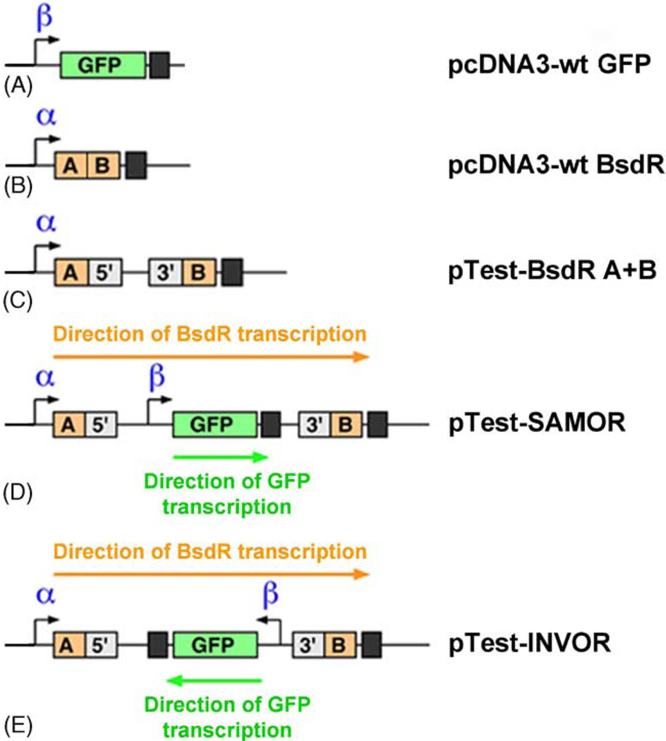
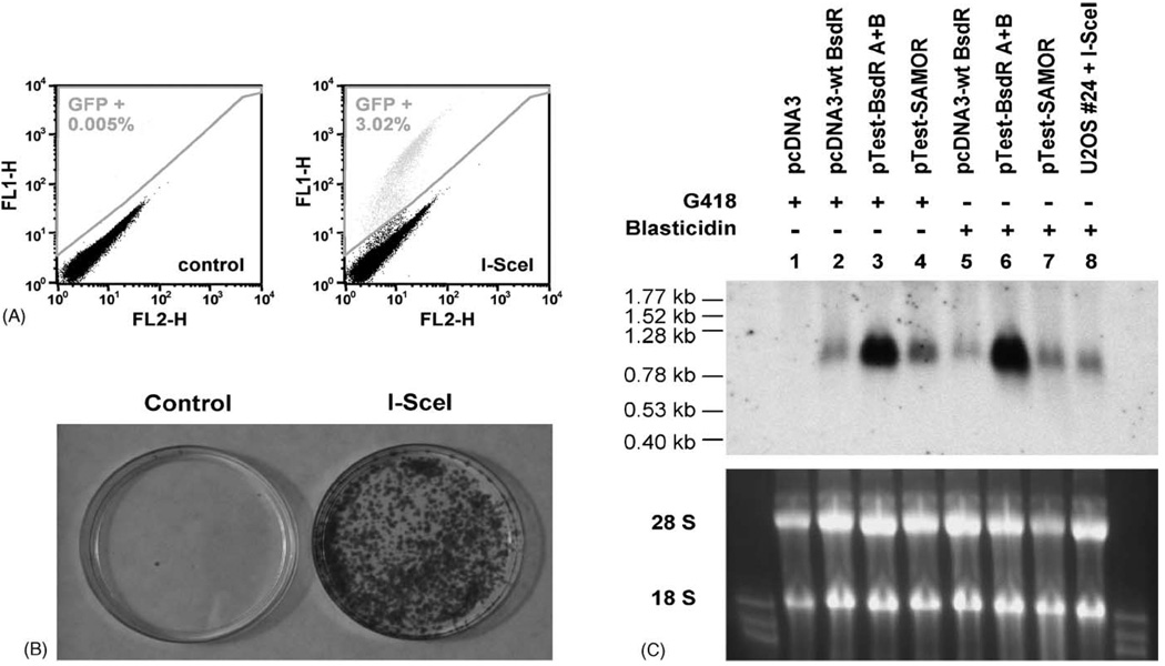
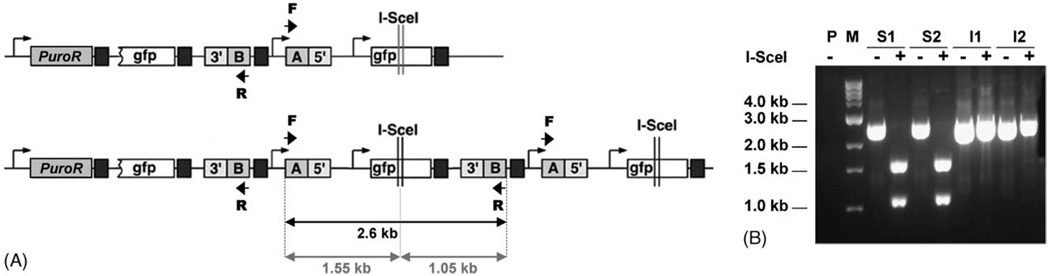
Sister chromatid recombination (SCR) is a potentially error-free pathway for the repair of double-strand breaks arising during replication and is thought to be important for the prevention of genomic instability and cancer. Analysis of sister chromatid recombination at a molecular level has been limited by the difficulty of selecting specifically for these events. To overcome this, we have developed a novel "nested intron" reporter that allows the positive selection in mammalian cells of "long tract" gene conversion events arising between sister chromatids. We show that these events arise spontaneously in cycling cells and are strongly induced by a site-specific double-strand break (DSB) caused by the restriction endonuclease, I-SceI. Notably, some I-SceI-induced sister chromatid recombination events entailed multiple rounds of gene amplification within the reporter, with the generation of a concatemer of amplified gene segments. Thus, there is an intimate relationship between sister chromatid recombination control and certain types of gene amplification. Dysregulated sister chromatid recombination may contribute to cancer progression, in part, by promoting gene amplification.


Figures

Fig. 1
Fig. 1
Construction of the sister chromatid recombination reporter. Open boxes: mutant gfp genes; green boxes: wtGFP. Black arrow: human CMV promoter; black boxes: SV40 polyadenylation signals for GFP genes. Double red lines identify the restriction site cut by I-SceI. (A) Repair of an I-SceI-induced DSB by STGC, whether inter- or intra-chromatid, generates wtGFP but does not duplicate the intervening sequence between the two GFP copies (marked by red star). (B) Repair of the I-SceI-induced DSB by LTGC between sister chromatids, or by crossing over (not shown) can generate “GFP triplication”. The middle GFP gene becomes wild-type and the red star is duplicated. Depending on the length of the gene conversion tract, other LTGC products are also possible (see text). (C) Schematic of the BsdR cassette, inserted at the site of the red star, and of the final reporter, “HRsub”. Artificial BsdR exons A and B are orange; fragments of the rabbit β-globin intron II are grey. Each intron-exon boundary constitutes a consensus splicing site, as shown. The SV40 early promoter (black arrow) is located 5′ of exon A and the BGH polyadenylation signal (black box/pA) is 3′ of exon B. The murine PGK promoter (black arrow) drives expression of the puromycin resistance gene, PuroR (blue box), for selection of cells having integrated the HRsub reporter. An HSV TK polyadenylation signal is 3′ of PuroR. (D) Selection for LTGC events. In the “GFP triplication” outcome, the BsdR cassette is duplicated. Promoter α (SV40) drives expression of wtBsdR, and promoter β (CMV) drives expression of wtGFP. Expected RNA transcripts are shown.
Fig. 2
Fig. 2
Test constructs mimicking the “nested intron” rearrangement. See Fig. 1 legend for key to gene/intron fragments and other elements. (A) Plasmid encoding a wtGFP gene (β: CMV promoter). (B) Plasmid encoding a wtBsdR gene (α: SV40 promoter). (C) Test construct with artificially split exons A and B of the BsdR gene, separated by the intact rabbit β-globin intron II. (D) Test construct “SAMOR” contains a wtGFP gene “nested” within the BsdR intron, both GFP and BsdR genes being transcribed in the same orientation. (E) Test construct “INVOR” contains a wtGFP gene “nested” within the BsdR intron, but with GFP gene transcription in the opposite orientation to that of the BsdR gene. See Table 1 and text for more details.
Fig. 3
Fig. 3
Induction of GFP+ cells and BsdR+ colonies by I-SceI. (A) FACS analysis after transfection of recombination reporter Clone U2OS #24 with control plasmid or with I-SceI plasmid. FL1-H, green fluorescence; FL2-H, red fluorescence. GFP+ cells lie above the diagonal, percentages as indicated. (B) Crystal violet staining of BsdR+ colonies after transfection of U2OS Clone #24 with control plasmid or with I-SceI plasmid. For each transfection, 1.2 × 10⁶ cells were cultured in blasticidin for 2 weeks before staining. (C) Northern blot analysis of BsdR transcripts. RNA from U2OS cells transfected with a series of test constructs (lanes 1–7) or from pooled I-SceI-induced BsdR+ colonies of reporter Clone #24 (lane 8). The cells were selected either in G418 (resistance given by the plasmid backbone for the test constructs) or in Blasticidin. Top panel: Northern blot probed with BsdR cDNA (400 bp) reveals a ~1 kb mRNA in all cell lines expressing BsdR (lanes 2–7). BsdR mRNA abundance is greatly increased by the presence of the β-globin intron between BsdR exons A and B (pTest-BsdR A+B, lanes 3 and 6; see Fig. 2C) but this effect is reduced when the wtGFP gene is “nested” within the intron (pTest–SAMOR, lanes 4 and 7; see Fig. 2D). Pooled I-SceI-induced BsdR+ colonies from U2OS Clone #24 contain a processed BsdR transcript (lane 8). Bottom: 18S and 28S rRNA from the same gel revealed by ethidium bromide staining.
Fig. 4
Fig. 4
PCR analysis of the reporter locus in BsdR+ colonies. (A) Scheme for PCR analysis, showing parental reporter (top) and “GFP triplication” outcome (bottom). Duplication of the BsdR cassette in the rearranged reporter allows a 2.6 kb fragment to be amplified from genomic DNA of BsdR+ clones with the indicated primers F and R (short black arrows). The presence of an I-SceI site in the PCR product allows I-SceI digestion to generate two smaller fragments, as indicated. (B) Representative results of the PCR analysis in Clone #24. P: genomic DNA from parental #24 clone does not give rise to a PCR product, since the BsdR cassette is not duplicated; S1, S2: 2.6 kb PCR products from spontaneously arising GFP− BsdR+ colonies contain an I-SceI site; I1, I2: 2.6 kb PCR products from I-SceI-induced GFP+ BsdR+ colonies lack an I-SceI site; M: 1 kb ladder molecular weight marker.
Fig. 5
Fig. 5
Southern blot analysis of spontaneously arising BsdR+ colonies. (A) PstI and HindIII maps of the reporter, restriction fragment sizes indicated. Figure shows the parental reporter (top) and the predicted rearrangement in the “GFP triplication” outcome (bottom). (B–E) Southern blotting performed on U2OS Clones #18 (B, D) and #24 (C, E), using PstI (B, C) or HindIII (D, E) digested genomic DNA. The probe was the 700 bp GFP cDNA. P: parental Clone #18 or #24 indicates the unrearranged reporter; 1–7: for each clone, #18 and #24, seven spontaneously-arising BsdR+ colonies are shown.
Fig. 6
Fig. 6
Southern blot analysis of I-SceI-induced BsdR+ colonies. (A–D) Southern blotting was performed on U2OS reporter Clones #18 (A, C) and #24 (B, D) using PstI (A, B) or HindIII (C, D) digested genomic DNA. The probe was the 700 bp GFP cDNA. See restriction maps in Fig. 5A. P: parental Clone #18 or #24 indicates the unrearranged reporter; 1–7: representative rearrangements of the reporter resulting from sister chromatid recombination events. Lanes 1–3 for Clone #18 and lanes 1–2 for Clone #24 show the most frequent LTGC products (“GFP triplication”). Lanes 4–6 for Clone #18 and lanes 3–7 for Clone #24 show aberrant patterns of expansion (see text). Note the large intensely hybridizing PstI fragment in Panel A (Clone #18), lane 4, and the correspondingly increased intensity of the 3.2 kb band in Panel C (Clone #18), lane 4. (E) Schematic of the gene amplification event in Clone #18, lane 4 (Panels A and C)—not to scale. Stars indicate positions of BsdR cassettes.

References

    1. Kadyk LC, Hartwell LH. Sister chromatids are preferred over homologs as substrates for recombinational repair in Saccharomyces cerevisiae. Genetics. 1992;132:387–402. - PMC - PubMed
    1. Johnson RD, Jasin M. Sister chromatid gene conversion is a prominent double-strand break repair pathway in mammalian cells. EMBO J. 2000;19:3398–3407. - PMC - PubMed
    1. Gonzalez-Barrera S, Cortes-Ledesma F, Wellinger RE, Aguilera A. Equal sister chromatid exchange is a major mechanism of double-strand break repair in yeast. Mol. Cell. 2003;11:1661–1671. - PubMed
    1. Dronkert ML, Beverloo HB, Johnson RD, Hoeijmakers JH, Jasin M, Kanaar R. Mouse RAD54 affects DNA double-strand break repair and sister chromatid exchange. Mol. Cell. Biol. 2000;20:3147–3156. - PMC - PubMed
    1. Cox MM. The non-mutagenic repair of broken replication forks via recombination. Mut. Res. 2002;510:107–120. - PubMed

Publication types

Substances