. 2005 Feb 18;280(7):6197-203.
doi: 10.1074/jbc.M412911200. Epub 2004 Dec 7.

Dmp1-deficient mice display severe defects in cartilage formation responsible for a chondrodysplasia-like phenotype

Affiliations

Dmp1-deficient mice display severe defects in cartilage formation responsible for a chondrodysplasia-like phenotype

Ling Ye et al. J Biol Chem. .

Abstract

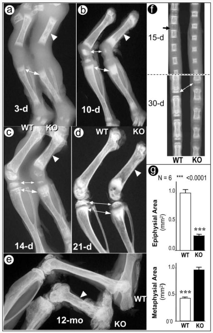
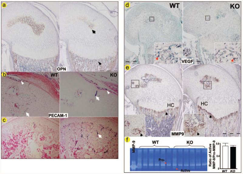
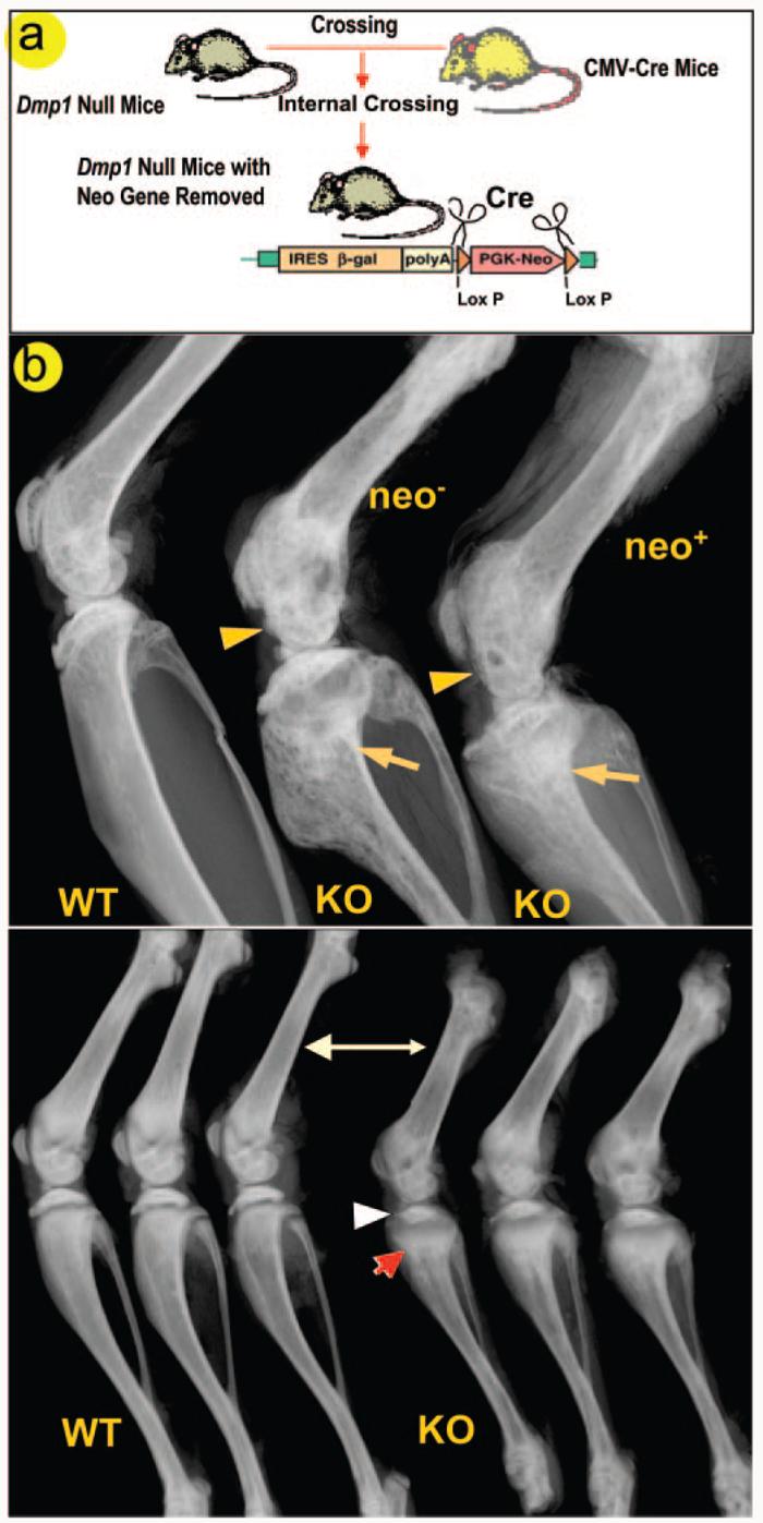
Understanding the molecular mechanisms by which cartilage formation is regulated is essential toward understanding the physiology of both embryonic bone development and postnatal bone growth. Although much is known about growth factor signaling in cartilage formation, the regulatory role of noncollagenous matrix proteins in this process are still largely unknown. In the present studies, we present evidence for a critical role of DMP1 (dentin matrix protein 1) in postnatal chondrogenesis. The Dmp1 gene was originally identified from a rat incisor cDNA library and has been shown to play an important role in late stage dentinogenesis. Whereas no apparent abnormalities were observed in prenatal bone development, Dmp1-deficient (Dmp1(-/-)) mice unexpectedly develop a severe defect in cartilage formation during postnatal chondrogenesis. Vertebrae and long bones in Dmp1-deficient (Dmp1(-/-)) mice are shorter and wider with delayed and malformed secondary ossification centers and an irregular and highly expanded growth plate, results of both a highly expanded proliferation and a highly expanded hypertrophic zone creating a phenotype resembling dwarfism with chondrodysplasia. This phenotype appears to be due to increased cell proliferation in the proliferating zone and reduced apoptosis in the hypertrophic zone. In addition, blood vessel invasion is impaired in the epiphyses of Dmp1(-/-) mice. These findings show that DMP1 is essential for normal postnatal chondrogenesis and subsequent osteogenesis.


Figures

Fig. 1
Fig. 1. Progressive changes of long bone structure in the Dmp1−/− mice
Representative radiographs of hind limbs from 3- (a), 10- (b), 14- (c), and 21-day-old (d) and 12-month-old animals (e). The shorter long bones with wider shafts of Dmp1−/− bones (KO) femurs are indicated by arrowheads, the delayed epiphyses are indicated by small arrows, and the expanded metaphyses are indicated by large arrows. Radiographs of tail vertebrae from 15- and 30-day-old mice show delayed formation of epiphyses and shortened vertebral bodies (f). Quantitative data for the extent of epiphyseal and metaphyseal ossification show a reduction in epiphysial area but an increase in metaphyseal area in Dmp1−/− mice (g).
Fig. 2
Fig. 2. Analyses of deformed tibias in Dmp1−/− mice
Photographs (a) and quantitative (b) analyses of tibias from 3-week-old and 3- and 5-month-old animals. A significant difference is observed between the WT and the mutant (KO) tibiae in both length and width. Representative three-dimensional micro-CT reconstruction of 1-month-old tibiae with whole mount view (c) and sagittal section view (d) are shown. The secondary ossification center is delayed and malformed (arrows), and the growth plate is dramatically expanded (arrowheads) in the tibia from Dmp1−/− mice.
Fig. 3
Fig. 3. Malformed growth plates in the Dmp1−/− mice
Images from backscattered scanning electron microscopy of 6-week-old and 6- and 12-month-old mice. Well formed growth plate (arrows), epiphyses and metaphyses can be easily seen in the WT mice (a, upper panel). The growth plate of Dmp1−/− mice however, is expanded and disorganized with malformed epiphyses and metaphyses (a, lower panel, arrows). Representative sections from decalcified femurs, stained with Safranin-O show a striking expansion of the growth plate at the age of 3 weeks and irregularities at 3 months and complete disruption at 5 months in the Dmp1−/− null mice (b, lower panel) compared with the WT growth plates (b, upper panel). Visually, histology shows an increased proliferation and hypertropic zone (c). Quantitative histomorphometry shows a significant increase in the proliferation zone of almost a doubling and in the hypertrophic zone of a 3-fold increase in femurs of 3-month-old Dmp1−/− mice compared with wild type controls (d). PZ, proliferation zone; HZ, hypertrophic zone.
Fig. 4
Fig. 4. Increased proliferation, reduced apoptosis, and reduced calcification of the cartilaginous matrix and trabecular bone in the Dmp1−/− mice
A significant increase in cell proliferation as detected using bromodeoxyuridine labeling was observed in tibial growth plate of 2-month-old Dmp1−/− mice (a, arrows). No apparent difference in intensity of type II (b, arrowheads) and type X (c) collagen expression was observed in femora of 3-week-old Dmp1−/− mice. Caspase 3 staining showed a significant decrease in apoptosis of hypertrophic chondrocytes in 3-week-old Dmp1−/− mice (d). Goldner's Masson Trichrome staining of a tibial growth plate displays an expanded, disorganized growth plate, a poorly calcified cartilage matrix, and reduced trabecular bone formation in a 3-month-old Dmp1−/− mouse (e). PC, proliferating chondrocyte; HC, hypertrophic chondrocyte.
Fig. 5
Fig. 5. Delayed blood vessel invasion in Dmp1−/− epiphyses
Polyclonal antibody against OPN (a), a marker of new bone formation is reduced in the KO epiphysis (arrow) but increased in the KO metaphysis (arrowhead) from a 3-week-old animal. Monoclonal antibody against PECAM-1 was used on sections of day 10 (b) and 17 (c) femurs. The positively stained endothelial cells (in blue purple, arrows) are significantly reduced in the Dmp1−/− epiphysis at day 10, although the stained endothelial cells in surrounding areas appear normal (arrowhead). By day 17, more endothelial cells stained positive for PECAM-1 in the Dmp1−/− epiphysis compared with the control with well formed bone marrow (c, arrow). In situ hybridization was performed to examine changes in mRNA expression of VEGF (d), and immunostaining was performed to examine protein expression of MMP9 (f), and enzyme activity of MMP9 (e) were measured by genetin zymography in 10-day-old Dmp1−/− mice and the age-matched control mice (right panels). Both VEGF and MMP9 are expressed at sites of neovascularization (arrows) with no apparent difference in VEGF and MMP9 expression in Dmp1−/− mice compared with control littermates. HC, hypertrophic chondrocyte.
Fig. 6
Fig. 6. Neither the mouse strain nor the neo cassette has an effect on the Dmp1−/− phenotype
To exclude the potential effect of the neo gene that was inserted into the Dmp1 knock-out construct on the bone phenotype of Dmp1−/− mice, CMV-cre transgenic mice were crossed with Dmp1−/− mice to remove the neo gene (a). The presence or absence of the neo gene has no effect on either the malformed epiphyses (arrowheads) or metaphysis (arrows) in 4-month-old Dmp1−/− mice (b, upper panel). Because the mouse strain itself can have considerable effect on bone structure and bone mineral density (35), we compared radiographs of Dmp1 null long bone of 1-month-old animals on the C57/B6 and the CD-1 background and found no apparent differences (b, lower panel).

References

    1. Kronenberg HM. Nature. 2003;423:332–336. - PubMed
    1. de Crombrugghe B, Lefebvre V, Nakashima K. Curr. Opin. Cell Biol. 2001;13:721–727. - PubMed
    1. Ducy P, Zhang R, Geoffroy V, Ridall AL, Karsenty G. Cell. 1997;89:747–754. - PubMed
    1. Komori T, Yagi H, Nomura S, Yamaguchi A, Sasaki K, Deguchi K, Shimizu Y, Bronson RT, Gao YH, Inada M, Sato M, Okamoto R, Kitamura Y, Yoshiki S, Kishimoto T. Cell. 1997;89:755–764. - PubMed
    1. Ueta C, Iwamoto M, Kanatani N, Yoshida C, Liu Y, Enomoto-Iwamoto M, Ohmori T, Enomoto H, Nakata K, Takada K, Kurisu K, Komori T. J. Cell Biol. 2001;153:87–100. - PMC - PubMed

Publication types

MeSH terms