. 2004 Dec;3(6):1525-32.
doi: 10.1128/EC.3.6.1525-1532.2004.

Cellular localization and role of kinase activity of PMK1 in Magnaporthe grisea

Affiliations

Cellular localization and role of kinase activity of PMK1 in Magnaporthe grisea

Kenneth S Bruno et al. Eukaryot Cell. 2004 Dec.

Abstract

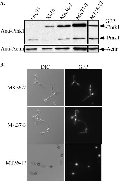
A mitogen-activated protein (MAP) kinase gene, PMK1, is known to regulate appressorium formation and infectious hyphal growth in the rice blast fungus Magnaporthe grisea. In this study, we constructed a green fluorescent protein gene-PMK1 fusion (GFP-PMK1) to examine the expression and localization of PMK1 in M. grisea during infection-related morphogenesis. The GFP-PMK1 fusion encoded a functional protein that complemented the defect of the pmk1 deletion mutant in appressorium formation and plant infection. Although a weak GFP signal was detectable in vegetative hyphae, conidia, and germ tubes, the expression of GFP-Pmk1 was increased in appressoria and developing conidia. Nuclear localization of GFP-Pmk1 proteins was observed in a certain percentage of appressoria. A kinase-inactive allele and a nonphosphorylatable allele of PMK1 were constructed by site-directed mutagenesis. Expression of these mutant PMK1 alleles did not complement the pmk1 deletion mutant. These data confirm that kinase activity and activation of PMK1 by the upstream MAP kinase kinase are required for appressorium formation and plant infection in M. grisea. When overexpressed with the RP27 promoter in the wild-type strain, both the kinase-inactive and nonphosphorylatable PMK1 fusion proteins caused abnormal germ tube branching. Overexpression of these PMK1 mutant alleles may interfere with the function of native PMK1 during appressorium formation.


Figures

FIG. 1.
FIG. 1.
Complementation of the pmk1 mutant with the GFP-PMK1 fusion construct. (A) Appressorium formation assay. Germ tubes from the wild-type strain (Guy11) developed appressoria by 24 h, but no appressorium formation was observed in the pmk1 mutant (nn78). Under the same conditions, a transformant of nn78 expressing the GFP-PMK1 fusion construct (Xh14) formed melanized appressoria that had GFP signals when examined under epifluorescence microscopy (Xh14-GFP). Bar = 10 μm. (B) Rice infection assay. Left to right, rice leaves were sprayed with conidia of nn78, Guy11, or Xh14 or 0.25% gelatin solution as the control. Typical leaves were photographed at 7 days postinoculation.
FIG. 2.
FIG. 2.
GFP-PMK1 was highly expressed in developing conidia. When blocks of Xh14 oatmeal cultures were examined in situ by epifluorescence microscropy, a strong fluorescence signal was observed in young developing conidia. Conidiophores bearing young conidia were also fluorescent, but the signal was much weaker. Mature conidia exhibited only weak fluorescence. CP, conidiophore; YC, young conidium; MC, mature conidium. Bar = 10 μm.
FIG. 3.
FIG. 3.
Expression and cellular localization of GFP-PMK1 during appressorium formation in transformant Xh14. (A) Xh14 conidia germinated on glass coverslips were removed at indicated times and examined under differential interference contrast (left panels) and epifluorescence microscopy (right panels). Fluorescence was observed in the conidia and germ tubes at the early time points, such as 2 or 4 h. By 12 h, the majority of the GFP signal was observed in appressoria. At 24 h, conidial cells exhibited no GFP signal or only a very weak GFP signal. (B) The GFP-Pmk1 signal localized to the nucleus in appressoria. A conidium of Xh14 incubated on a glass coverslip for 24 h was stained with Calcofluor and Hoechst 33258 (middle panel) to visualize cell walls and nuclei (arrow). GFP signal (right panel) appeared to be concentrated in the appressorium at the position of the nucleus. Bar = 10 μm.
FIG. 4.
FIG. 4.
GFP-PMK1 was expressed in infectious hyphae. Conidia from strain Xh14 were inoculated on onion epidermal cells and examined under differential interference contrast (left panels) or epifluorescence microscopy (right panels). At 24 h, the majority of appressoria formed by Xh14 had not penetrated plant cells yet and contained strong GFP fluorescence. At 48 h, appressoria of Xh14 that had successfully penetrated onion epidermal cells contained no GFP signal or weak GFP signal. Fluorescence was observed in infectious hyphae without any special cellular localization pattern. In some appressoria, fluorescent signals in underlying infectious hyphae could be observed through the collapsed appressoria. Bar = 10 μm. A, appressorium; IF, infectious hypha; C, conidium.
FIG. 5.
FIG. 5.
The K53R and AEF mutations abolished the function of PMK1 in appressorium formation and infectious growth. (A) Conidia of transformants expressing GFP-PMK1AEF (AEF-4) and GFP-PMK1K53R (K53R-110) were incubated on glass coverslips for 24 h and examined under DIC (left panels) or epifluorescence microscopy (right panels). GFP-Pmk1 expression was detectable in conidia and germ tubes of AEF-4 and K53R-110, but no appressorium formation was observed. (B) Leaves of rice cultivar CO-39 injected with conidia from the pmk1 mutant (nn78) or transformants of nn78 expressing the GFP-PMK1 (Xh14), GFP-PMK1AEF (AEF-4), or GFP-PMK1K53R (K53R-110) constructs. Spreading lesions were observed only in leaves injected with Xh14 but not AEF-4 or K53R-110. Photos were taken at 7 days after inoculation. DIC, differential interference contrast.
FIG. 6.
FIG. 6.
Western blot analysis of the expression of PMK1 and GFP-PMK1 constructs. Total protein was isolated from mycelia of the pmk1 deletion mutant (nn78), the wild-type strain (Guy11), or transformants of nn78 expressing GFP-PMK1 (Xh14), GFP-PMK1K53R (K53R-110), or GFP-PMK1AEF (AEF-4). When probed with the anti-Pmk1 antiserum (top panel), a 42-kDa Pmk1 band was observed in Guy11. In Xh14, AEF-4, and K53R-110, a larger protein of 68 kDa corresponding to the GFP-Pmk1 fusion was detected. When probed with an anti-GFP antibody (middle), a 68 kDa band was detected in Xh14, AEF-4, and K53R-110 but not in Guy11. The bottom panel was detection with an antiactin antibody to show that a similar amount of total protein was loaded in each lane.
FIG. 7.
FIG. 7.
Overexpression of PMK1 mutant alleles resulted in morphological defects during appressorium formation. (A) Western blot analyses with total proteins extracted from the wild-type strain (Guy11) or transformants expressing the PMK1K53R (MK36-2) or GFP-PMK1AEF (MK37-3) fusion construct under the RP27 promoter. When probed with the anti-Pmk1 antiserum, MK36-2 and MK37-3 had two bands corresponding to the native Pmk1 and GFP-Pmk1 fusion proteins. A separate blot containing total protein from strain MT37-12 probed with anti-Pmk1 contains similar levels of protein between the native PMK1 and the GFP fusion. Blots probed with antiactin antibody demonstrated the relative variance in total proteins loaded in each lane. (B) Branching germ tubes formed by MK36-2 and MK37-3. Conidia from 10-day-old cultures were incubated at room temperature for 24 h and examined with differential intererence contrast (DIC) or epifluorescence microscopy. MT36-17 expressing GFP-PMK1K53R fusion under the native Pmk1 promoter had no morphological defects during appressorium formation.

References

    1. Adachi, M., M. Fukuda, and E. Nishida. 1999. Two co-existing mechanisms for nuclear import of MAP kinase: passive diffusion of a monomer and active transport of a dimer. EMBO J. 18:5347-5358. - PMC - PubMed
    1. Bardwell, L., J. G. Cook, D. Voora, D. M. Baggott, A. R. Martinez, and J. Thorner. 1998. Repression of yeast Ste12 transcription factor by direct binding of unphosphorylated Kss1 MAPK and its regulation by the Ste7 MEK. Genes Dev. 12:2887-2898. - PMC - PubMed
    1. Bardwell, L., J. G. Cook, J. X. Zhu-Shimoni, D. Voora, and J. Thorner. 1998. Differential regulation of transcription: repression by unactivated mitogen-activated protein kinase Kss1 requires the Dig1 and Dig2 proteins. Proc. Natl. Acad. Sci. USA 95:15400-15405. - PMC - PubMed
    1. Blackwell, E., I. M. Halatek, H. J. N. Kim, A. T. Ellicott, A. A. Obukhov, and D. E. Stone. 2003. Effect of the pheromone-responsive G(alpha) and phosphatase proteins of Saccharomyces cerevisiae on the subcellular localization of the Fus3 mitogen-activated protein kinase. Mol. Cell. Biol. 23:1135-1150. - PMC - PubMed
    1. Bourett, T. M., J. A. Sweigard, K. J. Czymmek, A. Carroll, and R. J. Howard. 2002. Reef coral fluorescent proteins for visualizing fungal pathogens. Fungal Genet. Biol. 37:211-220. - PubMed

Publication types

Substances