. 2004 Dec 8;32(21):6454-67.
doi: 10.1093/nar/gkh981. Print 2004.

A novel E2 box-GATA element modulates Cdc6 transcription during human cells polyploidization

Affiliations

A novel E2 box-GATA element modulates Cdc6 transcription during human cells polyploidization

Nuria Vilaboa et al. Nucleic Acids Res. .

Abstract

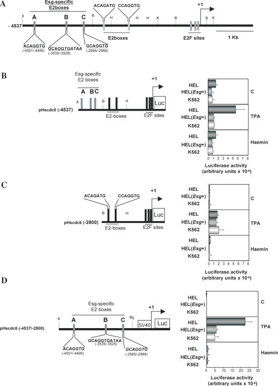
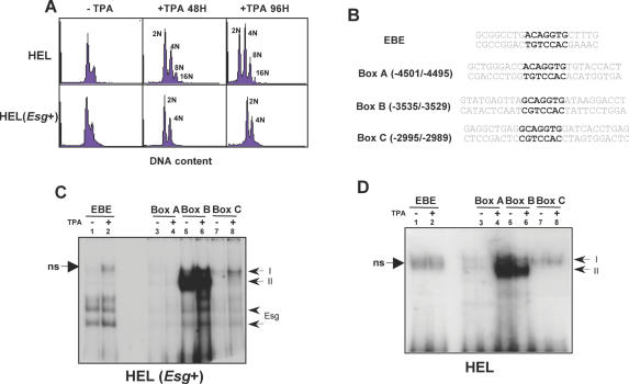
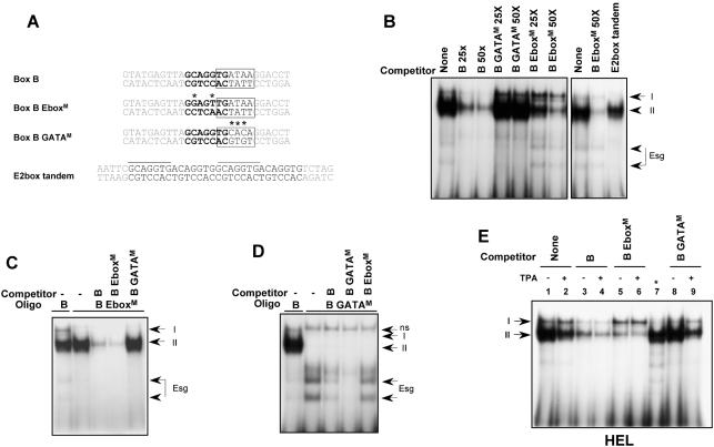
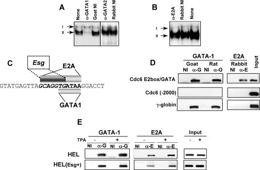
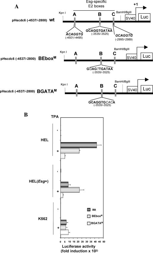
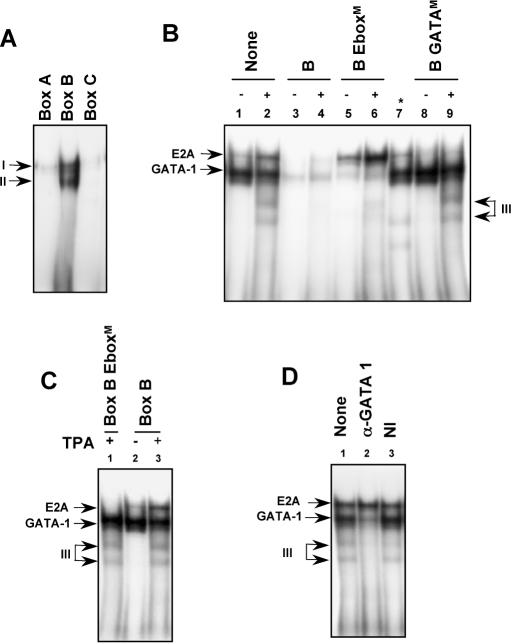
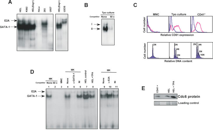
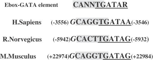
Cdc6 is a key regulator of the strict alternation of S and M phases during the mitotic cell cycle. In mammalian and plant cells that physiologically become polyploid, cdc6 is transcriptionally and post-translationally regulated. We have recently reported that Cdc6 levels are maintained in megakaryoblastic HEL cells, but severely downregulated by ectopic expression of transcriptional repressor Drosophila melanogaster escargot. Here, we show that cdc6 promoter activity is upregulated during megakaryocytic differentiation of HEL endoreplicating cells, and that Escargot interferes with such activation. Transactivation experiments showed that a 1.7 kb region located at 2800 upstream cdc6 transcription initiation site behaved as a potent enhancer in endoreplicating cells only. This activity was mainly dependent on a novel cis-regulatory element composed by an E2 box overlapping a GATA motif. Ectopic Escargot could bind this regulatory element in vitro and endogenous GATA-1 and E2A formed specific complexes in megakaryoblastic cells as well as in primary megakaryocytes. Chromatin Immunoprecipitation analysis revealed that both transcription factors were occupying the E2 box/GATA site in vivo. Altogether, these data suggest that cdc6 expression could be actively maintained during megakaryocytic differentiation through transcriptional mechanisms involving specific cis- and trans-regulatory elements.


Figures

Figure 1
Figure 1
cdc6 mRNA levels are downregulated in differentiated HEL(Esg+) cells. (A) HEL and HEL(Esg+) were cultured for 48 h in the absence (−) or in the presence (+) of 10−8 M TPA for 48 h. Total RNA was extracted and analyzed by northern blotting with full-length cdc6 and 28S rRNA-specific cDNA probes. Quantification of cdc6 mRNA signals normalized by 28S RNA is also shown (dark bars, 3.3 kb specie and light bars, 2.5 kb specie). (B) Cells were cultured as in (A), and then treated for the indicated times with 5 μg/ml Act D. RNA extraction and analysis was performed as in (A). (C) HEL and HEL(Esg+) cells were seeded in triplicate wells and cultured in the absence (closed circles) or in the presence (open triangles) of TPA. At the indicated times, cells were harvested and viable cells were counted as presented in Materials and Methods. (D) Cells were cultured as in (A) and nuclear extracts were isolated, subjected to SDS–PAGE and analyzed by western blotting with anti-Cdc6 antibody. A portion of total protein staining of transferred gel is shown as a loading control.
Figure 2
Figure 2
cdc6 upstream region containing Esg-binding E2 boxes is responsible for reduced cdc6 expression in non-endoreplicating cells. (A) Scaled diagram of the human cdc6 gene showing the E2 boxes with perfect match to Esg consensus binding sites (referred to as Esg-specific E2 Boxes A, B and C) and E2 boxes corresponding to much less specific Esg binding sites and E2F-binding elements. The arrow indicates the transcription initiation site. Relevant restriction sites are also shown: X, XhoI; B, BamHI; H, HinDIII; K, KpnI; and B, BglII. (B and C) HEL, HEL(Esg+) and K562cells were transfected with pHscdc6(−4537) (B) or pHscdc6(−2800) (C), cultured in the absence or in the presence of TPA or hemin, as indicated, collected and assayed for luciferase activity. (D) HEL, HEL(Esg+) and K562 and cells were transfected with pHscdc6 (−4537/−2800), cultured in the absence or in the presence of TPA or hemin, as indicated, collected and assayed for luciferase activity. The data represent the mean ± SD of at least three independent experiments.
Figure 3
Figure 3
Esg and endogenous factors preferentially bind the E2 box/GATA site located at −3535/−3525 upstream cdc6 gene. (A) HEL and HEL(Esg+) were cultured in the absence (−) or in the presence (+) of 10−8 M TPA for 48 or 96 h as indicated. DNA content pattern of these cells is shown as analyzed by flow cytometry. Vertical axis, relative number of cells; horizontal axis, relative red fluorescence (FL2) in a logarithmic scale, indicating DNA content per cell. (B) Synthetic oligonucleotides corresponding to the EBE, Box A, Box B and Box C used for EMSAs. (C) Nuclear extracts obtained from HEL(Esg+) cells cultured in the absence (−) or in the presence (+) of TPA were incubated with labeled EBE, Box A, Box B or Box C and analyzed using EMSA. (D) Nuclear extracts obtained from HEL cells cultured in the absence (−) or in the presence (+) of TPA were incubated with labeled EBE, Box A, Box B or Box C and analyzed using EMSA. Arrows indicate I, II and Esg complexes, as well a non-specific complex (ns).
Figure 4
Figure 4
Endogenous nuclear factors require both E2 box and GATA motifs to bind the −3535/−3525 E2 box/GATA site. (A) Synthetic oligonucleotides corresponding to Box B, Box B EboxM, Box B GATAM and the E2 box tandem used for EMSAs. E2 box appears in boldface and GATA site is boxed. Asterisks show nucleotide modified to obtain mutant E2 box and GATA elements. Esg-specific E2 boxes are marked with a line on the E2 box tandem oligonucleotide. (B) Nuclear extracts from HEL(Esg+) cells were incubated with labeled Box B in the absence (None) or the presence of an excess of unlabelled Box B (B), Box B EboxM (B EboxM), Box B GATAM (B GATAM) or E2 box tandem (25 and 50 times, as indicated) and analyzed using EMSA. (C and D) Binding reactions were carried out with the following labeled oligonucleotides: Box B EboxM (C) or Box B GATAM (D) in the absence (−) or the presence of 50-fold excess of unlabeled Box B, Box B EboxM or Box B GATAM. A reaction containing labeled Box B is shown in the first lane of both (C) and (D) as a control. Arrows indicate I, II and Esg complexes, as well a non-specific complex (ns). (E) Nuclear extracts from HEL cells cultured in the absence (−) or in the presence (+) of TPA were incubated with labeled Box B in the absence (None, lanes 1 and 2) or the presence of 50-fold excess of unlabeled Box B (lanes 3–4), Box B EboxM (lanes 5 and 6) or Box B GATAM (lanes 8 and 9) and analyzed using EMSA. For comparison purposes, nuclear extracts from HEL(Esg+) cells were incubated with labeled Box B (*, lane 7).
Figure 5
Figure 5
Endogenous GATA-1 and E2A bind the −3535/−3525 E2 box/GATA site both in vitro and in vivo. (A) Nuclear extracts from HEL(Esg+) cells were incubated with labeled Box B in the absence (None) or in the presence of goat anti-GATA-1 antibody (α-GATA1), non-immune goat serum (Goat NI), rabbit anti-GATA-2 antibody (α-GATA2) or non-immune rabbit serum (Rabbit NI), and analyzed using EMSA. (B) A similar experiment was performed in the presence of rabbit anti-E2A antibody (α-E2A) or non-immune rabbit serum (Rabbit NI). Arrows show complexes I and II. (C) Schematic representation of the binding requirements of GATA1, E2 box and Escargot complexes to E2 box/GATA motif. (D) ChIP assay was performed using fragmented cross-linked chromatin from HEL cells and anti-GATA-1 antibodies, αG (Goat C20, or Rat N6), anti-E2A antibody, αE (RabbitV18) or the corresponding non-immune immunoglobulins, NI. One-sixth of total purified DNA was amplified by PCR with primers specific for the fragment including the E2 box-GATA site (Cdc6 E2 box/GATA) as well as an unrelated fragment (Cdc6-2000) of cdc6 upstream region. A fragment in the A-γ-globin locus (γ-globin), is included as a positive control. PCRs were also performed using the fragmented chromatin purified prior to antibody incubation (Input). (E) ChIP assays were performed using fragmented cross-linked chromatin from HEL and HEL(Esg+) cells cultured in the absence (−) or in the presence (+) of TPA and non-immune goat or rabbit Ig (NI), goat anti-GATA-1 (α-G) or rabbit anti-E2A antibodies (α-E).
Figure 6
Figure 6
Enhancer activity of −4537 to −2800 cdc6 5′ upstream region in megakaryocytic endoreplicating cells is mainly mediated through the E2 box/GATA motif. (A) Diagrams of pHscdc6(−4537/−2800) (wt) and Ebox (BEboxM) and GATA (BGATAM) mutant constructs. (B) HEL, HEL(Esg+) and K562 cells were transfected with wild-type or mutant constructs. Transfected cells were cultured in the absence (−) or in the presence (+) of TPA, collected and assayed for luciferase activity. The data represent the mean ± SD of at least three independent experiments.
Figure 7
Figure 7
Non-endoreplicating K562 cells show additional GATA-1-containing complexes after megakaryocytic differentiation. (A) Nuclear extracts obtained from exponentially growing K562 cells were incubated with labeled Box A, Box B or Box C oligonucleotides and analyzed by EMSA. (B) Nuclear extracts from K562 cells cultured in the absence (−) or in the presence (+) of TPA were incubated with labeled Box B in the absence (None, lanes 1 and 2) or in the presence of 50-fold excess of unlabeled Box B (lanes 3 and 4), Box B EboxM (lanes 5 and 6) or Box B GATAM (lanes 8 and 9) and analyzed using EMSA. For comparison purposes, nuclear extracts from HEL(Esg+) cells were incubated with labeled Box B (*, lane 7). (C) Nuclear extracts from K562 cells cultured in the absence (−) or in the presence (+) of TPA were incubated with labeled Box B EboxM or Box B and analyzed using EMSA. (D) Nuclear extracts from TPA-treated K562 cells were incubated with labeled Box B in the absence (None) or the presence of goat anti-GATA-1 antibody (α-GATA1) or non-immune goat serum (NI) and analyzed using EMSA. Arrows indicate the already described E2A and GATA-1 containing complexes, as well as GATA-1-containing complex III.
Figure 8
Figure 8
GATA-1 and E2A containing complexes are characteristic of megakaryocytes. (A) Nuclear extracts from exponentially growing HEL, K562, HEL(Esg+), HeLa, 293 U937 and U2OS cells were incubated with labeled Box B oligonucleotide and analyzed using EMSA. Arrows indicate E2A and GATA-1 containing complexes. (B) Nuclear extracts from megakaryocytic-enriched primary human cells (Tpo culture) were incubated with labeled Box B in the absence (None) or the presence (50×) of a 50-times excess of unlabeled Box B and analyzed using EMSA. (C) MNC isolated from human Umbilical Cord Blood, megakaryocytic-enriched cells obtained after culture of MNC-derived CD34+ cells in the presence of Tpo (Tpo-culture) and immuno-selected megakaryocytes (CD41+) were labeled with FITC-conjugated anti-GpIIIa (CD61) antibodies (upper panel) or Propidium Iodide (lower panel) and analyzed using flow cytometry. (D) Nuclear Extracts from CD41+ cells (MK), MNC and HEL cells untreated (control) or TPA-treated (+TPA), were incubated with labeled Box B and analyzed using EMSA. Lane 2: labeling reaction was performed with MK extracts in the presence of 50 times excess unlabeled Box B. Lanes 4–6 and 9–11: labelling reaction was performed with MK extracts in the absence (None) or the presence of non-inmune serum (NI) or the indicated antibodies (α-GATA-1 or α-E2A). (E) Nuclear extracts were obtained from CD41+ and untreated or TPA-treated HEL cells. Proteins were subjected to SDS–PAGE and analyzed by western blotting with anti-cdc6 antibody. A portion of total protein staining of transferred gel is shown as a loading control.
Figure 9
Figure 9
A Ebox-GATA element is present in cdc6 locus of mouse and rat. In silico search for the presence of an E2box-GATA motif through ∼50 000 bp of rat chromosome10 (87 675 000–87 725 000 bp) and mouse chromosome 1 (98 530 000–98 610 000 bp) which spans cdc6 gene and surrounding upstream and downstream 15 000 bp. Both human and rat elements are located upstream first exon, whereas mouse element lies in the 3′-untranslated region. Numbers refer to the positions respective to reported first exon.

Similar articles

Cited by

References

    1. Edgar B.A. and Orr-Weaver,T.L. (2001) Endoreplication cell cycles: more for less, Cell, 105, 297–306. - PubMed
    1. Ravid K., Lu,J., Zimmet,J.M. and Jones,M.R. (2002) Roads to polyploidy: the megakaryocyte example. J. Cell Physiol., 190, 7–20. - PubMed
    1. Zimmet J. and Ravid,K. (2000) Polyploidy: occurrence in nature, mechanisms, and significance for the megakaryocyte-platelet system. Exp. Hematol., 1, 3–16. - PubMed
    1. Garcia P. and Cales,C. (1996) Endoreplication in megakaryoblastic cell lines is accompanied by sustained expression of G1/S cyclins and downregulation of cdc25C. Oncogene, 13, 695–703. - PubMed
    1. Zimmet J.M., Ladd,D., Jackson,C.W., Stenberg,P.E. and Ravid,K. (1997) A role for cyclin D3 in the endomitotic cell cycle. Mol. Cell. Biol., 17, 7248–7259. - PMC - PubMed

Publication types

MeSH terms