. 2004 Dec 8;24(49):11035-45.
doi: 10.1523/JNEUROSCI.3722-04.2004.

Interactions of postsynaptic density-95 and the NMDA receptor 2 subunit control calpain-mediated cleavage of the NMDA receptor

Affiliations

Interactions of postsynaptic density-95 and the NMDA receptor 2 subunit control calpain-mediated cleavage of the NMDA receptor

Yi Na Dong et al. J Neurosci. .

Abstract

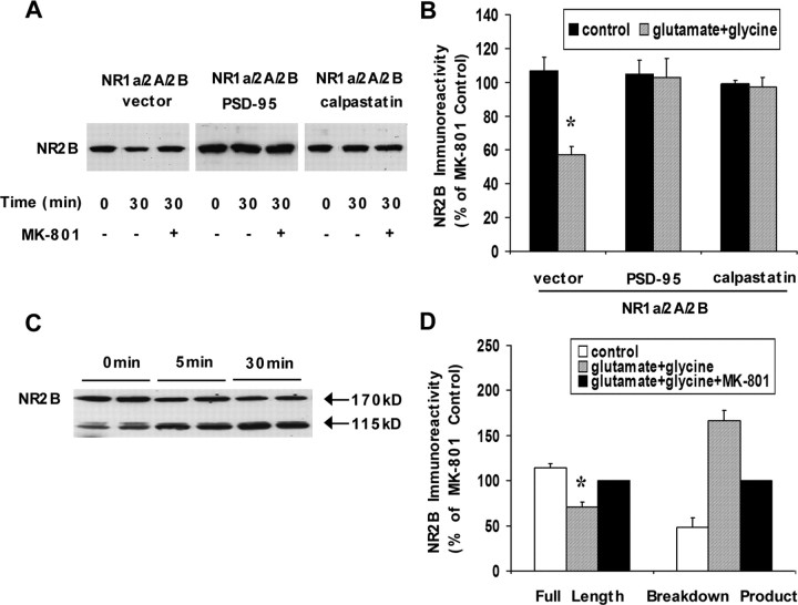
The calcium-dependent protease calpain cleaves the NMDA receptor 2 (NR2) subunit of the NMDA receptor both in vitro and in vivo and thus potentially modulates NMDA receptor function and turnover. We examined the ability of postsynaptic density-95 (PSD-95) protein to alter the calpain-mediated cleavage of NR2A and NR2B. Coexpression of PSD-95 with NMDA receptors in human embryonic kidney 293 cells blocked cleavage of NR2A and NR2B by NMDA receptor-activated calpain. NR2A cleavage by calpain occurred in the cell surface and intracellular fractions and required the presence of NR1 subunits. The blocking effect of PSD-95 did not result from decreased calpain activity, lowered intracellular calcium responses, or the blockade of internalization. Instead, this effect was eliminated by deletion of the C-terminal ESDV motif of NR2A or by overexpression of a palmitoylation-deficient PSD-95 mutant lacking the ability to cluster and to interact with NMDA receptors in situ, suggesting a role for association between the C terminus of NR2A and clustered PSD-95. Synapse-associated protein 102, a membrane-associated guanylate kinase interacting with NR2A but lacking palmitoylation motifs and the ability to cluster, did not protect NR2A from cleavage by calpain. Pharmacological inhibition of palmitoylation disrupted the interaction of PSD-95 with NMDA receptors in cortical neurons and allowed NR2A to be cleaved by calpain, whereas NR2A could not be cleaved in untreated neurons. These results indicate that PSD-95 clustering and direct association of NR2A and PSD-95 mediate the blocking effect of PSD-95 on calpain cleavage. PSD-95 could regulate the susceptibility of NMDA receptors to calpain-mediated cleavage during synaptic transmission and excitotoxicity.


Figures

Figure 1.
Figure 1.
Effects of PSD-95 on calpain-mediated cleavage of the NR2A subunit. HEK293 cells were transfected with NR1a, NR2A, and PSD-95, calpastatin, or vector control and treated with 100 μm glutamate and 100 μm glycine in the presence or absence of 100 μm MK-801 for 0 or 30 min. NMDA receptor activation resulted in a significant decrease in full-length NR2A immunoreactivity (A, representative Western blots of NR2A) as confirmed by quantification of NR2A levels (B) (N-terminal antibody A-6475; *p = 0.0008; n = 8). Coexpression of PSD-95 or calpastatin inhibited the reduction in NR2A immunoreactivity. No proteolysis of NR2A was observed when NMDA receptor activity was blocked by addition of MK-801. Quantitated data are expressed as a percentage of the MK-801 control condition. C, Calpain activity was measured after 30 min agonist treatment by assessment of a calpain-generated spectrin degradation product as detected by an antibody to this product (AB38); coexpression of PSD-95 had no effect on the production of calpain-cleaved spectrin compared with the vector control (p = 0.2441; n = 5). D, No difference was observed between intracellular calcium responses of NR1a/2A cells cotransfected with PSD-95 or vector control (p = 0.1709; n = 14). Data are shown as mean ± SEM (error bars).
Figure 2.
Figure 2.
Effects of PSD-95 on calpain-mediated cleavage of the NR2B subunit. A, Representative Western blots of NR2B from HEK293 cells cotransfected with NR1a, NR2A, NR2B, and PSD-95, calpastatin, or vector control and treated with 100 μm glutamate and 100 μm glycine in the presence or absence of 100 μm MK-801 for 0 or 30 min. B, Quantification of NR2B levels. Agonist treatment resulted in the significant degradation of NR2B (N-terminal antibody to NR2B; *p = 0.0157; n = 5). Coexpression of PSD-95 or calpastatin inhibited the reduction in NR2B immunoreactivity. No stable breakdown product of NR2B was observed subsequent to calpain activation. C, Representative Western blots of NR2B from cultured cortical neurons treated with 100 μm glutamate and 100 μm glycine for 5 or 30 min. D, Quantification of NR2B levels. Agonist treatment decreased the level of full-length NR2B and increased the level of an N-terminal breakdown product (115 kDa) compared with 0 min (N-terminal antibody to NR2B; n = 6; p = 0.0049 for full-length NR2B; 0 vs 30 min; p = 0.0057 for NR2B breakdown product; 0 vs 30 min). The inclusion of MK-801 prevented the loss of full-length NR2B and the increase in the NR2B breakdown product. NR2B immunoreactivity was quantitated as a percentage of the MK-801 control condition. Data are shown as mean ± SEM (error bars).
Figure 3.
Figure 3.
Effects of PSD-95 on the cleavage of shortened forms of NR2A by calpain. A, Representative Western blots of NR2A1400, NR2A1330, and NR2A1279 from HEK293 cells cotransfected with NR1a and PSD-95, calpastatin, or vector control and treated with 100 μm glutamate and 100 μm glycine in the presence or absence of 100 μm MK-801 for 0 or 30 min. B, Quantification of NR2A1400 levels. Agonist treatment of cells cotransfected with NR1a/NR2A1400 resulted in the significant degradation of NR2A1400 (N-terminal antibody A-6475; *p = 0.0001; n = 6). Coexpression of calpastatin inhibited the reduction in NR2A1400 immunoreactivity, but the presence of PSD-95 had no effect (*p = 0.0023; n = 4). C, Quantification of NR2A1330 levels. Agonist treatment of cells cotransfected with NR1a/NR2A1330 resulted in the significant degradation of NR2A1330 (N-terminal antibody A-6475; *p = 0.0037; n = 8). Coexpression of calpastatin inhibited the reduction in NR2A1330 immunoreactivity, but the presence of PSD-95 had no effect (*p = 0.0149; n = 7). D, Quantification of NR2A1279 levels. No detectable change in NR2A1279 immunoreactivity was observed after agonist treatment (p = 0.1767; n = 5). No difference was seen after coexpression of calpastatin or PSD-95. Quantitated data are expressed as a percentage of the MK-801 control condition. Data are shown as mean ± SEM (error bars).
Figure 4.
Figure 4.
Interaction of the ESDV motif with PSD-95 in NR2A cleavage by calpain. A, NR1a/2A1460 produced agonist-induced intracellular calcium transients similar to those of wild-type NR1a/2A (n = 10; p = 0.6632). B, Representative Western blots of NR2A1460 from HEK293 cells cotransfected with NR1a and PSD-95, calpastatin, or vector control and treated with 100 μm glutamate and 100 μm glycine in the presence or absence of 100 μm MK-801 for 0 or 30 min. C, Quantification of NR2A1460 levels. NMDA receptor activation resulted in the significant degradation of NR2A1460 (N-terminal antibody A-6475; *p = 0.0002; n = 5). Coexpression of calpastatin inhibited the reduction in NR2A1460 immunoreactivity, but the presence of PSD-95 had no effect (*p = 0.0002; n = 5). NR2A1460 immunoreactivity was quantitated as a percentage of the MK-801 control condition. Data are mean ± SEM (error bars). D, The histogram summarizes the different cleavage patterns of NR2A and NR2A mutants by calpain in HEK293 cells. One hundred micromolar glutamate and 100 μm glycine treatment for 30 min resulted in 46, 33, 35, and 24% decreases in NR2A, NR2A1460, NR2A1400, and NR2A1330 immunoreactivity, respectively. No significant cleavage in NR2A1279 was detected. Immunoreactivity of NR2A and NR2A mutants was quantitated as a percentage of the MK-801 control condition. Data are expressed as mean ± SEM (error bars).
Figure 5.
Figure 5.
Location of the calpain-mediated cleavage of NR2A in HEK293 cells. HEK293 cells cotransfected with the NR1a/2A combination were preincubated with 100 μm glutamate and glycine in the presence or absence of 100 μm MK-801 for 0 or 30 min (n = 8) and then labeled with NHS-biotin under nonlytic conditions. The extracellular and intracellular fragments were separated and subjected to Western blotting with an N-terminal antibody for NR2A (A-6475). The amount of immunoreactivity in the lysate (L), extracellular (E), and intracellular (I) fractions was quantified as a percentage of the MK-801 control condition. A representative Western blot and bar graph (A, B) demonstrate a reduced level of NR2A immunoreactivity in the intracellular fraction (50% decrease from the 0 min control) (n = 8; p = 0.0166) and extracellular fraction (45% decrease from the 0 min control) (n = 8; p = 0.0172). An anti-actin antibody confirmed that actin remains primarily in the intracellular fraction, with 90-95% of actin appearing in the intracellular fraction, as shown by a representative blot (A), showing that membrane integrity was preserved. Cotransfection with PSD-95 prevented the loss of NR2A in both the intracellular and extracellular fractions (n = 7; p = 0.4754 for intracellular fraction; p = 0.9247 for extracellular fraction) (C, D). Data are shown as mean ± SEM (error bars).
Figure 6.
Figure 6.
Assembly of NR1 and NR2 subunits is required for calpain-mediated cleavage. HEK293 cells cotransfected with different combinations of NMDA receptor subunits were treated with 1 μm ionomycin for 10 min to activate calpain and analyzed by Western blots. NR1 and NR2A immunoreactivities were quantitated as a percentage of the CalI3. Data are shown as mean ± SEM (error bars). NR1 was not cleaved by calpain whether it was present alone (A) or assembled with NR2A (B). In contrast, the level of NR2A significantly decreased after ionomycin treatment when associated with NR1 (31% decrease from 0 min) (n = 6; p = 0.0132). This was inhibited by the inclusion of CalI3 (n = 6; p = 0.018) (C). No degradation of NR2A was found in transfections including NR2A alone (n = 5; p = 0.31) (D), showing that only coassembled NMDA receptors are affected by calpain. E, HEK293 cells cotransfected with the NR1a/2A combination were treated with 100 μm glutamate and 100 μm glycine for 30 min and then subjected to immunoprecipitation. An anti-NR2A antibody (N-terminal antibody A-6475) was used for immunoprecipitation, and the resulting blots were probed with both NR2A and NR1 antibodies. Input, Total cell lysate; IP-NR2A, immunoprecipitated fraction. Representative Western blots show a significant decrease in NR1 and NR2A levels. F, Quantification of NR2A and NR1 levels in the immunoprecipitated fraction. Agonist treatment resulted in a 72% decrease in NR1 immunoreactivity (n = 3; p = 0.0088) and a 31% decrease in NR2A immunoreactivity compared with 0 min (n = 3; p = 0.012), suggesting that NR1 is also destroyed when associated with NR2A that is degraded by calpain. Data are shown as mean ± SEM (error bars).
Figure 7.
Figure 7.
Blockade of NMDA receptor internalization does not alter calpain-mediated cleavage. HEK293 cells cotransfected with the NR1a/2A combination and wild-type dynamin or K44A dynamin were treated with 100 μm glutamate and 100 μm glycine in the presence or absence of 100 μm MK-801 for 0 or 30 min and analyzed by Western blotting. The amount of NR2A immunoreactivity was quantified as a percentage of the MK-801 control condition. Agonist treatment caused a significant decrease in NR2A immunoreactivity (32% decrease from 0 min) in dynamin-cotransfected cells (n = 5; p = 0.034) (A), whereas cotransfection of the mutant dynamin form K44A had no effect on calpain-mediated breakdown of NR2A (31% decrease from 0 min) (n = 5; p = 0.038) (B) and did not result in appearance of novel products. C, D, NR1a/2A-cotransfected HEK293 cells were pretreated with concanavalin A (0.25 mg/ml) (D) or vehicle (C) for 30 min and exposed to 100 μm glutamate and 100 μm glycine in the presence or absence of 100 μm MK-801. No difference was found in the cleavage pattern of NR2A by calpain after concanavalin A pretreatment (n = 4; p = 0.0102; 0 vs 30 min) compared with the control group (n = 4; p = 0.0036). Data are shown as mean ± SEM (error bars).
Figure 8.
Figure 8.
PSD-95 clustering and its association with NMDA receptors are required for the PSD-95-mediated effect. HEK293 cells cotransfected with the NR1a/2A combination and wild-type (WT) PSD-95 or mutant PSD-95 (C3,5S) were treated with 100 μm glutamate and 100 μm glycine in the presence or absence of 100 μm MK-801 for 0 or 30 min and then labeled with NHS-biotin under nonlytic conditions. The extracellular and intracellular fragments were separated and subjected to Western blotting with an N-terminal antibody for NR2A (A-6475). The amount of immunoreactivity in the lysate (L), extracellular (E), and intracellular (I) fractions was quantified as a percentage of the MK-801 control condition. In cells transfected with PSD-95 (C3,5S), a representative Western blot and bar graph (A, B) demonstrate reduced levels of NR2A immunoreactivity after calpain activation in the intracellular fraction (47% decrease from the 0 min control) (n = 6; p = 0.021) and the extracellular fraction (34% decrease from the 0 min control) (n = 6; p = 0.0285), showing that the PSD-95 mutant (C3,5S) does not block calpain-mediated cleavage of NR2A in either the intracellular and extracellular fractions. An anti-actin antibody confirmed that actin remains primarily in the intracellular fraction. C, HEK293 cells cotransfected with the NR1a/2A combination and wild-type PSD-95 (WT) or mutant PSD-95 (C3,5S) were immunoprecipitated with an anti-NR2A antibody. Input, Total cell lysate; IPNR2A, immunoprecipitated fraction. The resulting blots were probed with both NR2A and PSD-95 antibodies. Representative Western blots show the disruption of the NR2A-PSD-95 interaction by transfection with PSD-95 (C3,5S). D, HEK293 cells cotransfected with the NR1a/2A combination and SAP102 were treated with 100 μm glutamate and 100 μm glycine in the presence or absence of 100 μm MK-801 for 0 or 30 min and analyzed by Western blotting. The amount of NR2A immunoreactivity was quantified as a percentage of the MK-801 control condition. Cotransfection with SAP102 did not protect NR2A from calpain-mediated cleavage (46% decrease from 0 min) (n = 5; p = 0.025). Data are expressed as mean ± SEM (error bars).
Figure 9.
Figure 9.
Inhibition of palmitoylation regulates the susceptibility of NR2A to be cleaved by calpain. A, Representative Western blot demonstrating the developmental decrease in NR2A cleavage by calpain in cortical neurons. Cortical neurons from different ages were exposed to NMDA receptor agonist stimulation over a 0 or 30 min time course and subjected to Western blots. NR2A was cleaved by calpain at 2 weeks (2W) and became uncleavable after 3 weeks (3W) of development. The inclusion of MK-801 prevented the loss of NR2A during agonist treatment. B, Representative Western blot showing coimmunoprecipitation of NR2A and PSD-95. Cortical neurons (≥22 DIV) were treated with 100 μm 2-bromopalmitate or control (vehicle only) for 10 hr. NR2A was immunoprecipitated, and samples were immunoblotted with PSD-95 antibody. 2-Bromopalmitate treatment disrupted the interaction of PSD-95 and NR2A (left lane, control; right lane, 2-bromopalmitate). Equivalent amounts of NR2A were precipitated in each condition. C, Representative Western blot showing the cleavage of NR2A after 30 min agonist stimulation after 100 μm 2-bromopalmitate treatment. D, Quantification of NR2A levels. Treatment with 2-bromopalmitate increased the amount of NR2A cleaved to 34% (of 0 min) (n = 6; p = 0.0092) compared with the control group (10% decrease from 0 min); the inclusion of MK-801 prevented the loss of NR2A. E, Identification of the protease involved in NR2A breakdown. Cortical neurons preincubated with 2-bromopalmitate were treated with 100 μm glutamate and glycine for 30 min in the presence of CalI3, CaspI2, or CathI1. Inclusion of CalI3 (p = 0.0002 by ANOVA; n = 6; p = 0.0090; CalI3 vs 30 min) but not inhibitors of caspase or cathepsin prevented the loss of NR2A (n = 6; p > 0.05 for both). Data are expressed as mean ± SEM (error bars).

Similar articles

Cited by

References

    1. Anegawa NJ, Guttmann RP, Grant ER, Anand R, Lindstrom J, Lynch DR (2000) N-Methyl-d-aspartate receptor mediated toxicity in nonneuronal cell lines: characterization using fluorescent measures of cell viability and reactive oxygen species production. Brain Res Mol Brain Res 77: 163-175. - PubMed
    1. Bi R, Bi X, Baudry M (1998a) Phosphorylation regulates calpain-mediated truncation of glutamate ionotropic receptors. Brain Res 797: 154-158. - PubMed
    1. Bi X, Rong Y, Chen J, Dang S, Wang Z, Baudry M (1998b) Calpain-mediated regulation of NMDA receptor structure and function. Brain Res 790: 245-253. - PubMed
    1. Ceresa BP, Kao AW, Santeler SR, Pessin JE (1998) Inhibition of clathrin-mediated endocytosis selectively attenuates specific insulin receptor signal transduction pathways. Mol Cell Biol 18: 3862-3870. - PMC - PubMed
    1. Cho KO, Hunt CA, Kennedy MB (1992) The rat brain postsynaptic density fraction contains a homolog of the Drosophila discs-large tumor suppressor protein. Neuron 9: 929-942. - PubMed

Publication types

LinkOut - more resources