. 2004 Dec 8;24(49):11070-8.
doi: 10.1523/JNEUROSCI.3695-04.2004.

Independent presynaptic and postsynaptic mechanisms regulate endocannabinoid signaling at multiple synapses in the ventral tegmental area

Affiliations

Independent presynaptic and postsynaptic mechanisms regulate endocannabinoid signaling at multiple synapses in the ventral tegmental area

Arthur C Riegel et al. J Neurosci. .

Abstract

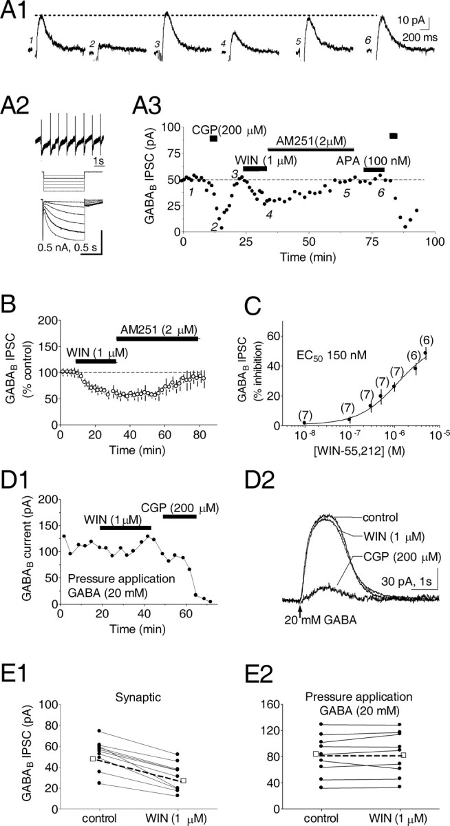
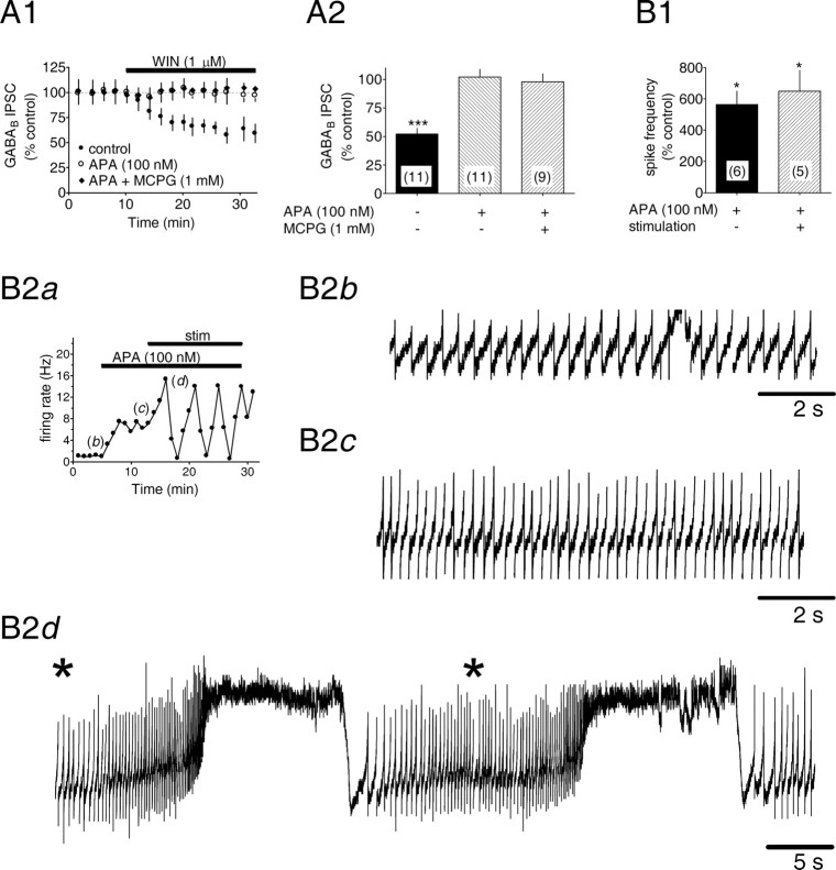
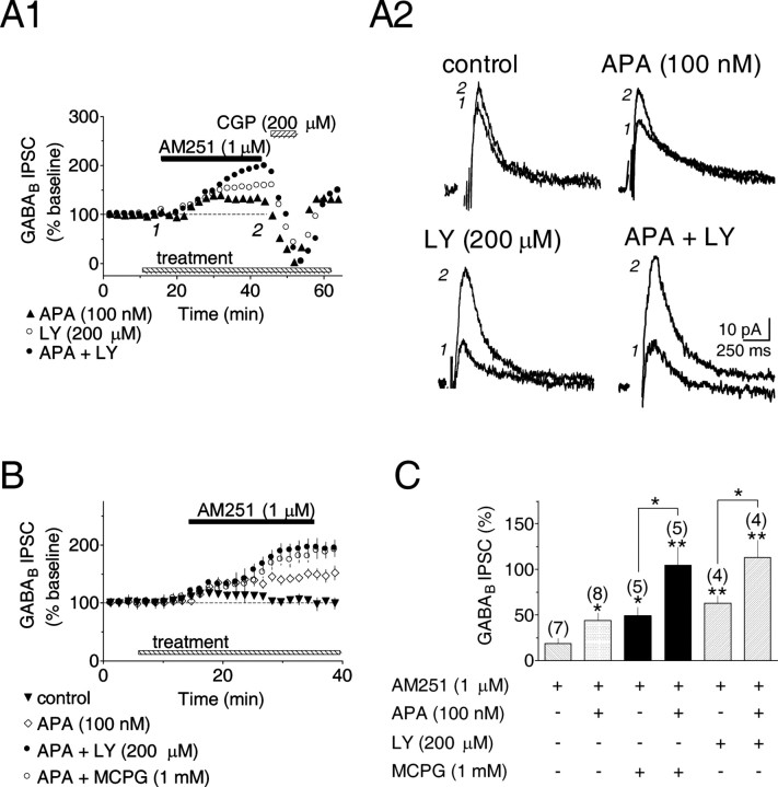
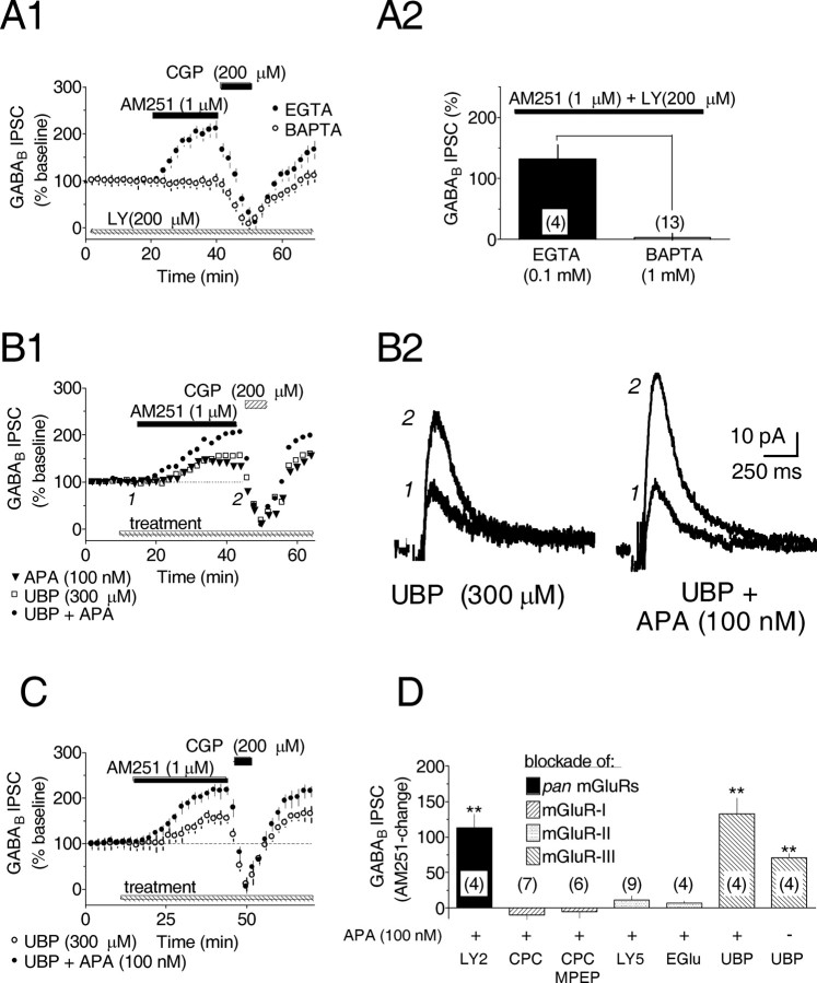
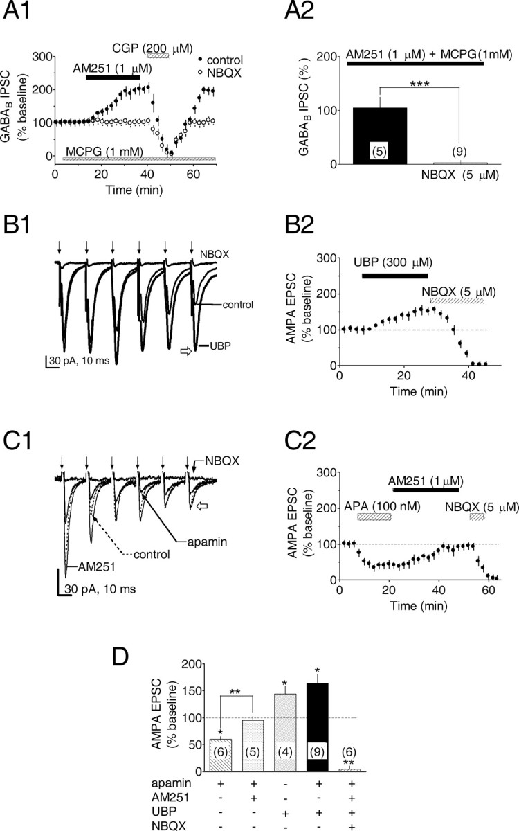
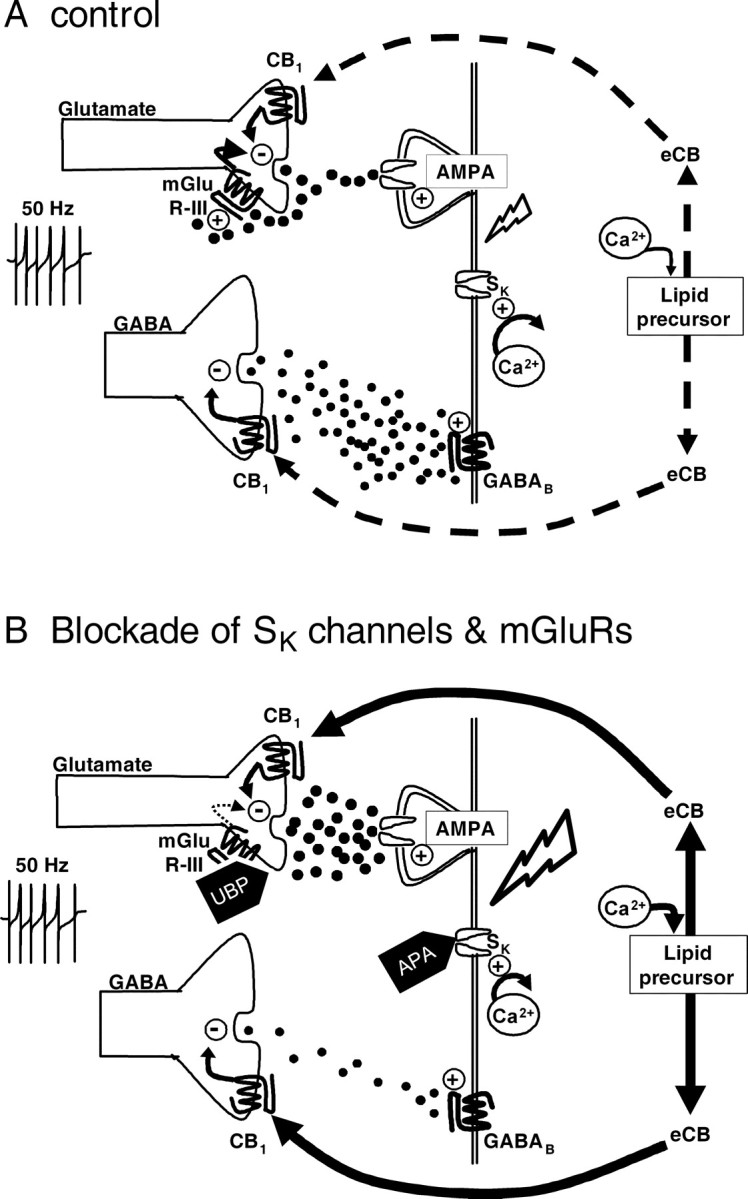
Dopamine (DA) neurons in the ventral tegmental area have been implicated in psychiatric disorders and drug abuse. Understanding the mechanisms through which their activity is regulated via the modulation of afferent input is imperative to understanding their roles in these conditions. Here we demonstrate that endocannabinoids liberated from DA neurons activate cannabinoid CB1 receptors located on glutamatergic axons and on GABAergic terminals targeting GABA(B) receptors located on these cells. Endocannabinoid release was initiated by inhibiting either presynaptic type-III metabotropic glutamate receptors or postsynaptic calcium-activated potassium channels, two conditions that also promote enhanced DA neuron excitability and bursting. Thus, activity-dependent release of endocannabinoids may act as a regulatory feedback mechanism to inhibit synaptic inputs in response to DA neuron bursting, thereby regulating firing patterns that may fine-tune DA release from afferent terminals.


Figures

Figure 1.
Figure 1.
Activation of presynaptic cannabinoid receptors diminishes GABAB IPSCs recorded in VTA DA neurons. A1, Averaged traces of GABAB IPSCs during conditions indicated in A3. In this and subsequent figures, stimulus artifacts have been removed. A2, Electrophysiological properties of the same VTA DA neuron include (1) regular pacemaker firing in the cell-attached configuration, (2) Ih (sag) current activated by hyperpolarizing voltage steps (-60 to -100 mV). A3, Time course showing effect of the cannabinoid receptor agonist WIN55,212-2 (WIN) and the CB1R antagonist AM251. IPSCs were blocked by the GABAB antagonist CGP35348 (CGP) but not APA. B, Time course showing the WIN55,212-2-induced inhibition of IPSCs and reversal by AM251. C, Concentration-response relationship for the effect of WIN on GABAB IPSCs. The number of experiments for each point is indicated. D1, Time course showing the peak amplitude of currents produced by pressure application of GABA to a VTA DA neuron. D2, Averaged traces demonstrate that GABA currents are inhibited by CGP but not by WIN. E, Summary of WIN effects on GABAB currents resulting from synaptic stimulation (E1) or pressure application of GABA (E2) in all DA neurons tested. (-□-) denotes the mean response. All experiments were conducted in the presence of picrotoxin (100 μm); Vh = -60 mV.
Figure 2.
Figure 2.
Blockade of SK channels prevents CB1R activation by WIN55,212-2 and increases DA neuron activity. A, Cannabinoid receptor-mediated inhibition of GABAB IPSCs is prevented by blockade of SK channels with APA. A1, Mean time courses showing that the WIN-mediated inhibition is blocked by APA and MCPG, a nonselective mGluR antagonist. A2, Summary of the effects of WIN under different conditions. The number of experiments is indicated. ***p < 0.001 versus control. B, Effects of APA and electrical stimulation on DA neuron activity. B1, Summary of the percentage increase in DA neuron spontaneous action potential frequency during the indicated conditions. B2a, Individual time course showing the increase in cell firing rate during control (b), APA (100 nm) (c), and apamin during electrical stimulation (d). Activity was assessed in the cell-attached configuration. Asterisk represents electrical stimulation.
Figure 3.
Figure 3.
Blockade of SK channels or mGluRs stimulates eCB production. A1, Three individual time courses during bath application of the CB1R-selective antagonist AM251. In the presence of APA or the nonselective mGluR antagonist LY341495 (LY), AM251 increased IPSC amplitudes. A2, Averaged GABAB IPSCs collected before and during AM251, as shown in A1. Calibration is identical in all traces. B, Mean time courses illustrating effects of SK channel or mGluR blockade on AM251-induced changes in GABAB IPSC amplitude. C, Summary showing combined blockade of both SK channels and mGluRs during AM251 additively increases IPSCs. Numbers above each bar indicate the number of experiments. *p < 0.01; **p < 0.001.
Figure 4.
Figure 4.
Endocannabinoid-mediated inhibition of GABA release involves intracellular Ca2+ and is initiated by blockade of presynaptic mGluR-IIIs. A1, Mean time courses during bath application of AM251, CGP, and LY. The AM251-induced increase in GABAB IPSC amplitude after mGluR antagonist treatment was prevented by substituting BAPTA for EGTA in the recording solution. A2, Summary of the AM251-induced change in GABAB IPSC amplitude with EGTA or BAPTA. The number of data points is indicated. p < 0.001; t test. All experiments were conducted in the presence of apamin (100 nm) and LY341495 (200 μm). B1, Individual time courses showing the AM251-induced increased in GABAB IPSCs during application of UBP1112 (UBP), a selective mGluR-III antagonist. For comparison, an additional time course for AM251 plus APA is also shown. B2, Averaged IPSC traces collected before and during AM251, as shown in B1. Calibration is identical in both traces. C, Mean time courses illustrating the AM251-induced increase in GABAB IPSCs during UBP1112 treatment. D, Summary of the effects of mGluR antagonists of various classes on the AM251-induced increase in GABAB IPSCs recorded in VTA DA neurons. The nonselective mGluR antagonists include LY341495 (LY2, 200 μm), the mGluR-I selective antagonist CPCCOET (CPC, 100 μm) and MPEP (30 μm), the mGluR-II selective antagonist LY341495 (LY5, 500 nm), and EGlu (200 μm). In all instances, apamin treatment occurred before mGluR antagonist application. **p < 0.01 versus control; one-way ANOVA and post hoc Newman-Keuls comparison.
Figure 5.
Figure 5.
Blockade of eCB production by AMPA-kainate receptor antagonism and CB1R-mediated inhibition of EPSCs via SK channel and mGluR-dependent eCB release. A1, Mean time courses showing that the AMPA-kainate receptor antagonist NBQX blocks the AM251-induced increase in the GABAB IPSC amplitude in the presence of the mGluR antagonist MCPG. A2, Summary of the effect of NBQX on the AM251-induced increase in IPSC amplitude during MCPG application. The number of experiments is indicated. B, C, During trains of electrical stimuli, application of UBP (B), a selective mGluR-III antagonist, or APA (C), a blocker of SK channels, facilitates or attenuates EPSCs in VTA DA neurons, respectively. Sample traces from a single experiment show averaged EPSCs evoked by the same electrical stimulation (↓) used to elicit GABAB IPSCs (50 Hz, 6 stimuli) during UBP (B1) or apamin (C1) application. In both B and C, EPSCs were blocked by NBQX, as determined as amplitude of the sixth EPSC of the series (B1, C1, open arrows). In C2, the apamin-induced inhibition was reversed by AM251, indicating that apamin acted via eCB release. D, Summary demonstrating the mean change in the EPSCs during the conditions described above. The number of treatments for each combination is indicated. *p < 0.05 and **p < 0.01 versus control; one-way ANOVA and post hoc Newman-Keuls comparison. All experiments were conducted in the presence of the GABAB antagonist CGP35348 (200 μm).
Figure 6.
Figure 6.
Proposed mechanism of CB1R activation by eCBs mediating the inhibition of GABA and glutamate release in the VTA. A, Under control conditions, electrical stimulation evokes the release of GABA from medium spiny terminals, which activates postsynaptic GABAB receptors on VTA DA neuron dendrites. Glutamate is also released by the stimulus. Glutamate then activates mGluR-IIIs located on glutamate terminals to limit further release and dampen DA neuron excitability. Under these conditions eCB release is absent, and cannabinoid agonists can activate CB1Rs on medium spiny neuron terminals to decrease GABA release onto DA neurons. B, Activation of glutamatergic afferents in the presence of apamin (which reduces AHPs and repolarization by blocking SK channels) increases the excitability of VTA DA neurons, leading to an increase in intracellular Ca2+ that then liberates eCBs from lipid precursors. The eCBs then bind to presynaptic CB1Rs on both GABAergic and glutamatergic axon terminals, decreasing the release of these neurotransmitters. Under these conditions, activation of CB1Rs with a synthetic agonist is occluded by the eCB; however, blockade of CB1Rs with AM251 blocks the eCB effect and increases GABAB IPSCs and AMPA EPSCs. Electrical stimulation in the presence of UBP, an mGluR-III antagonist, augments glutamate release, increasing activation of AMPA-kainate receptors on VTA DA neuron dendrites, which then increases excitability and intracellular Ca2+ levels. This leads to the production of eCBs, which then activate CB1Rs on axon terminals to decrease GABA and glutamate release. The combined application of UBP and apamin results in an additive increase in eCB production resulting in larger AM251 effects and the occlusion of CB1R agonist effects. Lightning bolt symbol represents membrane depolarization.

Similar articles

Cited by

References

    1. Alger BE (2002) Retrograde signaling in the regulation of synaptic transmission: focus on endocannabinoids. Prog Neurobiol 68: 247-286. - PubMed
    1. Bonci A, Grillner P, Siniscalchi A, Mercuri NB, Bernardi G (1997) Glutamate metabotropic receptor agonists depress excitatory and inhibitory transmission on rat mesencephalic principal neurons. Eur J Neurosci 9: 2359-2369. - PubMed
    1. Cameron DL, Wessendorf MW, Williams JT (1997) A subset of ventral tegmental area neurons is inhibited by dopamine, 5-hydroxytryptamine and opioids. Neuroscience 77: 155-166. - PubMed
    1. Charara A, Smith Y, Parent A (1996) Glutamatergic inputs from the pedunculopontine nucleus to midbrain dopaminergic neurons in primates: Phaseolus vulgaris-leucoagglutinin anterograde labeling combined with postembedding glutamate and GABA immunohistochemistry. J Comp Neurol 364: 254-266. - PubMed
    1. Cheer JF, Kendall DA, Mason R, Marsden CA (2003) Differential cannabinoid-induced electrophysiological effects in rat ventral tegmentum. Neuropharmacology 44: 633-641. - PubMed

Publication types

MeSH terms

LinkOut - more resources