. 2005 Feb 11;280(6):4013-6.
doi: 10.1074/jbc.C400433200. Epub 2004 Dec 9.

Maurocalcine and domain A of the II-III loop of the dihydropyridine receptor Cav 1.1 subunit share common binding sites on the skeletal ryanodine receptor

Affiliations

Maurocalcine and domain A of the II-III loop of the dihydropyridine receptor Cav 1.1 subunit share common binding sites on the skeletal ryanodine receptor

Xavier Altafaj et al. J Biol Chem. .

Abstract

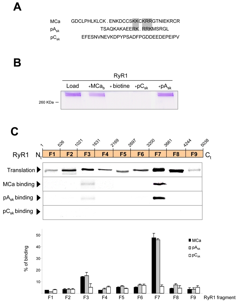
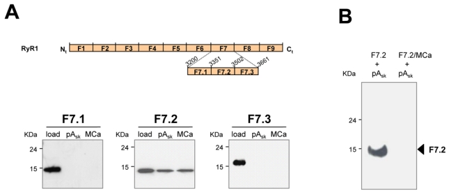
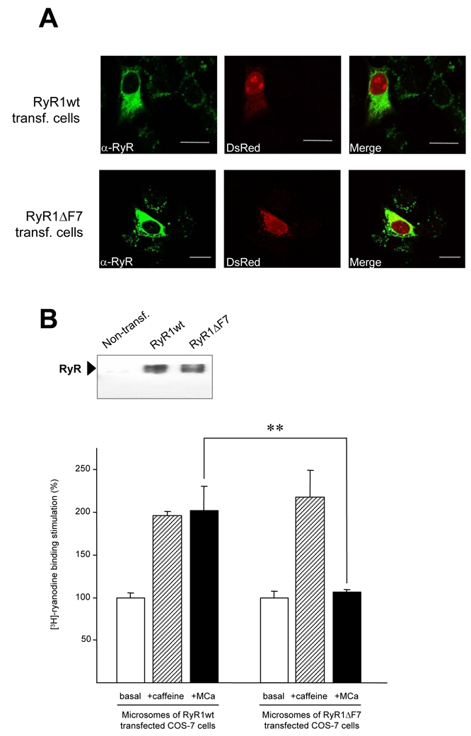
Maurocalcine is a scorpion venom toxin of 33 residues that bears a striking resemblance to the domain A of the dihydropyridine voltage-dependent calcium channel type 1.1 (Cav1.1) subunit. This domain belongs to the II-III loop of Cav1.1, which is implicated in excitation-contraction coupling. Besides the structural homology, maurocalcine also modulates RyR1 channel activity in a manner akin to a synthetic peptide of domain A. Because of these similarities, we hypothesized that maurocalcine and domain A may bind onto an identical region(s) of RyR1. Using a set of RyR1 fragments, we demonstrate that peptide A and maurocalcine bind onto two discrete RyR1 regions: fragments 3 and 7 encompassing residues 1021-1631 and 3201-3661, respectively. The binding onto fragment 7 is of greater importance and was thus further investigated. We found that the amino acid region 3351-3507 of RyR1 (fragment 7.2) is sufficient for these interactions. Proof that peptide A and maurocalcine bind onto the same site is provided by competition experiments in which binding of fragment 7.2 to peptide A is inhibited by preincubation with maurocalcine. Moreover, when expressed in COS-7 cells, RyR1 carrying a deletion of fragment 7 shows a loss of interaction with both peptide A and maurocalcine. At the functional level, this deletion abolishes the maurocalcine induced stimulation of [3H]ryanodine binding onto microsomes of transfected COS-7 cells without affecting the caffeine and ATP responses.


Figures

Figure 1
Figure 1. Identification of RyR1 fragments interacting with MCa and domain A
A, Sequence alignment of maurocalcine (MCa), domain A (pAsk) and domain C (pCsk) of the II-III loop of the α1 subunit of the DHPR. Grey boxes indicate the location of conserved clusters of basic amino acid residues in MCa and pAsk primary structures. In contrast, pCsk does not exhibit this repeat of positively charged residues. B, Physical interaction between purified RyR1 and MCa, domain A or domain C. Biotinylated-MCa (MCab, at 40 μM), biotinylated-domain A (pAsk, at 40 μM) and biotinylated-domain C (pCsk, at 40 μM), immobilized on streptavidine-coated magnetic beads, were used for pull-down experiments. In these conditions, MCab (lane 2) and domain A (lane 5) showed an interaction with full-length purified RyR1, whereas no interaction was observed with domain C-coated beads (lane 4) or biotin-coated beads (lane 3). C, Upper panel, Autoradiogram of MCab interaction with RyR1 fragments. After the generation of a set of clones spanning the full-length cDNA of RyR1 (dashed box indicates overlapping residues between fragments F8 and F9), in vitro translated fragments of RyR1 were assessed for their ability to interact either with 40 μM of MCab, pAsk or pCsk immobilized on streptavidine-magnetic beads. Lower panel, Histogram representing the percentage of binding of in vitro translated RyR1 fragments to MCab, estimated by densitometry and expressed as the means ± S.E.M. from at least three independent experiments.
Figure 2
Figure 2. Maurocalcine and domain A of the II-III loop of the α1 subunit bind onto the same site of RyR1
A, Fine dissection of RyR1 fragments that interact with MCa and pAsk. Upper panel, schematic representation of the RyR1-F7 sub-fragments (F7.1, F7.2, F7.3) cloned into pMR78-his tag vector for bacteria expression and affinity chromatography purification. Lower panel, Western blot with anti-his antibodies, showing the specific pull-down of the F7.2 fragment of RyR1 with both MCab and pAsk. B, Western blot resulting from a competition experiment, where the RyR1-F7.2 fragment was preincubated with an excess of MCa (1 μM) and then incubated with 16 nM of pAsk previously immobilized on streptavidine-magnetic beads. While the fragment F7.2 was able to bind to pAskb (left lane), the presence of MCa abolished this interaction (right lane), showing a competition of these two peptides for the same binding site on RyR1.
Figure 3
Figure 3. Deletion of the F7 region abolishes the effect of maurocalcine on [3H]-ryanodine binding onto RyR1 expressed in COS-7 cells
A, RyR expression pattern in COS-7 cells cotransfected either with DsRed and RyR1wt (upper panels) or with DsRed and RyR1-ΔF7 (lower panels) in COS-7 cells. Immunofluorescent signal in left panels (green signal) show the distribution pattern of RyR1 and red signal in central panels correspond to DsRed protein. Right panels correspond to merged images. B, Expression of RyR1 constructs in COS-7 cells. Western blot with anti-RyR1 (C-terminal) antibodies, showing a lack of endogenous RyR1 expression (lane 1) in control cells and equal protein amounts of RyR1 wt (lane 2) and RyR1-ΔF7 (lane 3) in 50 μg of microsomes, 48h after transfection. C, Functional effect of the F7 deletion on RyR1 binding properties. [3H]-ryanodine binding experiments on purified cell microsomes showed that, while both constructs responded to caffeine administration (6 mM), the deletion of the F7 fragment abolished MCa effect (30 nM).

References

    1. Chandler WK, Rakowski RF, Schneider MF. J Physiol. 1976;254:213–316. - PMC - PubMed
    1. Nakai J, Dirksen RT, Nguyen HT, Pessah IN, Beam KG, Allen PD. Nature. 1996;380:72–75. - PubMed
    1. Chavis P, Fagni L, Lansman JB, Bockaert J. Nature. 1996;382:719–722. - PubMed
    1. Catterall WA. Science. 1991;253:1499–1500. - PubMed
    1. Tanabe T, Beam KG, Adams BA, Nicodome T, Numa S. Nature. 1990;346:567–569. - PubMed

Publication types

MeSH terms