. 2005 Feb;16(2):997-1010.
doi: 10.1091/mbc.e04-10-0895. Epub 2004 Dec 9.

Phosphorylated BRCA1 is predominantly located in the nucleus and mitochondria

Affiliations

Phosphorylated BRCA1 is predominantly located in the nucleus and mitochondria

Elisabeth D Coene et al. Mol Biol Cell. 2005 Feb.

Abstract

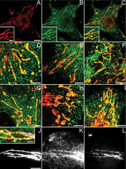
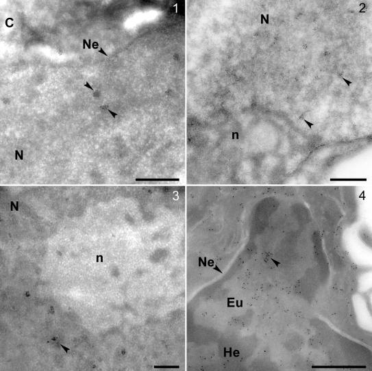
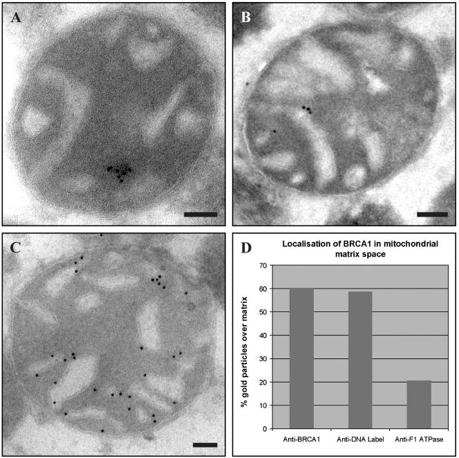
Multiple copies of the mitochondrial genome in eukaryotic cells are organized into protein-DNA complexes called nucleoids. Mitochondrial genome repair mechanisms have been reported, but they are less well characterized than their nuclear counterparts. To expand our knowledge of mitochondrial genome maintenance, we have studied the localization of the BRCA1 protein, known to be involved in nuclear repair pathways. Our confocal and immunoelectron microscopy results show that BRCA1 is present in mitochondria of several human cancer cell lines and in primary breast and nasal epithelial cells. BRCA1 localization in mitochondria frequently overlapped that of nucleoids. Small interfering RNA-mediated knockdown of BRCA1 in human cancer cells (confirmed by Western blot) results in decreased nuclear, cytoplasmic, and mitochondrial staining after immunofluorescence microscopy, establishing the specificity of the BRCA1 immunolabeling. Furthermore, using cell fractionation, dephosphorylation, and enzyme protection experiments, we show that a 220-kDa phosphorylated isoform of BRCA1 is enriched in mitochondrial and nuclear fractions but reduced in cytoplasmic subcellular fractions. Submitochondrial fractionation confirmed the presence of BRCA1 protein in isolated mitoplasts. Because phosphorylation of BRCA1 and subsequent changes in subcellular localization are known to follow DNA damage, our data support a universal role for BRCA1 in the maintenance of genome integrity in both mitochondria and nucleus.


Figures

Figure 1.
Figure 1.
Schematic diagram of the BRCA1 gene and polypeptide representing the multiple BRCA1 epitopes against which the different antibodies were raised. Exons are drawn to scale.
Figure 2.
Figure 2.
Confocal and wide-field microscopical data of ECV 304, HeLa, and SK-BR-3 cells labeled with MitoTracker (red) and anti-BRCA1-FITC/Alexa Fluor 488 antibodies (green). PFA-fixed ECV 304 cells stained with Ab-5 (B and C) and Ab-1 (E) and methanol-fixed ECV 304 cells stained with Ab-4 (D), Ab-C (F), and Ab-D (G) show granular BRCA1 cytoplasmic staining and colocalization of linear MitoTracker staining with linear BRCA1 staining results in yellow. (A) Single MitoTracker staining, overview. (B) Single Ab-5 staining, overview. (C) Merge of A and B. Insets in A–C represent a higher magnification. Double labeling of PFA-fixed HeLa (H) and SK-BR-3 (I) cells with MitoTracker and Ab-5 again shows yellow colocalization of both labels. Wide-field microscopical data shows colocalization of MitoTracker (J), Ab-5 (K), and mtDNA DAPI (L) on PFA-fixed ECV 304 cells, resulting in white foci (J, inset: higher magnification merge of J–L). Bars, 3 μm in insets; 5 μm in the other panels.
Figure 3.
Figure 3.
Thawed frozen section EM-immunocytochemistry by using antibodies Ab-1 and Ab-5 on human breast cells, HeLa cells, and normal primary human cells obtained by nasal brushing. BRCA1 gold clusters (arrowheads) in nuclei of HBL100 (1), HeLa (2), HMEC (3), and normal primary human cells (4) by using anti-BRCA1 antibodies Ab-1 (1–3) and Ab-5 (4). In normal primary human cells the clusters are only visible in the lighter euchromatin regions or at the boundary of eu- and heterochromatin (4). Bars, 500 nm (1–4). C, cytoplasm; Eu, euchromatin; He, heterochromatin; Ne, nuclear envelope; N, nucleus; and n, nucleolus.
Figure 4.
Figure 4.
Thawed frozen section EM-immunocytochemistry by using antibodies Ab-C, Ab-1, and Ab-5 on human breast cells and normal primary human cells obtained by nasal brushing. BRCA1 gold clusters in mitochondria of normal primary human cells (1), HBL100 (2), HMEC (3), and HeLa (4 and 5) by using anti-BRCA1 antibodies Ab-C (2), Ab-1 (3 and 4), and Ab-5 (1 and 5). Panel 6 shows a schematic representation of a higher mitochondrial magnification of 5; the selected region is depicted by a box. Bars, 100 nm. C, cytoplasm; Cr, cristae; IM, inner membrane; and M, matrix; circles emphasize gold clusters located in the matrix; OM, outer membrane.
Figure 9.
Figure 9.
Intramitochondrial localization of BRCA1. (A) EM analysis of rat liver mitochondria with anti-BRCA1 Ab-1 shows BRCA1 gold clusters in the matrix. (B) EM analysis of rat liver mitochondria with anti-DNA IgM shows IgM signal in the matrix. (C) EM analysis of rat liver mitochondria with anti-F1 ATPase shows that F1 ATPase is associated with the mitochondrial membrane. Bars, 100 nm. (D) Table shows localization of BRCA1 in mitochondrial matrix space. BRCA1 (60%) and IgM (59%) are both predominantly located in the mitochondrial matrix space; F1 ATPase is predominantly associated with the cristae and therefore only a minority (20%) is located over the matrix space.
Figure 5.
Figure 5.
Biochemical and confocal analysis of siRNA-mediated knockdown of BRCA1/Lamin B1 in ECV 304 cells. (A) Lamin B1 protein was knocked down in the cells transfected with specific Lamin B1 duplex (1), whereas the level remained high in the cells transfected with specific BRCA1 duplex (2). BRCA1 protein was knocked down in the cells transfected with specific BRCA1 duplex (2), whereas the level remained high in the cells transfected with specific Lamin B1 duplex (1) (arrowhead). Equal amounts of cells transfected with specific Lamin B1 duplex (1) and with specific BRCA1 duplex (2) were loaded, which was confirmed by a similar F1 ATPase expression in both lanes. (B) siRNA Lamin B1-only knockdown shows a diminished Lamin B1 expression at the nuclear envelope; the nuclear, cytoplasmic, and mitochondrial BRCA1 staining is not affected nor is the mitochondrial F1 ATPase staining pattern. (C) siRNA Lamin B1 and BRCA1 double knockdown shows a diminished expression of both nuclear, cytoplasmic, and mitochondrial BRCA1 and Lamin B1 proteins in the transfected cells. Mitochondrial F1 ATPase staining remains present. The untransfected cells still show Lamin B1 and BRCA1 nuclear, cytoplasmic, and mitochondrial staining as well as mitochondrial F1 ATPase expression. Bar, 10 μm.
Figure 6.
Figure 6.
Western blotting with anti-BRCA1 antibodies Ab-1, Ab-4, Ab-5, and Ab-C, and anti-CKBS, anti-Lamin A/C, and anti-Ki67 antibodies on NF, CF, WH, and MitF HeLa fractions. (A) Western blotting with Ab-1, Ab-4, and Ab-C on NF and CF HeLa fractions. A 220- and 200-kDa double BRCA1 band is visualized, the top 220-kDa band being strongly reduced or absent in the CF. Control Western blots with anti-CKBS, anti-Lamin A/C, and anti-Ki67 on NF and CF show only Lamin A/C and Ki67 present in NF and cytokeratin in CF. Control Western blots with anti-Ki67 on MitF versus WH show only Ki67 present in WH. Arrowheads, left, 210 kDa; top right, 41 kDa; middle right, 71 kDa; and two bottom right, 340 kDa. (B) Western blotting with antibodies Ab-1, Ab-4, Ab-5, and Ab-C on WH and MitF HeLa fractions detecting the BRCA1 protein.
Figure 7.
Figure 7.
Western blotting of both untreated and treated NF, CF, MitF, and WH HeLa samples with p-BRCA1 Ser 988, p-BRCA1 Ser 1497, Ab-4, and Ab-C. (A) p-BRCA1 Ser 988 and p-BRCA1 Ser 1497 on MitF HeLa fractions (1) and WH (2) give a 220-kDa BRCA1 band in both lanes. (B) Western blotting with Ab-C and Ab-4 on NF and CF before (1) and after (2) dephosphorylation with λ protein phosphatase. The 220-kDa band disappeared after λ protein phosphatase treatment, showing that it is this species that is phosphorylated. (C) Western blotting with Ab-C on mitochondrial fractions treated for enzyme protection assay. Mitochondrial HeLa untreated control fraction (1), treated with TX100 (2), treated with λ protein phosphatase (3), and treated with λ protein phosphatase and TX100 (4) are presented (top). The 220-kDa BRCA1 band only disappears after combined λ protein phosphatase and TX100 treatment. Mitochondrial HeLa untreated control fraction (1), treated with proteinase K (2), and treated with proteinase K and TX100 (3) are presented (bottom). The 200-kDa band disappears after proteinase K treatment alone, whereas the 220-kDa band only becomes vulnerable after combined proteinase K and TX100 treatment.
Figure 8.
Figure 8.
Biochemical analysis of rat liver mitoplasts. (A) Antibody-5 anti-BRCA1 antibody detects the protein in WH, MitF, and a MP-enriched fraction (arrowhead). After dephosphorylation, the top BRCA1 band is strongly reduced, whereas a lower band occurs (arrow). P-BRCA1 Ser 1497 anti-BRCA1 antibody detects the same protein in WH, MitF, and the MP-enriched fraction. After dephosphorylation, all p-BRCA1 Ser 1497 signal is absent. (B) Western blot of Percoll-purified IM and MP stained with Ab-5 showed an increase in signal for two hyperphosphorylated isoforms (* and **) after MP preparation from IM. Arrowhead, 160 kDa. Table of enrichment shows the MP/IM ratio for CCO and MLDH enzymes and the two asterisked BRCA1 protein isoforms. The graph displays the intensity of the BRCA1 protein forms versus the relative migration. Two hyperphosphorylated BRCA1 proteins (* and **) are enriched in the MP fraction versus IM fraction.

Similar articles

Cited by

References

    1. Anderson, S., et al. (1981). Sequence and organization of the human mitochondrial genome. Nature 290, 457-465. - PubMed
    1. Anson, R. M., Croteau, D. L., Stierum, R. H., Filburn, C., Parsell, R., and Bohr, V. A. (1998). Homogenous repair of singlet oxygen-induced DNA damage in differentially transcribed regions and strands of human mitochondrial DNA. Nucleic Acids Res. 26, 662-668. - PMC - PubMed
    1. Bianchi, N. O., Bianchi, M. S., and Richard, S. M. (2001). Mitochondrial genome instability in human cancers. Mutat. Res. 488, 9-23. - PubMed
    1. Bourgeois, C. A., Hemon, D., and Bouteille, M. (1979). Structural relationship between the nucleolus and the nuclear envelope. J. Ultrastruct. Res. 68, 328-340. - PubMed
    1. Chen, C. F., Li, S., Chen, Y., Chen, P. L., Sharp, Z. D., and Lee, W. H. (1996). The nuclear localization sequences of the BRCA1 protein interact with the importin-alpha subunit of the nuclear transport signal receptor. J. Biol. Chem. 271, 32863-32868. - PubMed

Publication types

MeSH terms

LinkOut - more resources