. 2005 Feb;16(2):689-700.
doi: 10.1091/mbc.e04-09-0828. Epub 2004 Dec 9.

The tricornered Ser/Thr protein kinase is regulated by phosphorylation and interacts with furry during Drosophila wing hair development

Affiliations

The tricornered Ser/Thr protein kinase is regulated by phosphorylation and interacts with furry during Drosophila wing hair development

Ying He et al. Mol Biol Cell. 2005 Feb.

Abstract

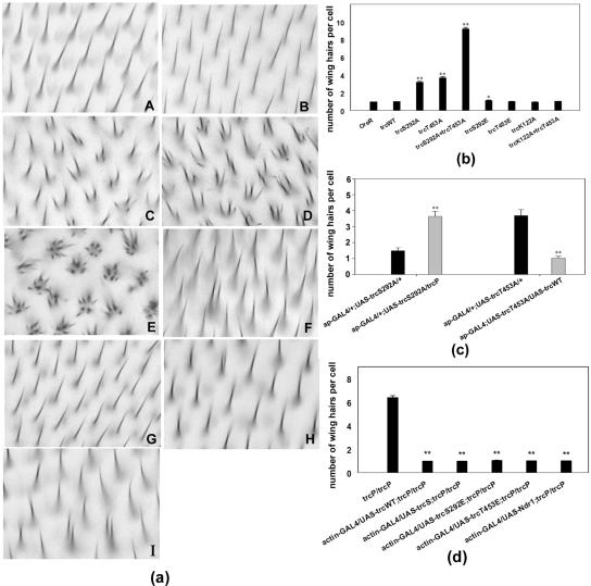
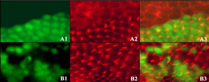
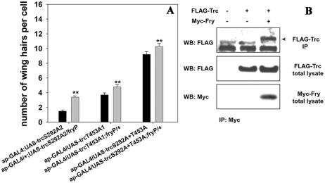
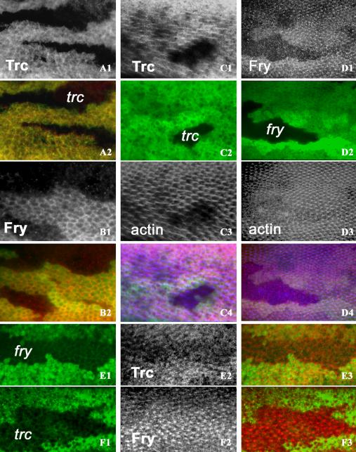
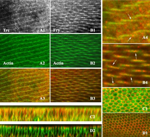
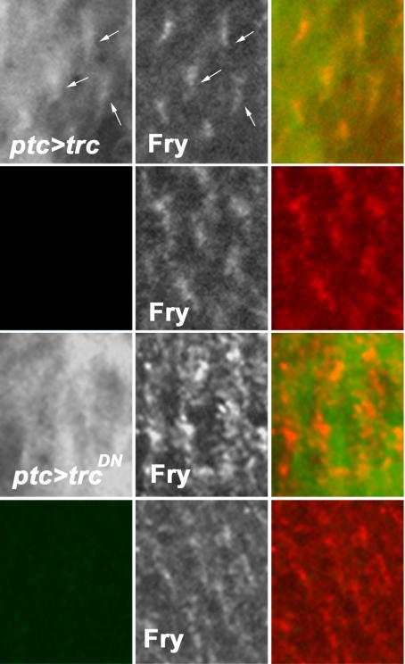
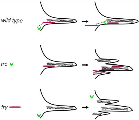
The Trc/Ndr/Sax1/Cbk1 family of ser/thr kinases plays a key role in the morphogenesis of polarized cell structures in flies, worms, and yeast. Tricornered (Trc), the Drosophila nuclear Dbf2-related (Ndr) serine/threonine protein kinase, is required for the normal morphogenesis of epidermal hairs, bristles, laterals, and dendrites. We obtained in vivo evidence that Trc function was regulated by phosphorylation and that mutations in key regulatory sites resulted in dominant negative alleles. We found that wild-type, but not mutant Trc, is found in growing hairs, and we failed to detect Trc in pupal wing nuclei, implying that in this developmental context Trc functions in the cytoplasm. The furry gene and its homologues in yeast and Caenorhabditis elegans have previously been implicated as being essential for the function of the Ndr kinase family. We found that Drosophila furry (Fry) also is found in growing hairs, that its subcellular localization is dependent on Trc function, and that it can be coimmunoprecipitated with Trc. Our data suggest a feedback mechanism involving Trc activity regulates the accumulation of Fry in developing hairs.


Figures

Figure 2.
Figure 2.
Directed expression of Trc mutants can result in multiple hair cells similar to those seen in trc loss of function mutants. (a) Wing phenotypes. A-J show bright-field micrographs of the same region of wings of the wild-type and tested genotypes: OreR (A), ap-GAL4/UAS-trcWT (B), ap-GAL4/UAS-trcS292A (C), ap-GAL4/UAS-trcT453A (D), ap-GAL4/UAS-trcS292A+T453A (E), ap-GAL4/UAS-trcS292E (F), ap- GAL4/UAS-trcT453E (G), ap-GAL4/UAS-trcK122A (H), and ap-GAL4/UAS-trcK122A+T453A (I). (b) Average number of hairs per cell is presented. The overexpression of wild-type, the single glu mutants (S292E and T453E), lys122ala, or lys122ala+thr453ala had little effect. Most cells formed a single hair. One or two cells shown in F, I, and J produced a double hair. Moderate multiple hair cell phenotypes were seen when either of the single ala mutants were expressed (S292A and T453A). In these wings, there was average of four hairs per cell. Dramatic, strong phenotypes resulted from the expression of the S292A T453A double mutant. Error bars represent the SE of the mean. In several cases, the error bars were too small to be visible. Each genotype was compared with Oregon R by using a t test, and significant differences were noted. (c) Evidence the mutants acted as dominant negatives. To determine whether the phosphorylation site ala mutant proteins decreased or increased Trc activity, we examined the consequences of a reduction in Trc dose or coexpression of trcWT. Shown are data for flies that were ap-GAL4/+; UAS-trcS292A/+ versus ap-GAL4/+; UAS-trcS292A/trcP, and ap-GAL4/+; UAS-trcT453A/+ versus ap-GAL4/+; UAS-trcT453A/UAS-trcWT. Note the large degree of rescue that resulted from the coexpression of the wild-type protein, implying that the ala mutants were acting as dominant negative proteins. Consistent with this interpretation, a mutation in the endogenous trc gene acted as a dominant enhancer of the ala mutant protein. Significant differences from t test comparisons are indicated. (d) Rescue of the trcP/trcP lethal mutant by driving expression of UAS-trcS292E, UAS-trcT453E, UAS-trcWT, UAS-trcshort (see Materials and Methods for description of trcshort) and UAS-ndr1 by using actin-GAL4. The importance of the phosphorylation sites was further assayed by testing the ability of various mutant proteins to provide trc rescue activity. We generated flies that were actin-GAL4/UAS-trcX; trcP/trcP. None of the Ala mutants showed rescue activity. The UAS constructs that contained trcWT, NDR1, trcS292E, and trcT453E completely rescued the lethality of trc mutants and also resulted in rescue of the wing phenotype. We compared the number of hairs per wing cell for each rescued wing sample and data for trcP/trcP mutant clones by using a t test. All results were highly significant (P ≪ 0.001). We also compared the rescue achieved by expression of trcL (trcWT) with the other rescuing constructs. These did not show highly significant differences (our unpublished data). Note **P value is ≪ 0.001, which means a highly significant difference; * means the P value between two samples is between 0.001 < P < 0.05, which represents a likely difference; and no * means the P value between the two samples is >0.05, which represents no significant difference.
Figure 1.
Figure 1.
trc Mutant phenotype in wing clones. Part of a pupal wing that contains a trc clone marked by a loss of GFP. (A1 and B1) GFP. (A2 and B2) Actin staining. (A3 and B3) Merged images. Top, clone where hair development is strongly delayed. Bottom, clone where the mutant cells have formed hairs. Note the enlarged and irregularly shaped clone cells compared with the wild-type cells. Apical actin staining is stronger than in neighboring wild-type cells. Note the multiple hair cells in the clones and the delay in hair morphogenesis by some clone cells. fry mutant clones show similar phenotypes (our unpublished data).
Figure 3.
Figure 3.
Genetic and physical interaction between trc and fry. (A) Flies that carried each of the trc ala single or double mutants with or without the presence of one copy of fryP. The expression of the UAS-trc transgenes was driven by ap-GAL4. We found that fry dominantly enhanced the phenotypes of each of the trc mutants. A similar enhancement is also seen with a Deficiency for the fry gene (our unpublished data). Statistical analyses as in Figure 2. (B) Trc and N-Fry (Fry1-1637) coimmunoprecipitate in an S2 cell lysate. Cells were transfected with UAS-FLAG-Trc and/or UAS-N-Fry-3 × Myc. Top, Western blotting using anti-FLAG antibody to detect Trc after immunoprecipitation with an anti-Myc antibody that brought down N-Fry-3 × Myc protein complex. The lower band in this panel is likely IgG and serves as a loading control. The bottom two panels show the results from Western blotting analysis of the whole cell lysate by using anti-FLAG or anti-Myc antibody.
Figure 4.
Figure 4.
Trc and Fry subcellular localizations in wing cells and their interdependency. Homozygous trc or fry clones on the wing were generated by FLP/FRT recombination. The clones are marked by loss of GFP fluorescence (green). Only antibody staining or actin staining are in grey scale to increase contrast. All samples are pupal collected at 26-30 h after white prepupae (before hair formation) or 32-36 h after white prepupae (after hair formation). (A1 and A2) A trc clone (labeled in A2) stained before hair formation. Note in the clone that there is a loss of Trc staining, showing the specificity of the antibody. Surrounding wild-type cells show both cytoplasmic and cell periphery Trc staining. Trc staining (A1) and merge of Trc (red) staining (A2) and GFP staining (green). (B1 and B2) A fry clone stained before hair formation. Fry staining is more prominently at the cell periphery than Trc. Staining is lost in the clone cells showing the specificity of the antibody. (B1) Fry staining. (B2) Merge of Fry (red) staining with GFP staining. (C1-C4) A trc clone stained after hair formation. Note in the clone there is a loss of the Trc accumulation and disruption of hair formation as reported on by actin staining. Surrounding wild-type cells show Trc in the hair in a punctuate pattern. (C1) Trc staining. (C2) GFP staining. (C3) Actin staining. (C4) Merge of Trc (red), GFP (green), and actin (blue). (D1-D4) A fry clone stained after hair formation. Note in the clone there is a loss of the Fry accumulation and disruption of actin cytoskeleton. The surrounding wild-type cells show Fry in the hairs. (D1) Fry staining. (D2) GFP staining. (D3) Actin staining. (D4) Merge of Fry (red), GFP (green), and actin (blue). The bottom two panels show the interdependency of Trc And Fry accumulation. Clones were marked by the loss of GFP (left), antibody staining (middle, in grey scale), and merged images (right, Trc or Fry in red). (E1-E3) A fry clone stained with anti-Trc antibody early in hair morphogenesis. Note the Trc signal is stronger in the wild-type cells than in the neighboring clone cells. (F1-F3) A trc clone stained with anti-Fry antibody after hair formation. Fry hair staining is substantially increased in the trc clone. In all panels, antibody staining is labeled as Trc or Fry and clones are labeled by trc or fry.
Figure 5.
Figure 5.
Trc and Fry partially overlapping with actin in wing hairs. (A1-A4 and B1-B4) OreR pupal wings double staining with anti-Trc and phalloidin (to stain F-actin) or anti-Fry antibody with phalloidin after hair formation. Anti-Trc staining (A1), actin staining (A2), and a merged image (A3). A4 is the blow up image showing the punctate pattern of Trc distribution. Arrowheads point to bright regions of staining. Anti-Fry staining (B1), actin staining (B2), and a merged image (B3). B4 is a blow up image illustrating the punctuate staining of Fry in hairs. Arrowheads point to the accumulations of Fry in the hairs. (C1-D2) Twenty-eight-hour OreR pupal wings double stained for Fry and Armadillo or Trc and Armadillo. Fry (or Trc) is in green and Armadillo in red. (C1) An XY section shows the strong cell outline staining of Arm and the broader staining of Trc. (C2) A z section from the same wing showing the relative restriction of Armadillo to the adherens junction and hemidesmosomes and that Trc staining is seen over a wide range of the lateral cell surface. (D1) An XY section shows the strong cell outline staining of Arm and the peripheral staining of Fry. (D2) A z section from the same wing showing the relative restriction of Arm to the adherens junction and that Trc staining is seen over a wide range of the lateral cell surface.
Figure 6.
Figure 6.
Fry staining pattern is dependent on active Trc. Thirty-six to 40 hr pupal wings from ptc-Gal4 UAS-trcX animals were stained with polyclonal anti-Fry (generated in rabbit) and monoclonal anti-FLAG (Sigma-Aldrich) antibodies (the trc transgene was tagged with a FLAG epitope at the N-terminal region). ptc-Gal4 drives expression in a stripe down the center of the wing. This domain is bounded by wing veins. In this composite, we compare the staining in the ptc domain and in neighboring cells just lateral to the vein. Top, cells within the ptc domain that are overexpressing wild-type trc. Left, overexpressed Trc. Hair staining (arrows) can be seen, but the background is high due to over expression. Middle, Fry staining, which seems normal. Right, the merge. The second row shows a neighboring region of cells that is outside of the ptc domain. We see no evidence of FLAG staining and Fry staining (middle) seems normal. The lower two rows of panels are equivalent micrographs except that a dominant negative Trc (TrcS292A+T453A) is being overexpressed. Note that hair staining is no longer seen for the overexpressed Trc (left) and Fry staining is increased (middle). The staining for FLAG is not even across the cells, but the brighter regions no longer line up with the brighter Fry staining in the hair. The bottom row shows a region of the same wing just outside of the ptc domain. Once again, we see no FLAG staining and Fry staining is normal.
Figure 7.
Figure 7.
Model for Trc and Fry interacting during hair Development. A cartoon of a developing hair is shown. Trc is in green, Fry in red, and actin in gray. In wild-type, Fry is recruited to the hair and it in turn recruits Trc. Trc activity locally inhibits further recruitment of Fry. This feedback mechanism ensures the proper level of these proteins in the hair. In the trc mutant, excess Fry is recruited to the hair due to the lack of the Trc-based negative feedback. Due to a lack of Trc activity hair morphogenesis is also abnormal. In the fry mutant, Fry protein is not present to be recruited to the hair, and this in turn results in decreased recruitment of Trc. The lack of Trc activity results in abnormal hair development.

References

    1. Bhattacharya, S., Large, E., Heizmann, C. W., Hemmings, B., and Chazin, W. J. (2003). Structure of the Ca2+/S100B/NDR1 kinase peptide complex: insights into S100 target specificity and activation of the kinase. Biochemistry 42, 14416-14426. - PubMed
    1. Bichsel, S. J., Tamaskovic, R., Stegert, M. R., and Hemmings, B. A. (2004). Mechanism of activation of nuclear Dbf2-related (NDR) kinase by the hMOB1 protein. J. Biol. Chem. 279, 35228-35235. - PubMed
    1. Bidlingmaier, S., Weiss, E. L., Seidel, C., Drubin, D. G., and Snyder, M. (2001). The Cbk1p pathway is important for polarized cell growth and cell separation in Saccharomyces cerevisiae. Mol. Cell. Biol. 21, 2449-2462. - PMC - PubMed
    1. Brand, A. H., and Perrimon, N. (1993). Targeted gene expression as a means of altering cell fates and generating dominant phenotypes. Development 118, 401-415. - PubMed
    1. Colman-Lerner, A., Chin, T. E., and Brent, R. (2001). Yeast Cbk1 and Mob2 activate daughter-specific genetic programs to induce asymmetric cell fates. Cell 107, 739-750. - PubMed

Publication types

MeSH terms