. 2005 Feb;16(2):902-17.
doi: 10.1091/mbc.e04-08-0687. Epub 2004 Dec 9.

Agonist-induced endocytosis of CC chemokine receptor 5 is clathrin dependent

Affiliations

Agonist-induced endocytosis of CC chemokine receptor 5 is clathrin dependent

Nathalie Signoret et al. Mol Biol Cell. 2005 Feb.

Abstract

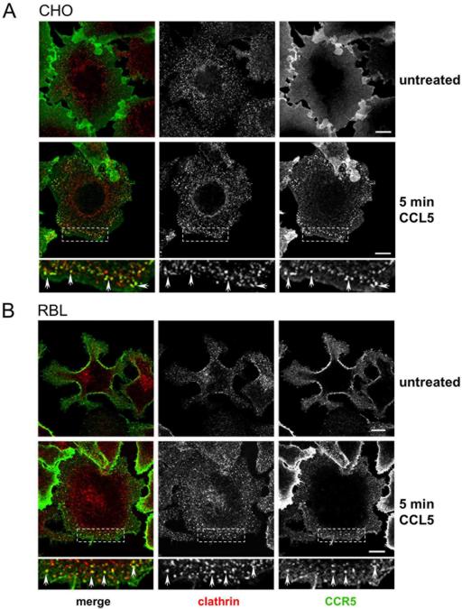
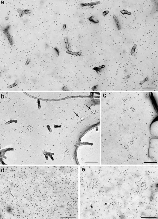
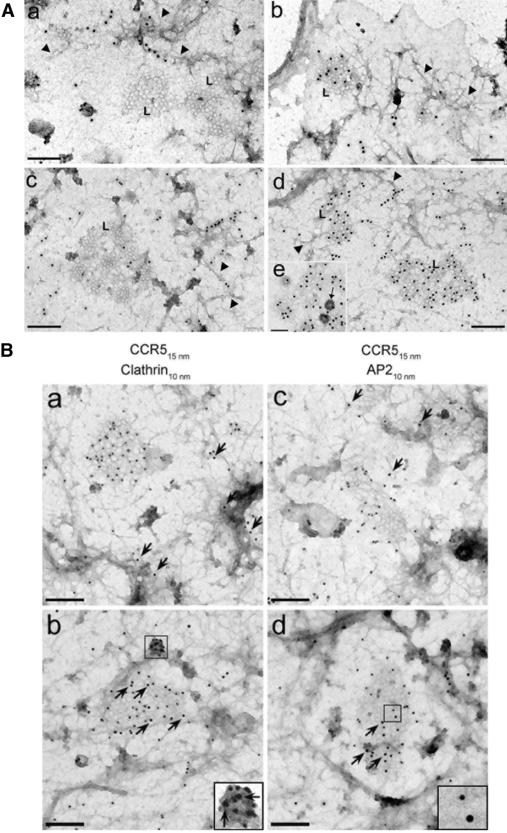
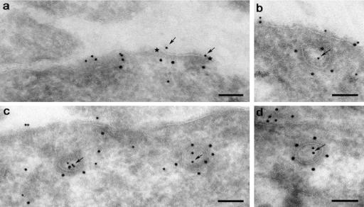
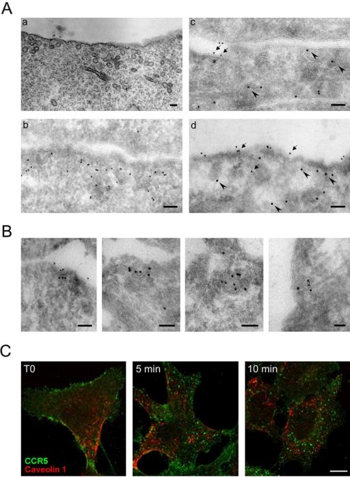
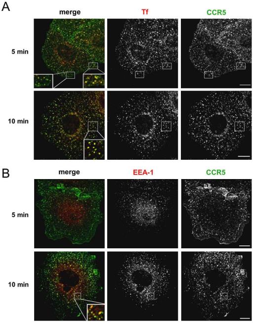
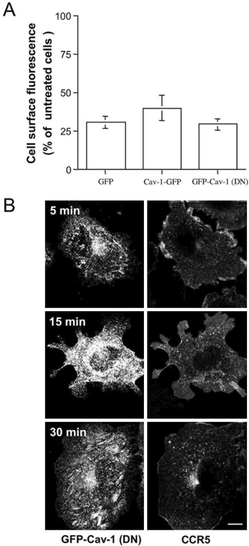
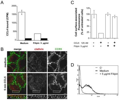
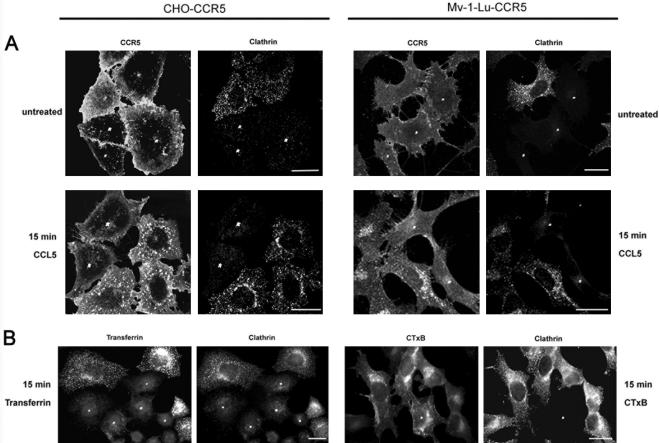
The signaling activity of several chemokine receptors, including CC chemokine receptor 5 (CCR5), is in part controlled by their internalization, recycling, and/or degradation. For CCR5, agonists such as the chemokine CCL5 induce internalization into early endosomes containing the transferrin receptor, a marker for clathrin-dependent endocytosis, but it has been suggested that CCR5 may also follow clathrin-independent routes of internalization. Here, we present a detailed analysis of the role of clathrin in chemokine-induced CCR5 internalization. Using CCR5-transfected cell lines, immunofluorescence, and electron microscopy, we demonstrate that CCL5 causes the rapid redistribution of scattered cell surface CCR5 into large clusters that are associated with flat clathrin lattices. Invaginated clathrin-coated pits could be seen at the edge of these lattices and, in CCL5-treated cells, these pits contain CCR5. Receptors internalized via clathrin-coated vesicles follow the clathrin-mediated endocytic pathway, and depletion of clathrin with small interfering RNAs inhibits CCL5-induced CCR5 internalization. We found no evidence for CCR5 association with caveolae during agonist-induced internalization. However, sequestration of cholesterol with filipin interferes with agonist binding to CCR5, suggesting that cholesterol and/or lipid raft domains play some role in the events required for CCR5 activation before internalization.


Figures

Figure 1.
Figure 1.
Agonist-induced accumulation of CCR5 in clathrin-positive plasma membrane domains. CHO-CCR5 cells (A) or RBL-CCR5 cells (B) were left untreated or treated with CCL5 for 5 min, fixed, and labeled intact for cell surface CCR5 with MC-5. Cells were permeabilized and stained with a rabbit anti-clathrin light chain and then with 488GAM and 594GAR secondary antibodies to detect CCR5 and clathrin, respectively. The figure shows single confocal sections from a representative experiment. Boxed areas are shown at higher magnification under each panel. Bars, 10 μm.
Figure 2.
Figure 2.
CCR5 cell surface distribution. CHO-CCR5 (a–c) and RBL-CCR5 (d and e) cells were treated in BM alone (a and d) or with CCL5 for 30 s (b) or 5 min (c and e) at 37°C before fixation. Surface CCR5 was detected by labeling with MC-5 and PAG15, and cell surface replicas were prepared as described in Materials and Methods. Agonist-treated CCR5 was occasionally seen associated with shadowed invaginations of the membrane (arrows). Bars, 500 nm.
Figure 3.
Figure 3.
Agonist-induced redistribution of CCR5 into clathrin lattices. (A) CHO-CCR5 (a and b) and RBL-CCR5 (c–e) cells were incubated in BM alone (a and c) or with CCL5 (b, d, and e) for 2 min at 37°C, rinsed at 4°C, and labeled with MC-5 and PAG15 on ice before being ripped open as described in Materials and Methods. The cortical filamentous network (arrowheads), large areas of flat lattice characterized by their polygonal coat (L), and small electron-dense coated invaginations (arrow) are visible on the inner face of the plasma membrane. (B) Membrane sheets from CHO-CCR5 cells treated for 2 min in BM alone (a and c) or with CCL5 (b and d) were prepared as described above and labeled on the cytoplasmic surface with antibodies against clathrin (a and b) or AP2 (c and d) followed by PAG10. Arrows indicate some of the large gold particles (PAG15) marking CCR5. A clathrin-labeled coated invagination containing CCR5 is enlarged in b. Bars, 200 nm except A, e, which is 100 nm.
Figure 4.
Figure 4.
CCR5 internalized in CCVs. Cryosections of CHO-CCR5 cells treated with CCL5 for 5 min were double labeled with MC-5 plus PAG10 and anti-clathrin plus PAG15, respectively. CCR5 (arrows) is seen in coated areas of the plasma membrane (a, between stars) and in coated vesicles (b–d) that are labeled for clathrin. Bars, 100 nm.
Figure 6.
Figure 6.
Distribution of agonist-treated CCR5 in Mv-1-Lu cells. (A) Caveolae in Mv-1-Lu-CCR5 cells. Uniform small membrane invaginations resembling caveolae were observed at the cell surface and beneath the plasma membrane on Epon-embedded Mv-1-Lu-CCR5 cells (a) and can be labeled for caveolin-1/PAG10 on cryosections (b). In c and d, CCL5-treated Mv-1-Lu-CCR5 cells (125 nM CCL5, 5 min) were double labeled for CCR5 (MC-5 and PAG10, arrows) and caveolin-1 (PAG15, arrowheads). Although some CCR5 can be seen in regions containing PAG15-labeled caveolae, it is not found associated with the invaginations. Some CCR5 is located near an electron-dense flat, presumably clathrin-coated, region of the plasma membrane (c, *). Bars, 100 nm. (B) Cryosections of Mv-1-Lu-CCR5 cells treated with CCL5 for 5 min were double labeled for CCR5 (MC-5 and PAG10) and clathrin (PAG15). CCR5 was seen in clathrin-coated pits and vesicles. Bars, 100 nm. (C) Mv-1-Lu-CCR5 cells prelabeled with MC-5 for cell surface CCR5 were treated with CCL5 for 5 or 10 min, fixed, permeabilized with saponin, and costained for caveolin-1. Samples were analyzed by confocal microscopy. Bars, 10 μm.
Figure 5.
Figure 5.
Internalized CCR5 follows the clathrin-mediated endocytic pathway. Internalization of prelabeled cell surface CCR5 on CHO-CCR5 cells overexpressing HTf-R. (A) Cells prelabeled with fluorescent 488MC-5 were allowed to take up 594Tf for 10 min, and CCL5 was then added for a further 5 or 10 min. Cells were fixed, and the distribution of the fluorescent signals was analyzed using a confocal microscope. (B) Cells prelabeled with MC-5 were treated with CCL5 for 5 or 10 min, fixed, permeabilized with saponin, and stained with a rabbit anti-EEA-1 antibody. CCR5 and EEA-1 were then detected using Alexa-Fluor–conjugated secondary antibodies. The figure shows single confocal sections from a representative experiment. Boxed areas are shown at higher magnification in the merged panels. Bars, 10 μm.
Figure 7.
Figure 7.
Effect of GFP-tagged caveolin-1 proteins on CCR5 endocytosis. (A) CHO-CCR5 cells transiently transfected with plasmids for GFP alone, Cav-1-GFP or GFP-Cav-1 (DN) were treated with CCL5 for 30 min at 37°C. Cell surface CCR5 down-modulation was monitored by staining with MC-5 and 647GAM and quantified by FACS analysis of samples gated on GFP-positive cells. The results show the percentage of the 647GAM fluorescence ± SD compared with untreated cells for a representative experiment performed in triplicate. (B) CCR5 internalization in cells expressing GFP-Cav-1 (DN) prelabeled with MC-5 and incubated with CCL5 for up to 30 min. CCR5 was detected on fixed and saponin-permeabilized cells by staining with 647GAM. The figure shows single confocal sections. Bars, 10 μm.
Figure 8.
Figure 8.
Filipin inhibits CCR5 agonist activation. (A) 125I-CCL4 was bound, at 4°C, in medium alone or with 5 μg/ml filipin, to CHO-CCR5 cells (▪) and CHO-K1 cells (▪) pretreated or not with filipin. Samples were washed extensively and the amount of cell-associated radioactivity was determined by gamma counting. The means and SD of quadruplicate samples from a representative experiment are shown. (B) CHO-CCR5 cells on coverslips pretreated with filipin were incubated for 5 min with or without CCL5 at 37°C. The cells were fixed and labeled intact for cell surface CCR5 with MC-5 and then permeabilized, stained for clathrin, and analyzed as in Figure 1. Bars, 10 μm. (C) Effect of filipin on CCL5-induced CCR5 down-modulation was monitored by FACS analysis. The graph shows the mean ± SD of a representative experiment performed in triplicate. (D) Filipin treatment also inhibits the uptake of fluorescent transferrin in CHO-CCR5 cells overexpressing HTf-R. The histogram overlay represents the cell-associated fluorescence intensity before (C-) or after a 30-min incubation with 647Tf of filipin-treated or untreated (medium) cells.
Figure 9.
Figure 9.
Clathrin-dependent CCR5 endocytosis in CHO and Mv-1-Lu cells. CHO-CCR5 and Mv-1-Lu-CCR5 cells were twice transfected with chc-2 siRNAs. Cells prelabeled with MC-5 were incubated in medium alone or treated with CCL5 (A). Some cells were incubated for 15 min with 594Tf or 488CTxB (B). All samples were then fixed, permeabilized with saponin, and costained for clathrin using a rabbit anti-clathrin light chain antibody. CCR5 and clathrin were detected with Alexa-Fluor–conjugated secondary antibodies. The figure shows single confocal sections (A) and digital images recorded with a Zeiss Axioscope (B). Clathrin knocked down cells are marked with asterisks. Bars, 20 μm.

References

    1. Aggeler, J., Takemura, R., and Werb, Z. (1983). High-resolution three-dimensional views of membrane-associated clathrin and cytoskeleton in critical-point-dried macrophages. J. Cell Biol. 97, 1452-1458. - PMC - PubMed
    1. Alkhatib, G., Locati, M., Kennedy, P. E., Murphy, P. M., and Berger, E. A. (1997). HIV-1 coreceptor activity of CCR5 and its inhibition by chemokines: independence from G protein signaling and importance of coreceptor down-modulation. Virology 234, 340-348. - PubMed
    1. Amara, A., LeGall, S., Schwartz, O., Salamero, J., Montes, M., Loetscher, P., Baggiolini, M., Virelizier, J. L., and Arenzana-Seisdedos, F. (1997). HIV coreceptor downregulation as antiviral principle: SDF-1 alpha-dependent internalization of the chemokine receptor CXCR4 contributes to inhibition of HIV replication. J. Exp. Med. 186, 139-146. - PMC - PubMed
    1. Aramori, I., Zhang, J., Ferguson, S. S., Bieniasz, P. D., Cullen, B. R., and Caron, M.G. (1997). Molecular mechanism of desensitization of the chemokine receptor CCR-5, receptor signaling and internalization are dissociable from its role as an HIV-1 co-receptor. EMBO J. 16, 4606-4616. - PMC - PubMed
    1. Blanpain, C., et al. (2001). Palmitoylation of CCR5 is critical for receptor trafficking and efficient activation of intracellular signaling pathways. J. Biol. Chem. 276, 23795-23804. - PubMed

Publication types

MeSH terms

LinkOut - more resources