. 2004 Dec 21;101(51):17669-74.
doi: 10.1073/pnas.0408236101. Epub 2004 Dec 10.

A vesicle bioreactor as a step toward an artificial cell assembly

Affiliations

A vesicle bioreactor as a step toward an artificial cell assembly

Vincent Noireaux et al. Proc Natl Acad Sci U S A. .

Abstract

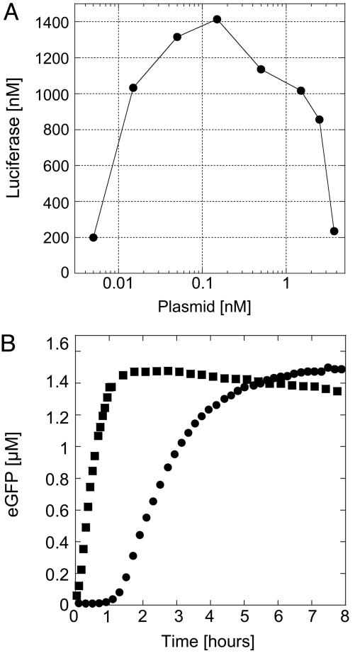
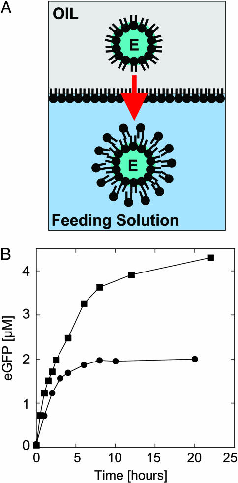
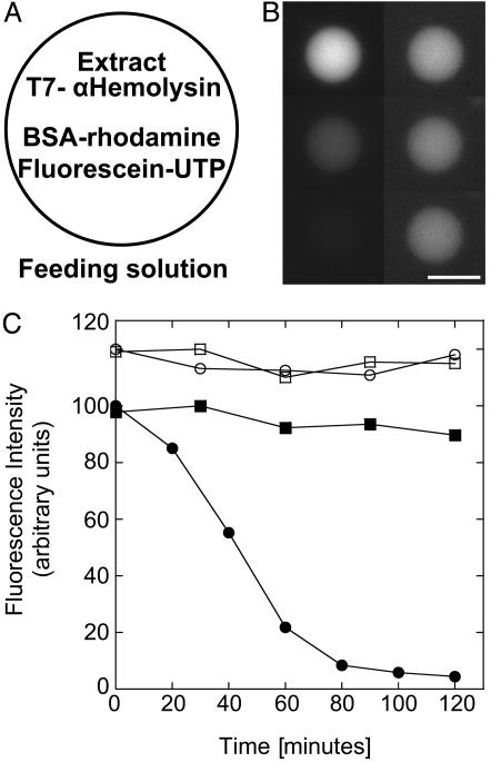
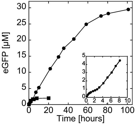
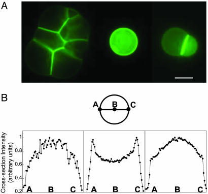
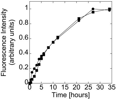
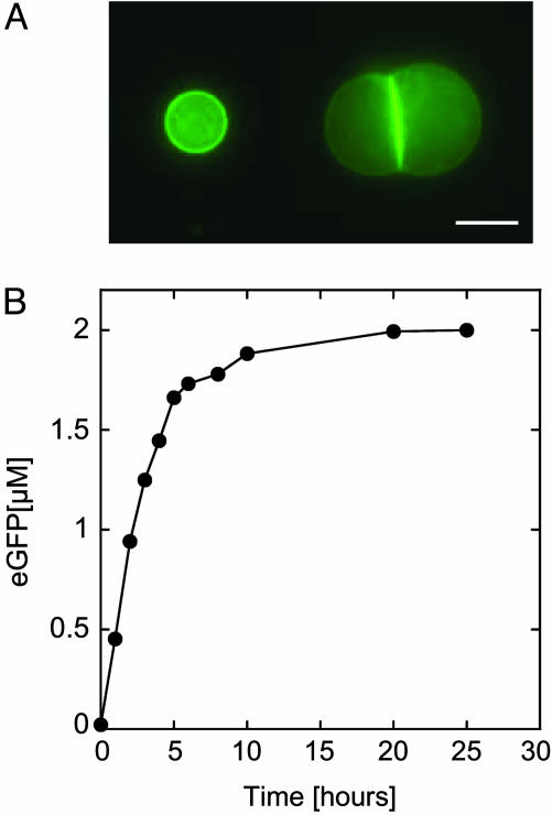
An Escherichia coli cell-free expression system is encapsulated in a phospholipid vesicle to build a cell-like bioreactor. Large unilamellar vesicles containing extracts are produced in an oil-extract emulsion. To form a bilayer the vesicles are transferred into a feeding solution that contains ribonucleotides and amino acids. Transcription-translation of plasmid genes is isolated in the vesicles. Whereas in bulk solution expression of enhanced GFP stops after 2 h, inside the vesicle permeability of the membrane to the feeding solution prolongs the expression for up to 5 h. To solve the energy and material limitations and increase the capacity of the reactor, the alpha-hemolysin pore protein from Staphylococcus aureus is expressed inside the vesicle to create a selective permeability for nutrients. The reactor can then sustain expression for up to 4 days with a protein production of 30 muM after 4 days. Oxygen diffusion and osmotic pressure are critical parameters to maintain expression and avoid vesicle burst.


Figures

Fig. 1.
Fig. 1.
Characterization of the extract. (A) Firefly luciferase production in the E. coli extract measured after2hasa function of the pIVEX2.3d-Luc plasmid concentration. (B) Time course of expression of eGFP in the extract, 0.5 nM pIVEX2.3d-eGFP plasmid. Twelve microliters of reaction was deposited between two glass coverslips, forming a droplet 6 mm in diameter. The fluorescence signal was measured at the air–sample interface (squares) and in the center of the sample (circles).
Fig. 2.
Fig. 2.
Encapsulation of a cell-free expression extract in a vesicle and expression of eGFP. (A) The extract–oil emulsion is added on top of the feeding solution; the microdroplets are stabilized with a monolayer of phospholipids while another monolayer forms at the interface of the biphasic solution. E, extract. Vesicles are formed after centrifugation through the interface (the arrow indicates the direction of centrifugation). (B) Expression of eGFP, 0.5 nM pIVEX2.3d-eGFP plasmid, inside a vesicle under different osmotic pressure: 100% extract encapsulated into the vesicles and feeding (squares), and 50% extract–50% feeding encapsulated into the vesicles and feeding supplemented with 4% extract (circles).
Fig. 3.
Fig. 3.
Selective permeability of the membrane with expression of α-hemolysin toxin inside the vesicle. (A) Schematic of a vesicle transferred in the feeding solution containing the extract, the plasmid pIVEX2.3d-α-hemolysin (0.5 nM), BSA-RITC (6 μM), and fluorescein-UTP (35 μM). (B) Time sequence of the vesicle fluorescence of BSA-RITC (Right) and fluorescein-UTP (Left) after 10, 70, and 120 min (Top to Bottom). (Scale bar, 20 μm.) (C) Kinetics of fluorescence of the vesicle: filled circles, fluorescein-UTP; filled squares, BSA-RITC. Negative control (data shifted up): expression of firefly luciferase instead of α-hemolysin in the same conditions (0.5 nM pIVEX2.3d-Luc plasmid), fluorescein-UTP (open circles) and BSA-RITC (open squares).
Fig. 4.
Fig. 4.
Kinetics of expression of α-hemolysin-eGFP inside a vesicle. Filled circles: 0.5 nM pIVEX2.3d-α-hemolysin-eGFP. (Inset) Blow up of the first 10 h of expression of α-hemolysin-eGFP. For comparison, the time course of expression of eGFP inside a vesicle without α-hemolysin under low osmotic pressure is shown (filled squares, curve from Fig. 2B; 0.5 nM pIVEX2.3d-eGFP).
Fig. 5.
Fig. 5.
α-Hemolysin-eGFP inside vesicles and fluorescence intensity along a cross section. (A) Expression of α-hemolysin fused to eGFP after a few tens of hours inside an aggregate of vesicles (Left), a single vesicle (Center), and a doublet (Right). The E. coli extract is encapsulated in the vesicles with the plasmid pIVEX2.3d-α-hemolysin-eGFP (0.5 nM) surrounded by a feeding solution. (Scale bar, 20 μm.) (B) Diameter section of a single-vesicle image schematic and sections after 70 min (Left), 550 min (Center), and 4,400 min (Right).
Fig. 6.
Fig. 6.
Coexpression of α-hemolysin-eCFP (squares; 0.25 nM pIVEX2.3d-α-hemolysin-eCFP) and eYFP (circles; 0.25 nM pIVEX2.3d-eYFP) inside a vesicle.
Fig. 7.
Fig. 7.
18L-eGFP inside vesicles and kinetics of expression. (A) Fluorescence images of a single vesicle and a doublet with 18L-eGFP after 5 h. The E. coli extract (50% extract–50% feeding) is encapsulated in the vesicles with the plasmid pIVEX2.3d-18L-eGFP (0.5 nM) surrounded by a feeding solution supplemented with 4% extract. (Scale bar, 15 μm.) (B) Kinetics of the expression.

References

    1. von Neumann, J. (1966) Theory of Self-Reproducing Automata (Univ. of Illinois Press, Champaign).
    1. Turing, A. M. (1936) Proc. London Math. Soc. Ser. 2 42, 230–265.
    1. Ganti, T. (2003) The Principles of Life (Oxford Univ. Press, New York).
    1. Jay, D. G. & Gilbert, W. (1987) Proc. Natl. Acad. Sci. USA 84, 1978–1980. - PMC - PubMed
    1. Pohorille, A. & Deamer, D. (2002) Trends Biotechnol. 20, 123–128. - PubMed

Publication types

MeSH terms