. 2004 Dec 21;101(51):17843-8.
doi: 10.1073/pnas.0408154101. Epub 2004 Dec 10.

A role for MHC class I molecules in synaptic plasticity and regeneration of neurons after axotomy

Affiliations

A role for MHC class I molecules in synaptic plasticity and regeneration of neurons after axotomy

Alexandre L R Oliveira et al. Proc Natl Acad Sci U S A. .

Abstract

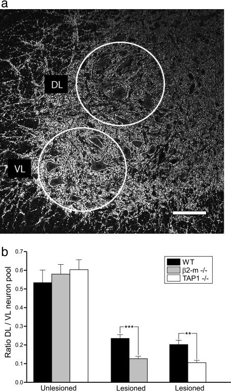
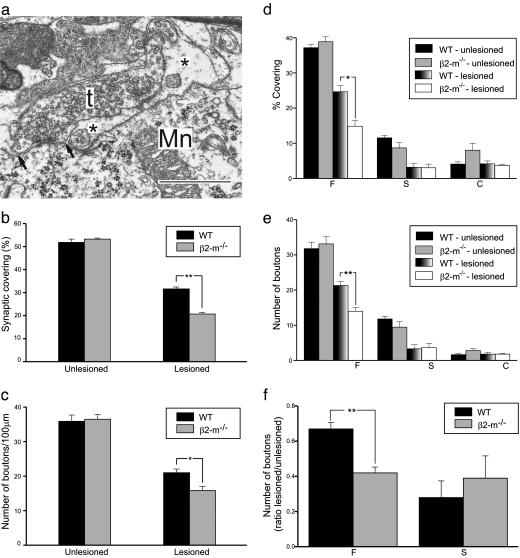
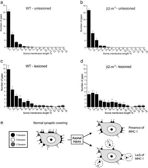
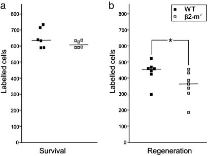
Recently, MHC class I molecules have been shown to be important for the retraction of synaptic connections that normally occurs during development [Huh, G.S., Boulanger, L. M., Du, H., Riquelme, P. A., Brotz, T. M. & Shatz, C. J. (2000) Science 290, 2155-2158]. In the adult CNS, a classical response of neurons to axon lesion is the detachment of synapses from the cell body and dendrites. We have investigated whether MHC I molecules are involved also in this type of synaptic detachment by studying the synaptic input to sciatic motoneurons at 1 week after peripheral nerve transection in beta2-microglobulin or transporter associated with antigen processing 1-null mutant mice, in which cell surface MHC I expression is impaired. Surprisingly, lesioned motoneurons in mutant mice showed more extensive synaptic detachments than those in wild-type animals. This surplus removal of synapses was entirely directed toward inhibitory synapses assembled in clusters. In parallel, a significantly smaller population of motoneurons reinnervated the distal stump of the transected sciatic nerve in mutants. MHC I molecules, which traditionally have been linked with immunological mechanisms, are thus crucial for a selective maintenance of synapses during the synaptic removal process in neurons after lesion, and the lack of MHC I expression may impede the ability of neurons to regenerate axons.


Figures

Fig. 1.
Fig. 1.
The reduction of synaptophysin immunoreactivity in axotomized motor pools is stronger in animals lacking functional MHC class I than in wild-type mice. (a) Immunofluorescence micrograph showing the studied areas in the spinal motor nuclei, the DL area representing the location of lesioned sciatic motoneurons, and the VL area intact motoneurons to the proximal hind limb. (Scale bar, 100 μm.) (b) Quantitative estimates of levels of immunoreactivity at 1 week after sciatic nerve transection show significantly stronger reductions in β2m-/- and TAP1-/- than in wild-type animals.
Fig. 2.
Fig. 2.
Synaptic detachment from cell bodies of axotomized motoneurons is stronger in animals lacking functional MHC class I than in wild-type mice with a selective surplus removal of putatively inhibitory type F nerve terminals. (a) Electron micrograph showing a partially detached nerve terminal (t) on an axotomized sciatic motoneuron (Mn) in the spinal cord at 1 week after lesion. Note the presence of a finger-like astrocytic process (*) between the motoneuron membrane and a large part of the terminal. The arrows denote the borders for the apposition of the terminal to the motoneuron. (Bar, 1 μm.) The detachment of synaptic terminals (b) and the reduction in number of nerve terminals apposing the motoneuron cell soma (c) are larger from motoneurons in β2m-/- mice than in wild-type animals at 1 week after sciatic nerve transection. Detailed analysis of the synaptic covering and number of synaptic terminals on the cell body of axotomized motoneurons reveals that the surplus removal of synapses in mutant mice is confined to type F nerve terminals with a putative inhibitory function (d and e). The extra removal of type F terminals in mutants abolishes the selective maintenance of a pool of type F terminals in wild-type animals (f).
Fig. 3.
Fig. 3.
The surplus removal of synaptic terminals in β2m-/- mice is preferentially directed toward clusters of nerve terminals. Graphs show the distribution of distances between nerve terminals along the cell soma membrane of motoneurons. The distances are calculated in percentage of total soma membrane length of the neuron in the nuclear plane. In unlesioned motoneurons, the intervals between nerve terminals are very similar in mutant and wild-type animals (a and b). In lesioned motoneurons at 1 week after axotomy, there is a distinct difference between the two groups of animals with a much more prominent loss of closely spaced terminals in β2m-/- mice compared with wild-type mice (c and d). The schematic drawing in e summarizes the main differences in the synaptic removal process between wild-type and β2m-/- mice.
Fig. 4.
Fig. 4.
Hampered axon regeneration, but no difference in motoneuron number in β2m-/- compared with wild-type mice at 3 weeks after a 1-mm resection of the sciatic nerve. The number of motoneurons retrogradely labeled with Fast Blue after application to the proximal stump is shown in a. The numbers are very similar in the two groups of animals. (b) The number of retrogradely labeled motonerons after application to the distal stump is significantly lower in β2m-/- animals. The numbers from individual animals are given, with horizontal lines denoting median values.

Comment in

References

    1. Huh, G. S., Boulanger, L. M., Du, H., Riquelme, P. A., Brotz, T. M. & Shatz, C. J. (2000) Science 290 2155-2158. - PMC - PubMed
    1. Corriveau, R. A., Huh, G. S. & Shatz, C. J. (1998) Neuron 21 505-520. - PubMed
    1. Lindå, H., Hammarberg, H., Piehl, F., Khademi, M. & Olsson, T. (1999) J. Neuroimmunol. 101 76-86. - PubMed
    1. Blinzinger, K. & Kreutzberg, G. (1968) Z. Zellforsch. Mikrosk. Anat. 85 145-157. - PubMed
    1. Sumner, B. E. H. (1975) Exp. Neurol. 49 406-417. - PubMed

Publication types

MeSH terms

Substances

LinkOut - more resources