

Review
doi: 10.1104/pp.104.052159.
The cytoskeleton as a regulator and target of biotic interactions in plants
Affiliations
- PMID: 15591444
- PMCID: PMC535820
- DOI: 10.1104/pp.104.052159
Item in Clipboard
Review
The cytoskeleton as a regulator and target of biotic interactions in plants
Plant Physiol.
2004 Dec.
No abstract available
Figures
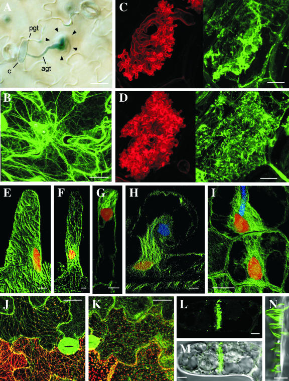
A, Accumulation of cytoplasm in an Arabidopsis epidermal cell around the attempted penetration site of the nonpathogen, B. graminis f. sp. hordei. Bar = 20 μm. B, GFP-tagged actin microfilaments focusing on the penetration site of B. graminis f. sp. hordei in an Arabidopsis epidermal cell. The fine actin microfilament network beneath the penetration site is likely to be indicative of active exocytosis. The asterisk indicates the attempted penetration site of the nonpathogen. Bar = 20 μm. pgt, Primary germ tube; agt, appressorial germ tube; c, conidum; n, plant nucleus. C and D, Arbuscules of G. versiforme developing in cortical cells of M. truncatula labeled with wheatgerm agglutinin (left) and with anti-tubulin (right). During early development (C), a diffuse fluorescence of anti-tubulin occurs around the developing arbuscular branches. Later in development (D), a dense array of short microtubules lines the perifungal membrane around the arbuscules. Bars = 10 μm. (Reproduced with permission from Blancaflor et al., 2001). E to I, Anti-tubulin labeling of microtubules in root hairs of M. truncatula before (E) and after (F–H) inoculation with rhizobia and in cortical cells (I). The helical array of cortical microtubules in the uninoculated hair (E) is replaced by a dense array connecting the nucleus with the tip of the hair (F and G) or the tip of the infection thread (H). Before the infection thread penetrates the cortical cells, a bridge of cytoplasm, the preinfection thread, forms in line with the advancing infection thread (I). Microtubules, nuclei, and infection threads are shown in green, red, and blue, respectively. E to H, Bars = 5 μm; I, bar = 15 μm. (Reproduced with permission from Timmers et al., 1999). J and K, Localization of wild-type TMV MP (J) and TMV MPR3 (K). Wild-type MP and MPR3 are visualized by tagging with DsRed (red) in transgenic plants expressing GFP-labeled microtubules (green). Bars = 25 μm. (Reproduced with permission from Gillespie et al., 2002). L to N, Fluorescent tubules in a young cross wall of a BY-2 suspension cell formed after expression of GFLV MP tagged with GFP. Bars = 5 μm. (Reproduced with permission from Laporte et al., 2003).
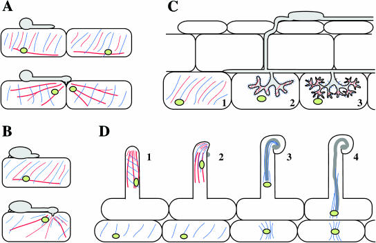
Diagrammatic representation of the organization of the plant cytoskeleton during different plant-microbe interactions. A, Interaction between a plant and a filamentous pathogen, such as that between Arabidopsis and P. parasitica. Top, Microtubules (blue), actin microfilaments (red), and nuclei (green) before appressorium formation or attempted penetration. Bottom, Actin microfilaments become focused on the penetration site and the nucleus is moved close to the invading pathogen. B, Interaction between a plant and a filamentous pathogen, such as that between barley and Erysiphe. Top, Cytoskeleton and nucleus before appressorium formation. Bottom, Microtubules and actin microfilaments become focused below the infection site and the nucleus also moves to this site. C, Cytoskeletal rearrangements during colonization by an arbuscular endomycorrhizal fungus. Before colonization, microtubules and actin microfilaments are aligned in ordered cortical arrays in cortex cells (C, subsection 1). During arbuscule development (C, subsection 2), actin microfilaments and apparently unpolymerized tubulin occur next to the perifungal membrane around the arbuscule. As the arbuscule matures (C, subsection 3), dense arrays of short microtubules and actin microfilaments line the perifungal membrane. The plant cell nucleus becomes positioned adjacent to the arbuscule. D, Infection thread formation during interaction of rhizobia with legume roots. In uninoculated root hairs (D, subsection 1), microtubules are axial or helically aligned and actin microfilaments are axially aligned in the cortex and endoplasm. The nucleus is positioned about 30 to 40 μm from the tip of the hair. After inoculation (D, subsection 2), dense arrays of microtubules and actin microfilaments form in the hair apex; the nucleus is moved closer to the hair tip and the cytoskeleton becomes focused on the site of growth as the hair begins to curl. As the infection thread grows along the root hair (D, subsections 3 and 4), microtubules line the plasma membrane surrounding the infection thread. A bridge of cytoplasm containing a parallel array of microtubules forms in cortical cells in alignment with the advancing infection thread (D, subsection 4).
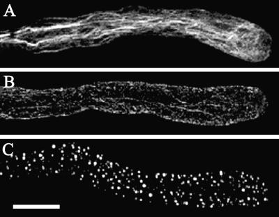
Depolymerization of actin microfilaments in a pollen tube triggered by a self-incompatibility response. A, Normally growing pollen tube. B, Incompatible pollen tube 5 min after treatment with S-protein. C, Incompatible pollen tube 60 min after treatment with S-protein. Actin filaments were visualized by Alexa-488-phalloidin staining. Bar = 10 μm. (Reproduced with permission from Snowman et al., 2002.)
Similar articles
-
The plant cytoskeleton.Curr Opin Cell Biol. 1994 Feb;6(1):10-5. doi: 10.1016/0955-0674(94)90110-4. Curr Opin Cell Biol. 1994. PMID: 8167014 Review.
-
The Role of Viruses in the Phytobiome.Annu Rev Virol. 2018 Sep 29;5(1):93-111. doi: 10.1146/annurev-virology-092917-043421. Epub 2018 Jul 26. Annu Rev Virol. 2018. PMID: 30048220 Review.
-
The cytoskeleton becomes multidisciplinary.Plant Physiol. 2004 Dec;136(4):3853-4. doi: 10.1104/pp.104.900130. Plant Physiol. 2004. PMID: 15591442 Free PMC article. No abstract available.
-
Getting sick may help plants overcome abiotic stress.New Phytol. 2008;180(4):738-41. doi: 10.1111/j.1469-8137.2008.02673.x. New Phytol. 2008. PMID: 19138229 No abstract available.
-
Endocytosis, actin cytoskeleton, and signaling.Plant Physiol. 2004 Jul;135(3):1150-61. doi: 10.1104/pp.104.040683. Plant Physiol. 2004. PMID: 15266049 Free PMC article. Review. No abstract available.
Cited by
-
Arabidopsis actin-depolymerizing factor AtADF4 mediates defense signal transduction triggered by the Pseudomonas syringae effector AvrPphB.Plant Physiol. 2009 Jun;150(2):815-24. doi: 10.1104/pp.109.137604. Epub 2009 Apr 3. Plant Physiol. 2009. PMID: 19346440 Free PMC article.
-
Rearrangement of actin cytoskeleton mediates invasion of Lotus japonicus roots by Mesorhizobium loti.Plant Cell. 2009 Jan;21(1):267-84. doi: 10.1105/tpc.108.063693. Epub 2009 Jan 9. Plant Cell. 2009. PMID: 19136645 Free PMC article.
-
RNA-Sequencing in Resistant (QL3) and Susceptible (Theis) Sorghum Cultivars Inoculated With Johnsongrass Isolates of Colletotrichum sublineola.Front Genet. 2021 Aug 11;12:722519. doi: 10.3389/fgene.2021.722519. eCollection 2021. Front Genet. 2021. PMID: 34456979 Free PMC article.
-
The cytoskeleton is disrupted by the bacterial effector HrpZ, but not by the bacterial PAMP flg22, in tobacco BY-2 cells.J Exp Bot. 2013 Apr;64(7):1805-16. doi: 10.1093/jxb/ert042. Epub 2013 Feb 13. J Exp Bot. 2013. PMID: 23408828 Free PMC article.
-
Plant actin depolymerizing factor: actin microfilament disassembly and more.J Plant Res. 2017 Mar;130(2):227-238. doi: 10.1007/s10265-016-0899-8. Epub 2017 Jan 2. J Plant Res. 2017. PMID: 28044231 Free PMC article. Review.
References
-
- Abdrakhamanova A, Wang QY, Khokhlova L, Nick P (2003) Is microtubule disassembly a trigger for cold acclimation? Plant Cell Physiol 44: 676–686 - PubMed
-
- Aist JR (1976) Papillae and related wound plugs of plant cells. Annu Rev Phytopathol 14: 145–163
-
- Armstrong L, Peterson RL (2002) The interface between the arbuscular mycorrhizal fungus Glomus intraradices and root cells of Panax quinquefolius: a Paris-type mycorrhizal association. Mycologia 94: 587–595 - PubMed
-
- Berg RH (1999) Cytoplasmic bridge formation in the nodule apex of actinorhizal root nodules. Can J Bot 77: 1351–1357
Publication types
MeSH terms
LinkOut - more resources
Full Text Sources
