. 2005 Jan;54(1):60-9.
doi: 10.1136/gut.2003.037663.

CD4+ T cell mediated intestinal immunity: chronic inflammation versus immune regulation

Affiliations

CD4+ T cell mediated intestinal immunity: chronic inflammation versus immune regulation

A M Westendorf et al. Gut. 2005 Jan.

Abstract

Background: Several studies have suggested that chronic inflammatory bowel disease may be a consequence of antigen specific recognition by appropriate T cells which expand and induce immunopathology.

Aims: We wished to investigate whether autoreactive CD4+ T cells can initiate the disease on recognition of enterocyte specific antigens directly and if induction of mucosal tolerance occurs.

Methods: Transgenic mice (VILLIN-HA) were generated that showed specific expression of haemagglutinin from influenza virus A exclusively in enterocytes of the intestinal epithelium. To investigate the impact of enterocyte specific haemagglutinin expression in an autoimmune environment, we mated VILLIN-HA mice with T cell receptor (TCR)-HA mice expressing an alpha/beta-TCR, which recognises an MHC class II restricted epitope of haemagglutinin, and analysed the HA specific T cells for induction of autoimmunity or tolerance.

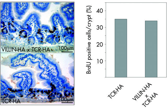
Results: In VILLIN-HAxTCR-HA mice, incomplete central deletion of HA specific lymphocytes occurred. Peripheral HA specific lymphocytes showed an activated phenotype and increased infiltration into the intestinal mucosa, but not into other organs of double transgenic mice. Enterocyte specific lamina propria lymphocytes showed a dose dependent proliferative response on antigen stimulation whereas the proliferative capacity of intraepithelial lymphocytes was reduced. Mucosal lymphocytes from VILLIN-HAxTCR-HA mice secreted lower amounts of interferon gamma and interleukin (IL)-2 but higher levels of tumour necrosis factor alpha, monocyte chemoattractant protein 1, and IL-6. Mucosal immune reactions were accompanied by broad changes in the gene expression profile with expression of proinflammatory genes, but strikingly also a remarkable set of genes discussed in the context of peripheral induction of regulatory T cells, including IL-10, Nrp-1, and Foxp3.

Conclusions: Enterocyte specific antigen expression is sufficient to trigger a specific CD4+ T cell response leading to mucosal infiltration. In our model, progression to overt clinical disease was counteracted most likely by induction of regulatory T cells.


Figures

Figure 1
Figure 1
Intestine specific haemagglutinin (HA) expression in VILLIN-HA transgenic mice. (A) Targeting construct. The 10.857bp Cla-I /SacII fragment comprising the VILLIN promoter followed by the HA protein (from influenza virus A/PR/8/34) was used for generation of (C57BL/6JxDBA/2) F1-transgenic mice. Mice were crossed on a BALB/c background. (B) HA mRNA expression analysis by reverse transcription-polymerase chain reaction in different organs of transgenic and control mice. The upper panel shows HA mRNA expression in BALB/c mice (negative control) and the lower panel in VILLIN-HA transgenic mice. DNA templates from transgenic and non-transgenic animals were used as positive and negative controls. (C) Semiquantitative HA mRNA expression analysis of intestinal epithelial cells from VILLIN-HA transgenic and BALB/c mice. (D) Western blot analysis of HA expression in the gut. As a positive control a lysate of HA producing recombinant Escherichia coli was used.
Figure 2
Figure 2
Antigen presenting capacity of intestinal epithelial cells from VILLIN-HA transgenic mice. Intestinal epithelial cells were isolated from VILLIN-HA transgenic and BALB/c mice and incubated for at least 72 hours with haemagglutinin (HA) specific CD4+ T cells. Proliferation was measured by 3[H] thymidine incorporation.
Figure 3
Figure 3
Haemagglutinin (HA) specific CD4+ T cells in the periphery of VILLIN-HA×T cell receptor (TCR)-HA mice and their proliferative capacity. (A) VILLIN-HA×TCR-HA and TCR-HA control mice were sacrificed, and spleen and mesenteric lymph node (MLN) cells were isolated and stained for 6.5 and CD4 expression to measure the percentage of transgenic T cells in the different compartments. (B) Proliferative capacity of HA specific CD4+ T cells from VILLIN-HA×TCR-HA mice in spleen and MLN. Splenic and lymph node cells from VILLIN-HA×TCR-HA and TCR-HA control mice were isolated and identical numbers of antigen specific 6.5+CD4+ T cells from the spleen and MLN were used for in vitro proliferation assays in the presence or absence of 10 µg/ml of HA peptide. Proliferation was measured by 3[H] thymidine incorporation.
Figure 4
Figure 4
Activation pattern of 6.5+ CD4+ T cells from double transgenic VILLIN-HA×T cell receptor (TCR)-HA mice compared with TCR-HA mice. 6.5+CD4+ T cells were isolated from the spleen and mesenteric lymph node (MLN) of VILLIN-HA×TCR-HA and TCR-HA mice, respectively. Lymphocytes were stained for 6.5 and CD4 expression on splenocytes (A) and MLN (B) as well as for CD25, CD45RB, CD62L, and CD69 antibodies. Cells were gated for 6.5 and CD4 expression and analysed regarding expression of the different activation markers by FACS.
Figure 5
Figure 5
VILLIN-HA×T cell receptor (TCR)-HA double transgenic mice were characterised by infiltration of lymphocytes into the lamina propria and intestinal epithelium. (A) Intestinal villi were distended by increased numbers of lymphocytes (left panel) compared with intestinal villi of TCR-HA transgenic mice (right panel). Similarly, the number of intraepithelial lymphocytes (IEL) was increased (insets). Insets show α-CD3 immunohistochemistry on paraffin embedded tissues. The avidin-biotin complex method with diaminobenzidine was used as substrate (brown colour) with haematoxylin counterstain (blue nuclei). Ileum: haematoxylin and eosin (H&E) stain; scale bar 80µm. (B) Increased number of lamina propria lymphocytes (LPL) and IEL. Lymphocytes were counted in H&E stained sections of 12–16 week old mice and are reported per 100 enterocytes. Individual animals are indicated for double transgenic VILLIN-HA×TCR-HA mice, TCR-HA mice, and for VILLIN-HA control mice.
Figure 6
Figure 6
No tissue damage occurred in the intestinal epithelial layer. T cell receptor (TCR)-HA and VILLIN-HA×TCR-HA mice were injected with 1 mg of BrdU via the intraperitonal route. After 24 hours the small intestine was harvested and processed for paraffin sections. Immunohistochemical staining of BrdU was performed using the BrdU In-situ Detection Kit (BD Bioscience). Proliferating cells in crypts that incorporated BrdU can be identified by the dark brown colour in their nuclei (left panel). Counted BrdU positive cell/crypt in TCR-HA and VILLIN-HA×TCR-HA transgenic mice are depicted in the right panel.
Figure 7
Figure 7
Reduced proliferative capacity of haemagglutinin (HA) specific intestinal epithelial cells (IEL) from VILLIN-HA×T cell receptor (TCR)-HA mice. Lamina propria lymphocytes (LPL) (A) and IEL (B) were isolated from VILLIN-HA×TCR-HA and TCR-HA control mice and stimulated in vitro with the corresponding haemagglutinin (HA) peptide. Thymidine incorporation was measured in cpm per 1000 6.5+CD4+ T cells.
Figure 8
Figure 8
Haemagglutinin (HA) specific CD4+ T cells from the infiltrated mucosa differed in cytokine profile. Lamina propria lymphocytes (LPL) (A) and intestinal epithelial cells (IEL) (B) from T cell receptor (TCR)-HA and VILLIN-HA×TCR-HA mice were stimulated in vitro with the HA110-120 peptide. Culture supernatants were analysed for several cytokines using the cytokine bead array from BD Bioscience. Cytokine quantities are depicted as pg/ml per 1000 6.5+CD4+ intestinal T cells. TNF-α, tumour necrosis factor α; MCP-1, monocyte chemoattractant protein 1; IFN-γ, interferon γ; IL, interleukin.
Figure 9
Figure 9
Global gene expression profiling of haemagglutinin (HA) specific CD4+ T cells. Cluster analysis of genes differentially expressed in 6.5+CD4+ T cells isolated from lamina propria (LPL) and epithelium (IEL) of infiltrated VILLIN-HA×T cell receptor (TCR)-HA as well as healthy TCR-HA mice. Red indicates induction of gene expression, green indicates repression. The brighter the colour the stronger the factor of gene regulation (+3, bright red; −3, bright green). Black indicates no changes. Inclusion into this heat map required at least a 1.5-fold difference in inducible gene expression. LPL (infil.) represents genes differentially expressed in 6.5+CD4+ T cells from the infiltrated lamina propria of VILLIN-HA×TCR-HA mice compared with the LPL of healthy TCR-HA donors. IEL (infil.) represents gene differentially expressed in 6.5+CD4+ T cells from the epithelium of VILLIN-HA×TCR-HA mice compared with TCR-HA. LPL v IEL characterises basal level expression of genes by LPL compared with IEL of healthy TCR-HA mice. Cluster (A): Genes upregulated in the LPL and IEL of VILLIN-HA×TCR-HA mice on mucosal infiltration. Cluster (B): Genes downregulated in LPL and IEL during infiltration. Cluster (C): Genes exclusively upregulated by LPL from double transgenic mice. Cluster (D): Genes downregulated in self reactive LPL CD4+ T cells in the infiltrated gut. Cluster (E): Genes exclusively upregulated by IEL from infiltrated tissue. Cluster (F): Genes downregulated by IEL from VILLIN-HA×TCR-HA mice. Results are from pooled individual mice (n>3).
Figure 10
Figure 10
Interleukin (IL)-10, Nrp-1, and Foxp3 expression pattern of 6.5+CD4+ lamina propria lymphocytes (LPL). 6.5+CD4+ LPL were sorted from T cell receptor (TCR)-HA and VILLIN-HA×TCR-HA transgenic mice. Total RNA was prepared, reverse transcribed, and mRNA expression levels determined in real time reverse transcription-polymerase chain reaction (RT-PCR) assays. Mean relative regulation is indicated. Results are from pooled individual mice (n>3) and obtained in duplicate real time RT-PCR assays. RPS9 mRNA expression served as a housekeeping gene control.

Comment in

Similar articles

Cited by

References

    1. Nagler-Anderson C . Man the barrier! Strategic defences in the intestinal mucosa. Nat Rev Immunol 2001;1:59–67. - PubMed
    1. Kisielow P , Teh HS, Bluthmann H, et al. Positive selection of antigen-specific T cells in thymus by restricting MHC molecules. Nature 1988;335:730–3. - PubMed
    1. Von Boehmer H , Aifantis I, Gounari F, et al. Thymic selection revisited: how essential is it? Immunol Rev 2003;191:62–78. - PubMed
    1. Melamed D , Friedman A. Direct evidence for anergy in T lymphocytes tolerized by oral administration of ovalbumin. Eur J Immunol 1993;23:935–42. - PubMed
    1. Monteleone I , Vavassori P, Biancone L, et al. Immunoregulation in the gut: success and failures in human disease. Gut 2002;50 (suppl 3) :III60–4. - PMC - PubMed

Publication types

MeSH terms

Substances