. 2005 Jan;54(1):142-51.
doi: 10.1136/gut.2004.042127.

Human hepatic stellate cell lines, LX-1 and LX-2: new tools for analysis of hepatic fibrosis

Affiliations

Human hepatic stellate cell lines, LX-1 and LX-2: new tools for analysis of hepatic fibrosis

L Xu et al. Gut. 2005 Jan.

Abstract

Background: Hepatic stellate cells (HSCs) are a major fibrogenic cell type that contributes to collagen accumulation during chronic liver disease. With increasing interest in developing antifibrotic therapies, there is a need for cell lines that preserve the in vivo phenotype of human HSCs to elucidate pathways of human hepatic fibrosis. We established and characterised two human HSC cell lines termed LX-1 and LX-2, and compared their features with those of primary human stellate cells.

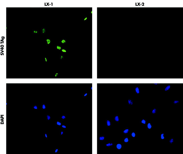
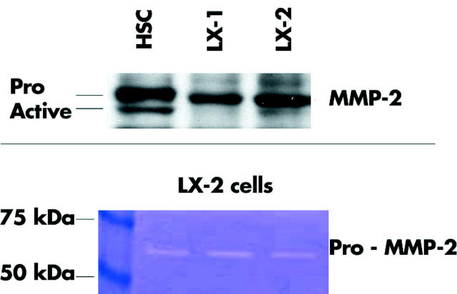
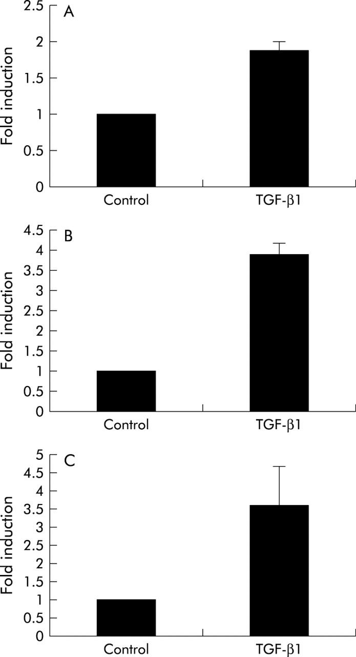
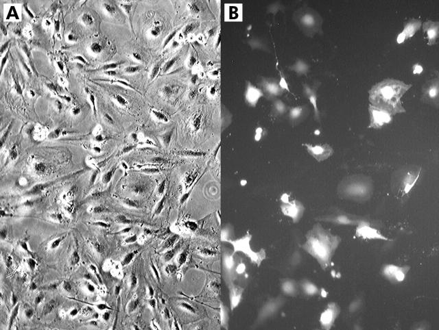
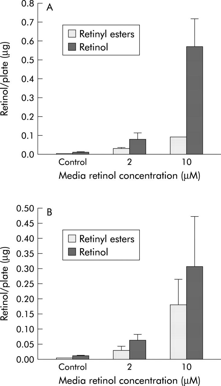
Methods and results: LX-1 and LX-2 were generated by either SV40 T antigen immortalisation (LX-1) or spontaneous immortalisation in low serum conditions (LX-2). Both lines express alpha smooth muscle actin, vimentin, and glial fibrillary acid protein, as visualised by immunocytochemistry. Similar to primary HSCs, both lines express key receptors regulating hepatic fibrosis, including platelet derived growth factor receptor beta (betaPDGF-R), obese receptor long form (Ob-RL), and discoidin domain receptor 2 (DDR2), and also proteins involved in matrix remodelling; matrix metalloproteinase (MMP)-2, tissue inhibitor of matrix metalloproteinase (TIMP)-2, and MT1-MMP, as determined by western analyses. LX-2 have reduced expression of TIMP-1. LX-2, but not LX-1, proliferate in response to PDGF. Both lines express mRNAs for alpha1(I) procollagen and HSP47. Transforming growth factor beta1 stimulation increased their alpha1(I) procollagen mRNA expression, as determined by quantitative reverse transcription-polymerase chain reaction. LX-2, but not LX-1, cells are highly transfectable. Both lines had a retinoid phenotype typical of stellate cells. Microarray analyses showed strong similarity in gene expression between primary HSCs and either LX-1 (98.4%) or LX-2 (98.7%), with expression of multiple neuronal genes.

Conclusions: LX-1 and LX-2 human HSC lines provide valuable new tools in the study of liver disease. Both lines retain key features of HSCs. Two unique advantages of LX-2 are their viability in serum free media and high transfectability.


Figures

Figure 1
Figure 1
Expression of SV40 large T antigen (TAg) in the LX-1 cell line. LX cells were subjected to immunocytochemical analysis (see methods). The SV40 large T antigen was detected in the nucleus of LX-1 cells but not in LX-2 cells. Cells were costained with DAPI to identify nuclei.
Figure 2
Figure 2
Expression of intermediate filaments by LX-1 and LX-2. LX cells were subjected to immunocytochemical analysis (see methods). Both LX-1 and LX-2 cells expressed and formed a cytoskeletal network of α-smooth muscle actin (α-SMA), vimentin, and glial fibrillary acid protein (GFAP). Cells were costained with DAPI to identify nuclei.
Figure 3
Figure 3
Expression of the platelet derived growth factor receptor β (βPDGF-R) and response of LX-2 cells to BB-PDGF. (Top) Immunoprecipitation/western analysis (see methods) detected expression of the βPDGF-R subunit in both LX-1 and LX-2 cells. NIH3T3 cells and passaged human hepatic stellate cells (HSC) were used as controls. (Bottom) BB-PDGF ligand (1 and 10 ng/ml) stimulated proliferation of LX-2 cells in serum free medium, as determined by 3H-thymidine incorporation (cpm).
Figure 4
Figure 4
Expression of receptors and matrix remodelling proteins detected by western analyses. LX cell lines and primary passaged hepatic stellate cells (HSC) were compared side by side for their expression of the receptors discoidin domain receptor 2 (DDR2) and obese receptor long form (Ob-RL), and also for the matrix remodelling proteins MT1-MMP, tissue inhibitor of matrix metalloproteinase (TIMP)-2 and TIMP-1 (see methods): 40 μg of protein from cell lysates were loaded per sample.
Figure 5
Figure 5
Matrix metalloproteinase (MMP)-2 protein expression and activity. (Top) Western analysis detected the pro- and active form of MMP-2 in primary hepatic stellate cells. In LX cells, pro-MMP-2 was mostly detected: 40 μg of protein from cell lysates were loaded per sample. (Bottom) Gelatin zymography of conditioned media from LX-2 cells indicated secretion of pro-MMP2.
Figure 6
Figure 6
HSP47 (A) and α1(I) procollagen (B) mRNA expression. Primary hepatic stellate cells (HSC), LX cells, and the hepatoma cells Huh7 and HepG2 were examined and compared for their expression of HSP47 (A) and α1(I) procollagen (B) mRNA. using quantitative real time reverse transcription-polymerase chain reaction (see methods). Expression levels of primary HSC were used as a reference point set at 1 and the fold differences are indicated. Error bars represent mean (SEM) (n = 3). All cells were cultured in Dulbecco’s modified Eagle’s medium (DMEM)/10% fetal bovine serum (FBS) except for LX-2 cells which were cultured in DMEM with only 1% FBS.
Figure 7
Figure 7
Transforming growth factor β1 (TGF-β1) increases α1(I) procollagen mRNA in hepatic stellate cells (HSC) (A), and LX-1 (B) and LX-2 (C) cells. Primary HSCs and LX cells were examined for their responsiveness to TGF-β1, as measured by its effects on α1(I) procollagen mRNA levels using quantitative real time reverse transcription-polymerase chain reaction (see methods). Untreated controls were used as a reference point set at 1 and the fold induction after TGF-β1 treatment (2.5 ng/ml for 20 hours) is shown. Error bars represent mean (SEM) (n = 3).
Figure 8
Figure 8
Transfection efficiency of LX-2 cells. LX-2 cells plated to 80% confluence in six well culture dishes were transfected with Fugene and 2.5 μg of pEGFP-C2 (Clontech), a plasmid that expresses enhanced green fluorescent protein. (A) Bright field image of transfected LX-2 cells. (B) Fluorescence image of the same field of cells.
Figure 9
Figure 9
LX-1 and LX-2 cells accumulate and esterify retinol. LX-1 (A) and LX-2 (B) cell culture media were supplemented with 0 (control), 2, or 10 μM retinol for 24 hours. Cells were then washed with ice cold phosphate buffered saline and harvested. Cell levels of retinol and retinyl esters were measured by high pressure liquid chromatography, as described in the methods section.

References

    1. Friedman SL. Molecular regulation of hepatic fibrosis, an integrated cellular response to tissue injury. J Biol Chem 2000;275:2247–50. - PubMed
    1. Schuppan D , Ruehl M, Somasundaram R, et al. Matrix as modulator of stellate cell and hepatic fibrogenesis. Semin Liver Dis 2001;21:351–72. - PubMed
    1. Rockey DC. Hepatic blood flow regulation by stellate cells in normal and injured liver. Semin Liver Dis 2001;21:337–50. - PubMed
    1. Friedman SL, Roll FJ, Boyles J, et al. Maintenance of differentiated phenotype of cultured rat hepatic lipocytes by basement membrane matrix. J Biol Chem 1989;264:10756–62. - PubMed
    1. Schnabl B , Choi YH, Olsen JC, et al. Immortal activated human hepatic stellate cells generated by ectopic telomerase expression. Lab Invest 2002;82:323–33. - PubMed

Publication types