. 2005 Jan;24(1):21-34.
doi: 10.1002/hbm.20068.

Fast robust subject-independent magnetoencephalographic source localization using an artificial neural network

Affiliations

Fast robust subject-independent magnetoencephalographic source localization using an artificial neural network

Sung Chan Jun et al. Hum Brain Mapp. 2005 Jan.

Abstract

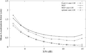
We describe a system that localizes a single dipole to reasonable accuracy from noisy magnetoencephalographic (MEG) measurements in real time. At its core is a multilayer perceptron (MLP) trained to map sensor signals and head position to dipole location. Including head position overcomes the previous need to retrain the MLP for each subject and session. The training dataset was generated by mapping randomly chosen dipoles and head positions through an analytic model and adding noise from real MEG recordings. After training, a localization took 0.7 ms with an average error of 0.90 cm. A few iterations of a Levenberg-Marquardt routine using the MLP output as its initial guess took 15 ms and improved accuracy to 0.53 cm, which approaches the natural limit on accuracy imposed by noise. We applied these methods to localize single dipole sources from MEG components isolated by blind source separation and compared the estimated locations to those generated by standard manually assisted commercial software.


Figures

Figure 1
Figure 1
Sensor surface and training region. The center of the spherical head model was varied within the given region. Diamonds denote sensors.
Figure 2
Figure 2
Mean localization error versus epoch for training of 100,000 exemplars with real brain noise. Testing used 25,000 patterns contaminated by real brain noise.
Figure 3
Figure 3
Mean localization errors of the trained MLP as a function of correct dipole location, binned into regions. All units are in cm. Left, coronal cross section; right, sagittal cross section.
Figure 4
Figure 4
Mean localization errors of the trained MLP for dipole noisy signals as a function of distance of the center of the spherical head model to the center of the helmet.
Figure 5
Figure 5
Mean localization error vs. SNR. MLP, MLP‐start‐LM, and optimal‐start‐LM were tested on signals from 25,000 random dipoles, contaminated by real brain noise.
Figure 6
Figure 6
MEG source localization procedure.
Figure 7
Figure 7
Dipole source localization results of Neuromag software (XFIT), our MLP, and MLP‐start‐LM for four real BSS‐separated MEG signal components obtained in the trump card task of Subject 1. Left, axial view; center, coronal view; right, sagittal view. The outer surface denotes the sensor surface, and diamonds on this surface denote sensors. The inner surface denotes a spherical head model fit to the subject. The center of a fitted spherical head model is (0.335, 0.698, 3.157). All units are in cm. Each localized dipole source triple is denoted by an acronym: PV, primary visual source; SV, secondary visual source; RA, auditory source in the right hemisphere; LA, auditory source in the left hemisphere.
Figure 8
Figure 8
Dipole source localization results of Neuromag software (XFIT), our MLP, MLP‐start‐LM for real BSS‐separated MEG signal components obtained by transverse patterning task of Subject 1. The center of a fitted spherical head model is at (0.373, 0.642, 3.205). Layout as in Figure 7. Each localized dipole source triple is denoted by an acronym: PV, primary visual source; SV, secondary visual source; RS, somatosensory source in the right hemisphere.
Figure 9
Figure 9
Dipole source localization results of Neuromag software (XFIT), our MLP, and MLP‐start‐LM, for four BSS‐separated primary visual MEG signal components from Subject S01, taken from four tasks. Dipole locations are shown full‐scale in the top row, whereas graphs on the bottom zoom in on the rectangular regions shown. Layout as in Figure 7.
Figure 10
Figure 10
Dipole source localization results of Neuromag software (XFIT), our MLP, and MLP‐start‐LM for four BSS‐separated secondary visual MEG signal components from Subject S01, over four tasks. Dipole locations are shown full‐scale in the top row, whereas graphs on the bottom zoom in on the rectangular regions shown. Layout as in Figure 7.
Figure 11
Figure 11
Dipole source localization results of Neuromag software (XFIT), our MLP, and MLP‐start‐LM for three real BSS‐separated auditory MEG signal components from Subject S01, over the trump card (TCT) and elemental discrimination (EDT) tasks. Dipole locations are shown full‐scale in the top row, whereas graphs on the bottom zoom in on the rectangular regions shown. Layout as in Figure 7.
Figure 12
Figure 12
Dipole source localization results of Neuromag software (XFIT), our MLP, and MLP‐start‐LM for three BSS‐separated somatosensory MEG signal components from the transverse patterning task (TPT) over three subjects (S01, S02, S03). Dipole locations are shown full‐scale in the top row, whereas graphs on the bottom zoom in on the rectangular regions shown. Layout as in Figure 7.
Figure 13
Figure 13
Training datasets were loaded into this MLP structure. Note that the input includes the coordinates of the center of a sphere fit to the subject's head.

References

    1. Abeyratne UR, Kinouchi Y, Oki H, Okada J, Shichijo F, Matsumoto K (1991): Artificial neural networks for source localization in the human brain. Brain Topogr 4: 3–21. - PubMed
    1. Ahonen AI, Hämäläinen MS, Knuutila JET, Kajola MJ, Laine PP, Lounasmaa OV, Parkkonen LT, Simola JT, Tesche CD (1993): 122‐channel SQUID instrument for investigating the magnetic signals from the human brain. Physica Scripta 49: 198–205.
    1. Baillet S, Riera JJ, Marin G, Mangin JF, Aubert J, Garnero L (2001): Evaluation of inverse methods and head models for EEG source localization using a human skull phantom. Phys Med Biol 46: 77–96. - PubMed
    1. Balish M, Sato S, Connaughton P, Kufta C (1991): Localization of im‐planted dipoles by magnetoencephalography. Neurology 41: 1072–1076. - PubMed
    1. Barth DS, Sutherling W, Broffman J, Beatty J (1986): Magnetic localization of a dipolar current source implanted in a sphere and a human cranium. Electroencephalogr Clin Neurophysiol 63: 260–273. - PubMed

Publication types