Review
. 2004 Dec 21;43(50):15643-56.
doi: 10.1021/bi047907k.

Oligomerization of G protein-coupled receptors: past, present, and future

Affiliations
Review

Oligomerization of G protein-coupled receptors: past, present, and future

Paul S-H Park et al. Biochemistry. .

Abstract

G protein-coupled receptor (GPCR)-mediated signal transduction has been studied for more than a century. Despite the intense focus on this class of proteins, a molecular understanding of what constitutes the functional form of the receptor is still uncertain. GPCRs have traditionally been conceptualized as monomeric proteins, and this view has changed little over the years until relatively recently. Recent biochemical and biophysical studies have challenged this traditional concept, and point instead to a mechanistic view of signal transduction wherein the receptor functions as an oligomer. Cooperative interactions within such an oligomeric array may be critical for the propagation of an external signal across the cell membrane and to the G protein, and may therefore underlie the mechanistic basis of signaling.
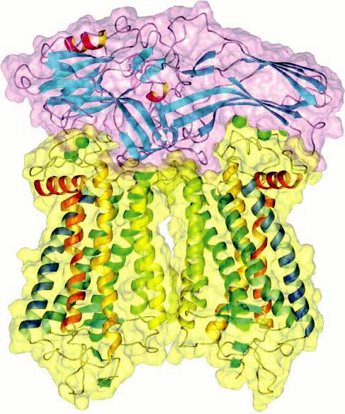

Figures

F<sc>igure</sc> 1
Figure 1
Evolving molecular concept of a G protein-coupled receptor. The molecular understanding of GPCRs during different periods in the history of the field is represented for GPCRs acting via AC and for the visual pigment rhodopsin. In the AC branch, agonists (yellow) were initially thought to bind to some part of the cell or tissue (A). The receptor was next thought to be a part of AC (violet) itself (B). It was then discovered that the receptor (blue) was a distinct molecular entity (C). The receptor has been conceptualized as a monomeric protein until relatively recently. Our current understanding is that the receptor exists as an oligomer (D). In the rhodopsin branch, the initial detection of rhodopsin (red) came from its characteristic reddish-purple color in the rod outer segments (E). The color was then determined to derive from a chromophore conjugated to a protein. Early biophysical methods suggested that rhodopsin was monomeric (F). More recent studies using atomic force microscopy point to an oligomeric arrangement of rhodopsin (G).
F<sc>igure</sc> 2
Figure 2
Schemes used to describe the interaction between an agonist and its receptor. Scheme 1 describes Clark's occupancy theory (). In Clark's theory, an agonist (A) will combine with the receptor (R) to form an agonist–receptor complex (AR). The level of the AR complex present at thermodynamic equilibrium can be defined by the equilibrium dissociation constant (KA). A physiological response (Q) will result only when the AR complex is formed, and a maximal response (Qmax) will occur when the agonist occupies all receptor sites. Scheme 2 describes the Stephenson–Furchgott model of efficacy (). Stephenson postulated that the activity of the agonist is determined by the affinity of the drug for the receptor (KA) and efficacy (e). Since the occupancy of receptors by the agonist did not appear to be linearly proportional to the response (Q), a dimensionless quantity called the stimulus (S) was introduced. The stimulus would lead to a response in a relationship that was not apparent at the time and was therefore left undefined [i.e., Q = f(S)]. The stimulus is defined explicitly as the product of efficacy and the fraction of receptors occupied by the agonist (i.e., S = e[AR]/[R]t). This model was refined later by Furchgott, who defined efficacy as the product of the concentration of the total active receptor ([R]t) and a quantity specific to the agonist–receptor complex designated as the intrinsic efficacy (ε) [i.e., e = ε[R]t, S = ε[AR] = e[A]/(KA + [A])] (). An explicit expression for the response is given for the simple case in which the dose–response relationship is in the form of a rectangular hyperbola. Scheme 3 describes the multisite model (), where an agonist (A) can bind to multiple classes of sites (Rj, j = 1, 2, 3, …, n) that are mutually independent and non-interconverting. The agonist can bind to each of the sites with distinct affinities (KAj). An equation for the binding isotherm is provided where Y is the fractional occupancy of the receptor by the agonist (i.e., Y=j=1n[ARj][R]t) and fj is the fraction of total receptor that is of type j (i.e., fj = [Rj]t/[R]t, where [R]t=j=1n[Rj]t. Scheme 4 describes the ternary complex model (, ), where the receptor (R) and G protein (G) are freely mobile within the plane of the membrane and interact through random collisions. Agonists bind to the free receptor (R) or receptor coupled to the G protein (RG) with dissociation constants KA and KAG, respectively. The G protein binds to the free receptor or receptor occupied by agonist (AR) with dissocation constants KG and KGA, respectively. Agonists promote coupling (KAG < KA) and therefore favor a ternary complex (ARG) (KA/KAG = KG/KGA). There are three conditions that need to be met for the ternary complex model to account for the dispersion of affinities observed in the binding of agonists. First, the agonist must differ in its affinity for the coupled and uncoupled states of the receptor (i.e., KAKAG). Second, the total concentration of the receptor must be bracketed by the affinity of the receptor for the G protein in the absence and presence of the agonist (i.e., KGA < [R]t < KG). Third, the total number of G proteins must be equal to or less than the total number of receptors (i.e., [G]t ≤ [R]t). If these conditions are not met, then the ternary complex model predicts that agonist binding curves will have the form of a rectangular hyperbola (i.e., nH = 1) (, , , , ). Scheme 5 describes cooperativity within a tetravalent receptor (n = 4) that interconverts spontaneously between two states (R and T) (). The distribution of sites in the absence of agonist is defined by the constant KRT. Binding occurs in a sequential manner where the first equivalent of agonist binds the vacant oligomer with the microscopic dissociation constant KAR or KAT (aR1 = 1 and aT1 = 1; A0R ≡ R and A0T ≡ T). The degree of cooperativity exerted on the binding of subsequent equivalents of the agonist will be reflected in cooperativity factors (aRj and aTj, j = 2, 3, or 4). Positive cooperativity occurs when aRj or aTj is less than 1; negative cooperativity occurs when aRj or aTj is greater than 1, and no cooperativity occurs when aRj or aTj is equal to 1. The oligomer is assumed to be symmetric, in that two or more vacant sites within either R or T are indistinguishable (1 ≤ j ≤ 3).
F<sc>igure</sc> 3
Figure 3
Ribbon and space-filling model of a complex between a rhodopsin dimer and a monomer of arrestin. Arrestin (purple) has a bipartite structure that can accommodate two molecules of rhodopsin (yellow). Phosphorylation sites on rhodopsin are represented by green spheres (adapted from ref ).
F<sc>igure</sc> 4
Figure 4
Ribbon and space-filling model of a complex between two rhodopsin dimers and a dimer of G. Information obtained from atomic force microscopy and from the crystal structures of rhodopsin and transducin suggests a 2:1 stoichiometry between rhodopsin (blue) and G (orange). Signaling may proceed via oligomers of rhodopsin and oligomers of G (adapted from ref ).

Similar articles

  • Monomeric G-protein-coupled receptor as a functional unit.
    Chabre M, le Maire M. Chabre M, et al. Biochemistry. 2005 Jul 12;44(27):9395-403. doi: 10.1021/bi050720o. Biochemistry. 2005. PMID: 15996094 Review.
  • The rhodopsin-arrestin-1 interaction in bicelles.
    Chen Q, Vishnivetskiy SA, Zhuang T, Cho MK, Thaker TM, Sanders CR, Gurevich VV, Iverson TM. Chen Q, et al. Methods Mol Biol. 2015;1271:77-95. doi: 10.1007/978-1-4939-2330-4_6. Methods Mol Biol. 2015. PMID: 25697518 Free PMC article.
  • Understanding the GPCR biased signaling through G protein and arrestin complex structures.
    Zhou XE, Melcher K, Xu HE. Zhou XE, et al. Curr Opin Struct Biol. 2017 Aug;45:150-159. doi: 10.1016/j.sbi.2017.05.004. Epub 2017 May 27. Curr Opin Struct Biol. 2017. PMID: 28558341 Review.
  • Crystal structure of rhodopsin bound to arrestin by femtosecond X-ray laser.
    Kang Y, Zhou XE, Gao X, He Y, Liu W, Ishchenko A, Barty A, White TA, Yefanov O, Han GW, Xu Q, de Waal PW, Ke J, Tan MH, Zhang C, Moeller A, West GM, Pascal BD, Van Eps N, Caro LN, Vishnivetskiy SA, Lee RJ, Suino-Powell KM, Gu X, Pal K, Ma J, Zhi X, Boutet S, Williams GJ, Messerschmidt M, Gati C, Zatsepin NA, Wang D, James D, Basu S, Roy-Chowdhury S, Conrad CE, Coe J, Liu H, Lisova S, Kupitz C, Grotjohann I, Fromme R, Jiang Y, Tan M, Yang H, Li J, Wang M, Zheng Z, Li D, Howe N, Zhao Y, Standfuss J, Diederichs K, Dong Y, Potter CS, Carragher B, Caffrey M, Jiang H, Chapman HN, Spence JC, Fromme P, Weierstall U, Ernst OP, Katritch V, Gurevich VV, Griffin PR, Hubbell WL, Stevens RC, Cherezov V, Melcher K, Xu HE. Kang Y, et al. Nature. 2015 Jul 30;523(7562):561-7. doi: 10.1038/nature14656. Epub 2015 Jul 22. Nature. 2015. PMID: 26200343 Free PMC article.
  • The orientation and stability of the GPCR-Arrestin complex in a lipid bilayer.
    Wang D, Yu H, Liu X, Liu J, Song C. Wang D, et al. Sci Rep. 2017 Dec 5;7(1):16985. doi: 10.1038/s41598-017-17243-y. Sci Rep. 2017. PMID: 29209002 Free PMC article.

Cited by

References

    1. Takeda S, Kadowaki S, Haga T, Takaesu H, Mitaku S. Identification of G protein-coupled receptor genes from the human genome sequence. FEBS Lett. 2002;520:97–101. - PubMed
    1. Ma P, Zemmel R. Value of novelty? Nat. Rev. Drug Discovery. 2002;1:571–572. - PubMed
    1. Wise A, Gearing K, Rees S. Target validation of G-protein coupled receptors. Drug Discovery Today. 2002;7:235–246. - PubMed
    1. Bockaert J, Pin JP. Molecular tinkering of G protein-coupled receptors: An evolutionary success. EMBO J. 1999;18:1723–1729. - PMC - PubMed
    1. Okada T, Ernst OP, Palczewski K, Hofmann KP. Activation of rhodopsin: New insights from structural and biochemical studies. Trends Biochem. Sci. 2001;26:318–324. - PubMed

Publication types