. 2004 Dec 21;43(50):15702-19.
doi: 10.1021/bi048364h.

Expanding the functional repertoire of CTD kinase I and RNA polymerase II: novel phosphoCTD-associating proteins in the yeast proteome

Affiliations

Expanding the functional repertoire of CTD kinase I and RNA polymerase II: novel phosphoCTD-associating proteins in the yeast proteome

Hemali P Phatnani et al. Biochemistry. .

Abstract

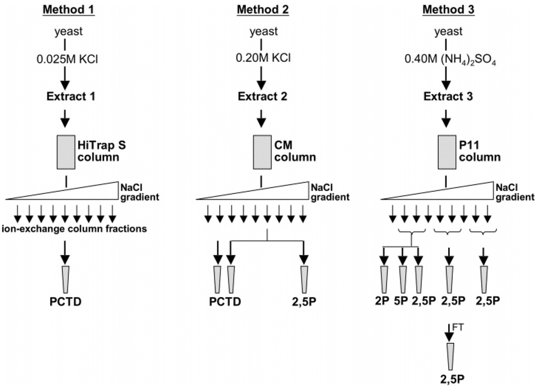
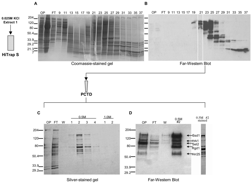
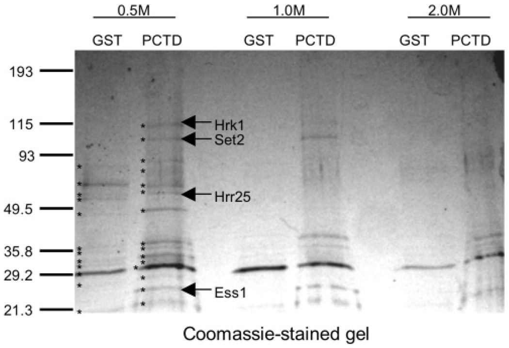
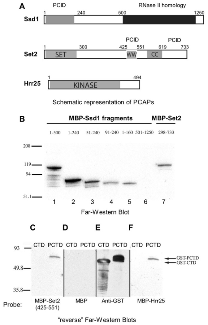
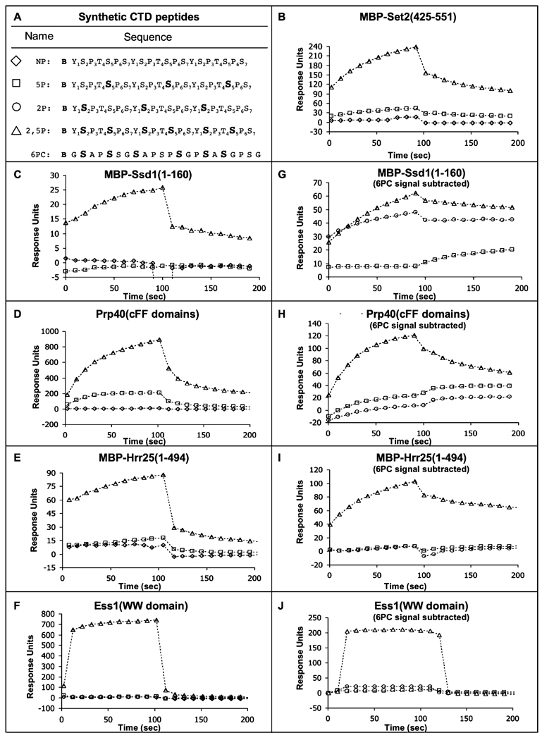
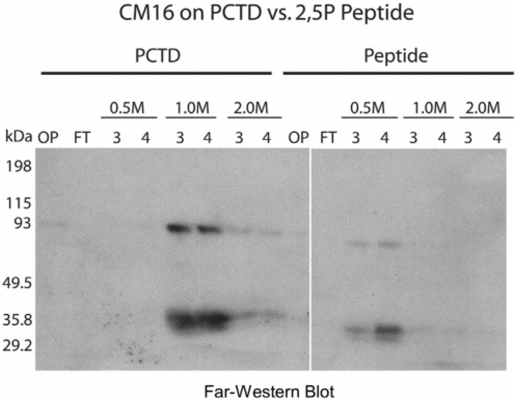
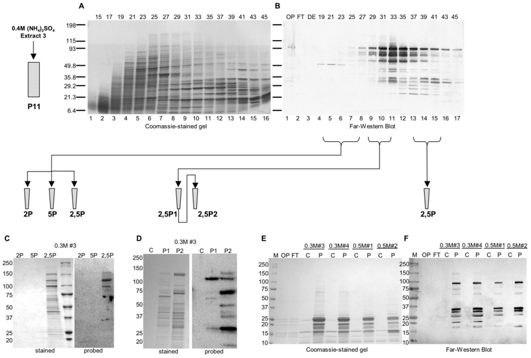
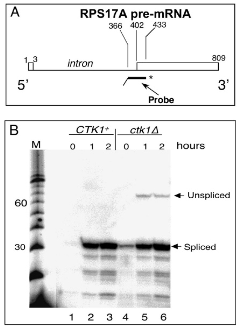
CTD kinase I (CTDK-I) of Saccharomyces cerevisiae is required for normal phosphorylation of the C-terminal repeat domain (CTD) on elongating RNA polymerase II. To elucidate cellular roles played by this kinase and the hyperphosphorylated CTD (phosphoCTD) it generates, we systematically searched yeast extracts for proteins that bound to the phosphoCTD made by CTDK-I in vitro. Initially, using a combination of far-western blotting and phosphoCTD affinity chromatography, we discovered a set of novel phosphoCTD-associating proteins (PCAPs) implicated in a variety of nuclear functions. We identified the phosphoCTD-interacting domains of a number of these PCAPs, and in several test cases (namely, Set2, Ssd1, and Hrr25) adduced evidence that phosphoCTD binding is functionally important in vivo. Employing surface plasmon resonance (BIACORE) analysis, we found that recombinant versions of these and other PCAPs bind preferentially to CTD repeat peptides carrying SerPO(4) residues at positions 2 and 5 of each seven amino acid repeat, consistent with the positional specificity of CTDK-I in vitro [Jones, J. C., et al. (2004) J. Biol. Chem. 279, 24957-24964]. Subsequently, we used a synthetic CTD peptide with three doubly phosphorylated repeats (2,5P) as an affinity matrix, greatly expanding our search for PCAPs. This resulted in identification of approximately 100 PCAPs and associated proteins representing a wide range of functions (e.g., transcription, RNA processing, chromatin structure, DNA metabolism, protein synthesis and turnover, RNA degradation, snRNA modification, and snoRNP biogenesis). The varied nature of these PCAPs and associated proteins points to an unexpectedly diverse set of connections between Pol II elongation and other processes, conceptually expanding the role played by CTD phosphorylation in functional organization of the nucleus.


Figures

FIGURE 1
FIGURE 1
Schematic overview of methods used to purify phosphoCTD-associating proteins (PCAPs) from yeast. Rectangles represent ion-exchange columns; triangles represent salt elution gradients; trapezoids represent affinity columns.
FIGURE 2
FIGURE 2
PCAP purification by method 1 (details in Materials and Methods). HiTrap S fractions subjected to SDS–PAGE were stained with Coomassie Blue (A) or analyzed by far-western blotting (B) with β-gal-[32P]PCTD fusion protein as probe [OP )onput, FT ) flow-through]. Proteins in HiTrap S fraction 23 were further fractionated on a 100 µL phosphoCTD affinity column. Aliquots of the onput (OP), flow-through (FT), wash (W), and 0.5 and 1.0 M salt step elution fractions were analyzed by SDS–PAGE and stained with silver (C). Corresponding aliquots were analyzed by far-western blotting with GST-PCTD as probe (D) (detected using affinity-purified anti-GST antibody). On the right is shown a silver-stained lane of the 0.5 M #2 sample. The identities of PCAPs 1 – 5 are listed alongside the respective bands. Symbols are as in Figure 1.
FIGURE 3
FIGURE 3
Example of PCAP purification by method 2 (details in Materials and Methods). CM fractions were passed through a control GST column and then applied to a GST-PCTD affinity matrix. Both columns were eluted with salt steps, and the eluted proteins were analyzed by SDS–PAGE. All stained bands (dots) were excised from the gel and analyzed by mass spectrometry. Proteins specifically eluted from the PCTD matrix are labeled.
FIGURE 4
FIGURE 4
A) Schematic representation of domain architecture of PCAPs Ssd1, Set2, and Hrr25. (B) Confirmation of the Ssd1– phosphoCTD interaction and identification of the binding domain. Fragments of Ssd1 and Set2 were expressed as recombinant MBP fusion proteins and analyzed by far-western blotting (amino acids defining the fragments are shown above their respective lanes). Approximately equal amounts of protein were run in each lane. The proteins were transferred to nitrocellulose and probed with GST-[32P]PCTD fusion protein followed by autoradiography. (C) A second PCID in Set2. GST-CTD and GST-phosphoCTD were subjected to SDS–PAGE, transferred to nitrocellulose, and probed with purified recombinant MBP–Set2 (amino acids 425–551) in a reverse far-western assay. The MBP fusion protein was detected with an anti-MBP antibody. (D) Control: the blot in panel C was stripped and probed with purified recombinant MBP. (E) The blot in panel F was stripped and probed with an anti-GST antibody to demonstrate the presence of the GST–CTD fusion proteins. (F) Confirmation of the Hrr25–phosphoCTD interaction. A blot identical to the one in panel C was probed with purified recombinant MBP–Hrr25. The MBP fusion protein was detected with an anti-MBP antibody.
FIGURE 5
FIGURE 5
Specificity of binding of various PCAPs to (phospho)CTD peptides using BIACORE (surface plasmon resonance) analysis. (A) Sequences of biotinylated peptides that were immobilized on streptavidin sensor chips with symbols used in graphs B–J. (B) Sensorgrams of binding of MBP–Set2(425–551) (2 µM) to the 2,5P, 5P, and NP CTD peptides, after subtracting signal from an empty flow cell (nonspecific binding). (C) Sensorgrams of binding of MBP–Ssd1(1–160) (2 µM) to CTD peptides as in (B). (D) Sensorgrams of binding of Prp40 (cFF domains) [amino acids 267–583 (34)] (2 µM) to CTD peptides as in (B). (E) Sensorgrams of binding of MBP–Hrr25(1– 494) (2 µM) to CTD peptides as in (B). (F) Sensorgrams of binding Ess1 (WW domain) (2 µM) to CTD peptides as in (B). (G) Sensorgrams of binding of MBP–Ssd1(1–160) (2 µM) to the 2,5P, 5P, and 2P CTD peptides, after subtracting signal from the scrambled 6PC peptide channel [sequences of peptides shown in (A)]. (H) Sensorgrams of binding of Prp40 (cFF domains) [amino acids 267–583 (34)] (2 µM) to CTD peptides as in (G). (I) Sensorgrams of binding of MBP–Hrr25(1–494) (2 µM) to CTD peptides as in (G). (J) Sensorgrams of binding of Ess1 (WW domain) (2 µM) to CTD peptides as in (G).
FIGURE 6
FIGURE 6
Synthetic 2,5P peptide column mimics PCTD column. Each half of a CM fraction was subjected to affinity chromatography on either a PCTD matrix or a 2,5P peptide matrix. Both columns were eluted with salt steps and the eluted proteins analyzed by far-western blotting using GST-[32P]PCTD as probe, followed by autoradiography.
FIGURE 7
FIGURE 7
PCAP purification by method 3. P11 fractions subjected to SDS-PAGE were stained with Coomassie Blue (A) or analyzed by far-western blotting (B) with GST-PCTD fusion protein as probe [OP ) onput, FT ) flow-through, DE ) proteins stepped off DEAE column with 0.35 M NaCl]. (C) Pooled early P11 fractions were passed through a control (no peptide) column and then applied to either a 2P, a 5P, or a 2,5P peptide column. Bound proteins were eluted with salt steps, electrophoresed in duplicate SDS gels, and either stained (Coomassie) or transferred to nitrocellulose and probed in a far-western blot with GST-[32P]PCTD as probe (0.3 M salt step fraction #3 is shown; see Materials and Methods). (D) Pooled middle P11 fractions were passed through a control column and then applied to a lowconcentration 2,5P peptide column (P1; see Materials and Methods), and the flow-through was applied to a high-concentration 2,5P peptide column (P2). Bound proteins were eluted and analyzed as in (C). (E) Pooled late P11 fractions were passed through a control column and then applied to a 2,5P peptide column. Bound proteins were eluted and analyzed as in (C) except the GST-PCTD probe was detected by western blotting. Two fractions each of the 0.3 and 0.5 M salt steps, as indicated, are shown. All stained bands from (C), (D), and (E) were excised and analyzed by mass spectrometry. Proteins so identified are listed in Figure 8. Symbols are as in Figure 1.
FIGURE 8
FIGURE 8
Proteins, from pooled P11 early, middle, and late fractions, affinity-purified on 2,5P peptide columns (see Figure 7 and Materials and Methods).
FIGURE 9
FIGURE 9
Cells lacking CTDK-I are defective in splicing. (A) Diagram of the nuclease protection assay components; the asterisk indicates radiolabeled phosphate at the 5′ end of the probe. (B) Nuclease-protected RNA fragments were analyzed by denaturing PAGE and visualized by autoradiography. Size markers (M) are multiples of 10 nucleotides. Hours indicate time after return from starvation.
FIGURE 10
FIGURE 10
Speculative model of PCAPs and associated proteins bound to the PCTD of elongating Pol II (see Discussion).

Similar articles

Cited by

References

    1. Dahmus ME. Reversible phosphorylation of the C-terminal domain of RNA polymerase II. J. Biol. Chem. 1996;271:19009–19012. - PubMed
    1. Dahmus ME. Phosphorylation of the C-terminal domain of RNA polymerase II. Biochim. Biophys Acta. 1995;1261:171–182. - PubMed
    1. Komarnitsky P, Cho EJ, Buratowski S. Different phosphorylated forms of RNA polymerase II and associated mRNA processing factors during transcription. Genes DeV. 2000;14:2452–2460. - PMC - PubMed
    1. Ahn SH, Kim M, Buratowski S. Phosphorylation of serine 2 within the RNA polymerase II C-terminal domain couples transcription and 3′ end processing. Mol. Cell. 2004;13:67–76. - PubMed
    1. Boehm AK, Saunders A, Werner J, Lis JT. Transcription factor and polymerase recruitment, modification, and movement on dhsp70 in vivo in the minutes following heat shock. Mol. Cell. Biol. 2003;23:7628–7637. - PMC - PubMed

Publication types

MeSH terms