. 2004 Dec 13:4:15.
doi: 10.1186/1471-213X-4-15.

The product of the split ends gene is required for the maintenance of positional information during Drosophila development

Affiliations

The product of the split ends gene is required for the maintenance of positional information during Drosophila development

Kimberly Mace et al. BMC Dev Biol. .

Abstract

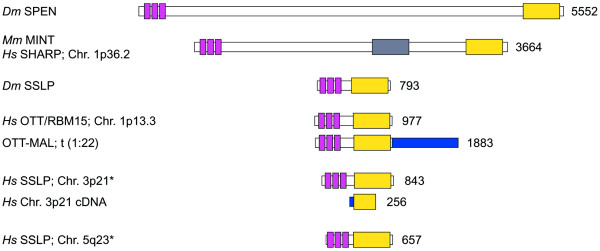
Background: The Drosophila split ends (spen) gene encodes a large nuclear protein containing three RNP-type RNA binding motifs, and a conserved transcriptional co-repressor-interacting domain at the C-terminus. Genetic analyses indicate that spen interacts with pathways that regulate the function of Hox proteins, the response to various signaling cascades and cell cycle control. Although spen mutants affect only a small subset of morphological structures in embryos, it has been difficult to find a common theme in spen mutant structural alterations, or in the interactions of spen with known signaling pathways.

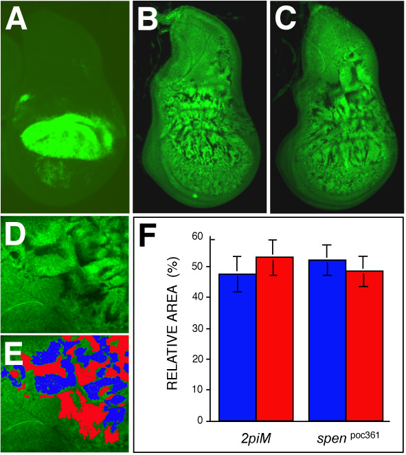
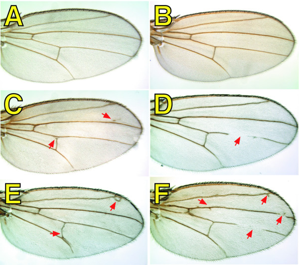
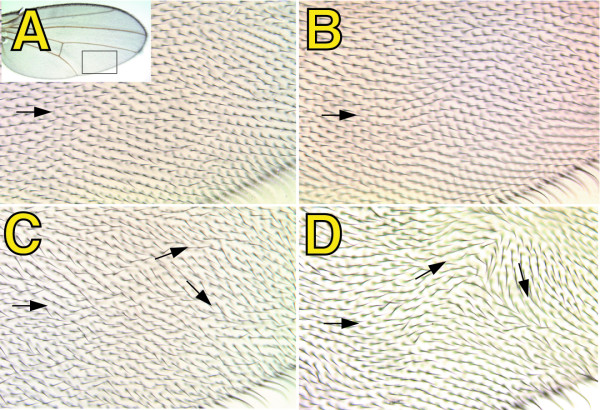
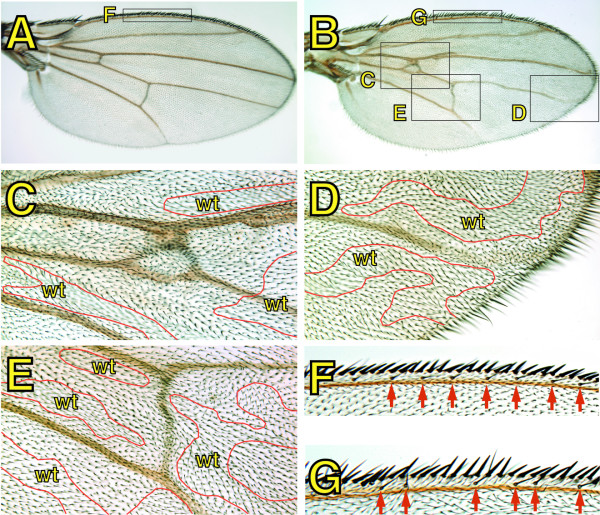
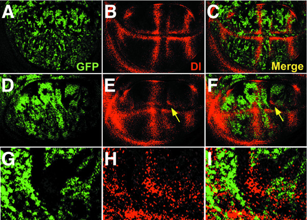
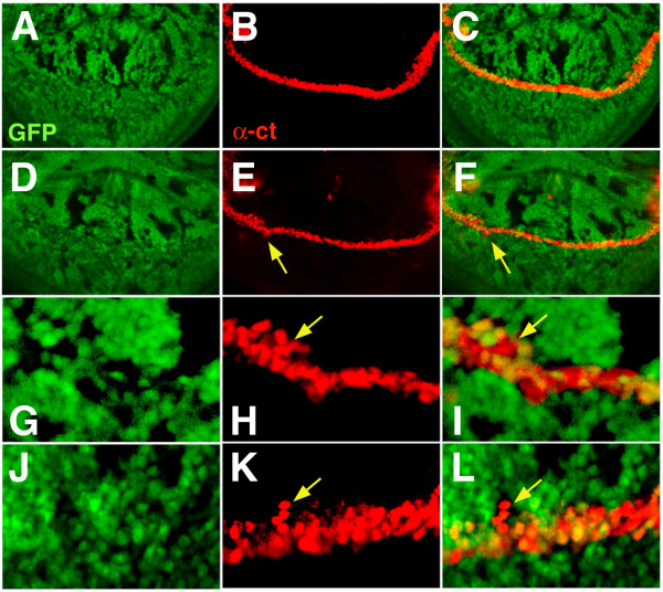
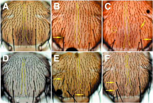
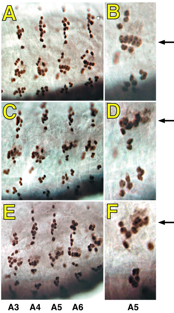
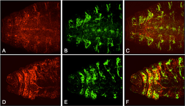
Results: By generating clones of spen mutant cells in wing imaginal discs, we show that spen function is required for the correct formation and positioning of veins and mechanosensory bristles both on the anterior wing margin and on the notum, and for the maintenance of planar polarity. Wing vein phenotypic alterations are enhanced by mutations in the crinkled (ck) gene, encoding a non-conventional myosin, and correlate with an abnormal spatial expression of Delta, an early marker of vein formation in third instar wing imaginal discs. Positioning defects were also evident in the organization of the embryonic peripheral nervous system, accompanied by abnormal E-Cadherin expression in the epidermis.

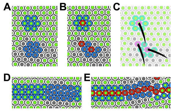
Conclusions: The data presented indicate that the role of spen is necessary to maintain the correct positioning of cells within a pre-specified domain throughout development. Its requirement for epithelial planar polarity, its interaction with ck, and the abnormal E-Cadherin expression associated with spen mutations suggest that spen exerts its function by interacting with basic cellular mechanisms required to maintain multicellular organization in metazoans. This role for spen may explain why mutations in this gene interact with the outcome of multiple signaling pathways.


Figures

Figure 1
Figure 1
Generation of spen mutant clones in wing imaginal discs. (A) Expression of a UAS-GFP transgene driven by MS1096 GAL4 in third instar wing imaginal discs. As described previously [22], the transgene is expressed mainly in the dorsal wing pouch, with weaker expression in the ventral side and in the prospective notum. (B, C) Virgin females with the genotype {MS1096 GAL4; 40 2piM FRT40A}, or {MS1096 GAL4; spenpoc361 FRT40A/ ln (2LR) CyO}, were crossed to {w*; GFP FRT40A; UAS-Flp1/TM6b} males. Shown are wing imaginal discs isolated from the resulting third instar larvae with the genotypes {MS1096 GAL4/+; 40 2 piM FRT40A/GFP FRT40A; UAS Flp1/ +} (B), and {MS1096 GAL4/+; spenpoc361 FRT40A/GFP FRT40A; UAS-Flp1/ +}(C). Green fluorescence reveals either compound heterozygous cells (not subjected to mitotic recombination) or GFP homozygotes (brightest), while dark spots indicate 2 piM (B) or spenpoc361 (C) homozygous clones. Dorsal is up and anterior is to the left. To know the relative area covered by either 2 piM or spenpoc361 homozygous clones versus wt clones, regions of equal intensity within the images were artificially colored in Adobe Photoshop using the Paint Bucket tool (D, E). Colors corresponding to mutant (red) or wt (blue) areas were extruded independently, and the total number of pixels contained within the regions of interest were calculated using the Kodak 1D Image Analysis Software (Eastman Kodak Company, Rochester, NY). The results are represented as the fraction covered by each genotype for each cross in a total of three discs for the crosses involving 2 piM, and 5 discs for spenpoc361 (F).
Figure 2
Figure 2
The presence of spen mutant clones affects wing vein morphology. Virgin females with the genotype {MS1096 GAL4; 40 2piM FRT40A}, {MS1096 GAL4; spenpoc231 FRT40A/ ln (2LR) CyO}, or {MS1096 GAL4; spenpoc361 FRT40A/ ln (2LR) CyO}, were crossed to {w*; GFP FRT40A; UAS-Flp1/TM6b} males, and adult wings were isolated from progeny females with the following genotypes: {MS1096 GAL4/+; 40 2 piM FRT40A/GFP FRT40A; UAS Flp1/ +} (A), {MS1096 GAL4/+; spenpoc361 FRT40A/ GFP FRT 40A; TM6b/ +} (B), {MS1096 GAL4/+; spenpoc231 FRT40A/ GFP FRT40A; UAS-Flp1/ +} (C, D), and {MS1096 GAL4/+; spenpoc361 FRT40A/ GFP FRT40A; UAS-Flp1/ +} (E, F). Arrows indicate gain (C, E, F), loss (D, F), or misplacement (E) of vein material.
Figure 3
Figure 3
spen mutations affect wing hair polarity. Crosses were performed as described in Figure 1. Shown are details (see inset in A) of adult wings isolated from progeny females with the genotypes: {MS1096 GAL4/ +; 40 2piM FRT40A/ GFP FRT40A; UAS-Flp1/ +} (A), {MS1096 GAL4/ +; spenpoc361 FRT40A/ GFP FRT40A; TM6b/ +} (B), {MS1096 GAL4/ +; spenpoc231 FRT40A/GFP FRT40A; UAS-Flp1/ +} (C), and {MS1096 GAL4/+; spenpoc361 FRT40A/ GFP FRT40A; UAS-Flp1/ +} (D). Arrows indicate the direction of the bristles.
Figure 4
Figure 4
Incorrect positioning of wing elements in mosaic spen mutant wings is enhanced by mutations in the crinkled (ck) gene. Virgin females with the genotype {MS1096 GAL4; ck FRT40A/ ln (2LR) CyO }, or {MS1096 GAL4; spenpoc361ck FRT40A/ ln (2LR) CyO }, were crossed to {w*; GFP FRT40A; UAS-Flp1/ TM6b} males, and adult wings were isolated from progeny females with the following genotypes: {MS1096 GAL4/+; ck FRT40A/ GFP FRT40A; UAS-Flp1/ +} (A, F), {MS1096 GAL4/+; spenpoc361 ck FRT40A/ GFP FRT40A; TM6b/ +} (B-E, G). Red lines separate either wt or compound heterozygous cells (indicated as wt) from spen mutant cells.
Figure 5
Figure 5
Delta protein expression is abnormally distributed in the presence of spen mutant clones. Crosses were performed as described in Figure 1, and third instar imaginal disks were isolated from progeny female larvae with the following genotypes: {MS1096 GAL4/+; 40 2piM FRT40A/ GFP FRT40A; UAS-Flp1/ +} (A-C), and {MS1096 GAL4/ +; spenpoc361 FRT40A/ GFP FRT40A; UAS-Flp1/ +} (D-I). Delta expression (in red) was detected in by using a mouse anti-Delta MAb followed by a Cy3-conjugated anti mouse antibody. The area indicated by the arrows in E and F is shown at higher magnification in G to I. Dorsal is up and anterior is to the left.
Figure 6
Figure 6
Analysis of Cut protein expression in the presence of spen mutant clones. Crosses were performed as described in Figure 1, and third instar imaginal disks were isolated from progeny female larvae with the following genotypes: {MS1096 GAL4/+; 40 2piM FRT40A/ GFP FRT40A; UAS-Flp1/ +} (A-C), and {MS1096 GAL4/ +; spenpoc361 FRT40A/ GFP FRT40A; UAS-Flp1/ +} (D-L). Cut expression (in red) was detected with an anti-Cut MAb as described in Figure 4 for Dl, and the absence of GFP (in green) defines mutant cells as explained on Figure 4. G to H shows a magnification of the spot indicated by an arrow on E and F. Note that the Cut protein is present in spen mutant cells. Panels J to L show the margin of another disc not shown in the figure. The arrow indicates a group of heterozygous cells that are away from the margin, surrounded by a group on spen mutant cells. Dorsal is up and anterior is to the left.
Figure 7
Figure 7
Sensory bristle number and position is abnormal in notums containing spen mutant clones. Virgin females with the genotype {spenpoc231 FRT40A/ ln (2LR) CyO; dppDISK GAL4/ TM6b} or {spenpoc361 FRT40A/ ln (2LR) CyO; dppDISK GAL4/ TM6b}, were crossed to {w*; GFP FRT40A; UAS-Flp1/ TM6b} males, and adult notums were isolated from progeny females (A-C), or males (D-F) with the following genotypes: {spenpoc361 FRT40A/ GFP FRT40A; dppDISK GAL4/ TM6b} (A, D), {spenpoc231 FRT40A/ GFP FRT40A; dppDISK GAL4/ UAS-Flp1} (B, E), and {spenpoc361 FRT40A/ GFP FRT40A; dppDISK GAL4/ UAS-Flp1} (C, F). Lines delineate the bristles at the dorsal midline. Empty circles indicate loss of bristles (in C), and arrows indicate either gain, or abnormal location of macrochaetae (B, C, E, and F).
Figure 8
Figure 8
Abnormal positioning of PNS neurons in spen maternal and zygotic mutant embryos. Maternal and zygotic spen mutant embryos were obtained as described in Materials and Methods, and at stage 14–15 were stained for the neuron specific marker Elav, together with an anti β-galactosidase antiserum to reveal the presence of the CyO, wg-lacZ balancer in heterozygotes (not shown), followed by biotinylated secondary antibodies, and streptavidin conjugated horse radish peroxidase. Brown staining reveals the nuclei of PNS neurons in wt (A, B), maternal and zygotic spen231 (C, D), or spen361 (E, F) embryos.
Figure 9
Figure 9
Increased epidermal expression of E-cadherin correlates with abnormal positioning of embryonic PNS neurons. Stage 16 wild type embryos (A-C) or maternal and zygotic spen mutant embryos (D-F) were generated as described in Materials and Methods. The expression of the PNS neuronal marker 22C10 (green) and E-cadherin (red), were detected with specific MAbs followed by fluorescein conjugated anti-mouse antiserum and a Cy3 conjugated anti rat as described earlier in Figure 4, and in the Material and Methods section.
Figure 10
Figure 10
Human spen-related genes. The figure shows all Drosophila and human related sequences putatively encoding polypeptides with three RNP-type RNA binding motifs at the N terminus (purple boxes), and the SPOC domain at the C terminus (yellow boxes). The gray box on Hs SHARP indicates the region that contains motifs required for the interaction of SHARP/MINT with Msx-2 (residues 2070 to 2394 [14]), nuclear receptors (residues 2201 to 2707 [15]), and RBP-Jκ (residues 2803 to 2817 [17], and 2638 to 2777 [18]). The expected sizes for each peptide is shown on the right, while names and chromosomal localization in humans is on the left. Asterisks indicate that the peptides have been predicted from the genomic sequence, either because there are no known full length ESTs corresponding to the genomic regions analyzed (case for Hs SSLP at 3p21), or because there are no reported ESTs at all (Hs SSLP at 5q23). In the case of the SSLP at 5q23, there are also stop codons in frame with the putative ORF, so it is likely that this sequence represents a pseudogene. Accession numbers for the sequences likely to represent full length cDNAs are: AAF13218 (Dm SPEN), NP_055816 (Hs SHARP), AAF59160 (Dm SSLP), NP_073605 (OTT/RBM15), and CAC38829 (OTT-MAL fusion). The putative full length ORF for the Hs SSLP at 3p21 was predicted using GENESCAN [54] on the genomic sequence AC092037. The truncated cDNA arising from this gene is found under AAA72367 or NP_037418. The Hs SSLP at 5q23 was predicted with GENESCAN from the genomic segment AC005915, and the assembly was completed with ENSP322787, a predicted peptide from the Ensembl Database [42].
Figure 11
Figure 11
A mechanistic model for Spen function. The cartoon illustrates our conclusions to explain the defects seen in the presence of spen mutant clones. Wild type or heterozygote cells are depicted with green nuclei, and spen mutant cells with gray nuclei. The appearance of spen mutant cells in fields that will give rise to specific structures such as bristles or veins would imply the lack of an instructive signal to remain in place during growth of the disc. This will finally result in a progressive mis-localization of cells, ultimately leading to the abnormal positioning of structures after development is completed. Such a model would explain that, in some cases as in the proneural clusters, a change of fate is generated because negative instructive signals that depend on cell to cell contact are lost, resulting in the formation of two sensory organ precursor (SOP) cells within the same pro neural cluster (shown as red cells in B and C). The same situation may occur in the formation of wing veins, where similar Notch dependent regulatory mechanisms take place (E). Loss of or veins (or bristles) may occur when vein-forming cells move into intervein regions after their commitment has taken place, therefore leaving a hole where they should have been, which has been filled with cells that are unable to form vein at that spot.

Similar articles

Cited by

References

    1. Kolodziej PA, Jan LY, Jan YN. Mutations that affect the length, fasciculation, or ventral orientation of specific sensory axons in the Drosophila embryo. Neuron. 1995;15:273–286. doi: 10.1016/0896-6273(95)90033-0. - DOI - PubMed
    1. Gellon G, Harding KW, McGinnis N, Martin M, McGinnis W. A genetic screen for modifiers of Deformed homeotic function identifies novel genes required for head development. Development. 1997;124:3321–3331. - PubMed
    1. Wiellette EL, Harding KW, Mace KA, Ronshaugen MR, Wang FY, McGinnis W. spen encodes an RNP motif protein that interacts with Hox pathways to repress the development of head-like sclerites in the Drosophila trunk. Development. 1999;126:5373–5385. - PubMed
    1. Dickson BJ, Van Der Straten A, Domínguez M, Hafen E. Mutations modulating Raf signaling in Drosophila eye development. Genetics. 1996;142:163–171. - PMC - PubMed
    1. Therrien M, Morrison DK, Wong AM, Rubin GM. A genetic screen for modifiers of a kinase suppressor of Ras-dependent rough eye phenotype in Drosophila. Genetics. 2000;156:1231–1242. - PMC - PubMed

Publication types

MeSH terms