Comparative Study
. 2005 Mar;88(3):1828-37.
doi: 10.1529/biophysj.104.049817. Epub 2004 Dec 13.

Melittin-induced bilayer leakage depends on lipid material properties: evidence for toroidal pores

Affiliations
Comparative Study

Melittin-induced bilayer leakage depends on lipid material properties: evidence for toroidal pores

Daniel Allende et al. Biophys J. 2005 Mar.

Abstract

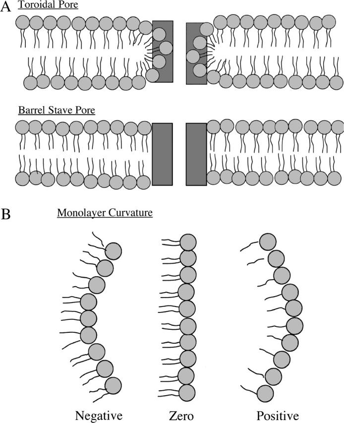
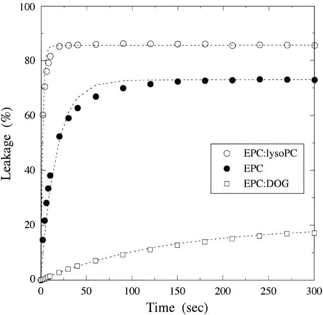
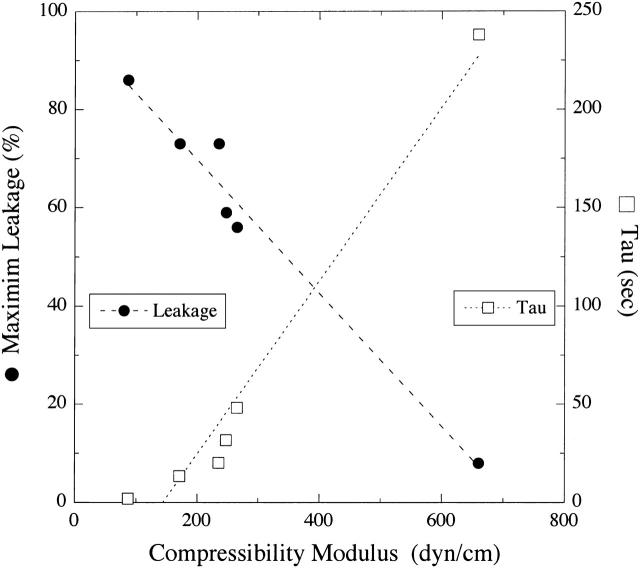
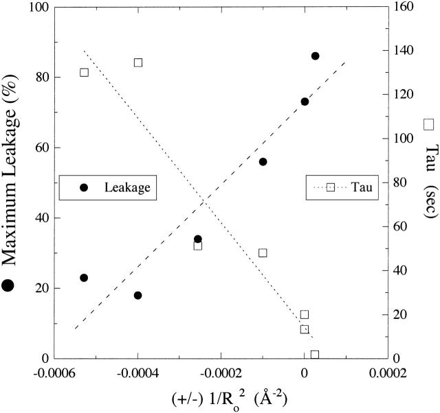
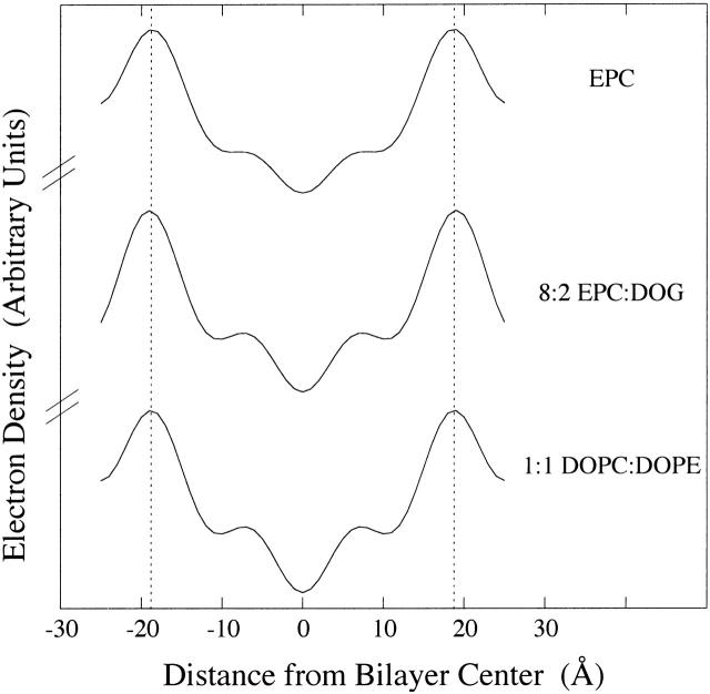
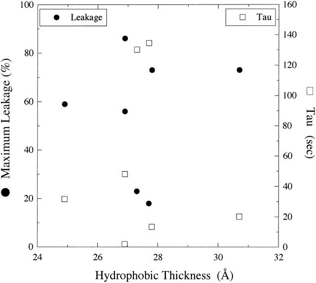
The membrane-lytic peptide melittin has previously been shown to form pores in lipid bilayers that have been described in terms of two different structural models. In the "barrel stave" model the bilayer remains more or less flat, with the peptides penetrating across the bilayer hydrocarbon region and aggregating to form a pore, whereas in the "toroidal pore" melittin induces defects in the bilayer such that the bilayer bends sharply inward to form a pore lined by both peptides and lipid headgroups. Here we test these models by measuring both the free energy of melittin transfer (DeltaG degrees ) and melittin-induced leakage as a function of bilayer elastic (material) properties that determine the energetics of bilayer bending, including the area compressibility modulus (K(a)), bilayer bending modulus (k(c)), and monolayer spontaneous curvature (R(o)). The addition of cholesterol to phosphatidylcholine (PC) bilayers, which increases K(a) and k(c), decreases both DeltaG degrees and the melittin-induced vesicle leakage. In contrast, the addition to PC bilayers of molecules with either positive R(o), such as lysoPC, or negative R(o), such as dioleoylglycerol, has little effect on DeltaG degrees , but produces large changes in melittin-induced leakage, from 86% for 8:2 PC/lysoPC to 18% for 8:2 PC/dioleoylglycerol. We observe linear relationships between melittin-induced leakage and both K(a) and 1/R(o)(2). However, in contrast to what would be expected for a barrel stave model, there is no correlation between observed leakage and bilayer hydrocarbon thickness. All of these results demonstrate the importance of bilayer material properties on melittin-induced leakage and indicate that the melittin-induced pores are defects in the bilayer lined in part by lipid molecules.


Figures

FIGURE 1
FIGURE 1
Schematic drawings of sections through (A) “toroidal pore” and “barrel stave pore” models and (B) models for monolayer spontaneous curvature. In A, peptides are depicted as rectangular dark shaded boxes, and lipids are shown with circular light shaded headgroups and wavy hydrocarbon chains. In B, lipids that have approximately the same excluded area in their headgroup and acyl chain regions (EPC or SOPC) form structures with near zero membrane curvature (middle image), lipids whose excluded headgroup area is smaller than the area of its acyl chains (DOPC, DOPE, or DOG) form structures with negative curvature (image on left), whereas lipids whose headgroup area is larger than its acyl chain area (lysoPC) have positive curvature (image on right).
FIGURE 2
FIGURE 2
Typical leakage curves for bilayers of EPC (○), 8:2 EPC/lysoPC (•), and 8:2 EPC/DOG (□). The addition of melittin at time t = 0 caused a rapid leakage of encapsulated SRB for the first minute or two, after which the leakage leveled off to a nearly constant value. For each lipid system, the dotted lines represent fits (R2 > 0.99) using Eq. 4, % Release = Lmax(1 − e−t/τ).
FIGURE 3
FIGURE 3
Plot of melittin-induced leakage (Lmax, •) and time constant (τ, □) versus area compressibility modulus (Ka) for EPC/lysoPC, EPC, SOPC, DOPC, DLinPC, and EPC/PS/cholesterol (35:15:50). Dashed and dotted lines represent least-square linear fits to the plots of Lmax versus Ka (R2 = 0.97) and τ versus Ka (R2 = 0.96), respectively.
FIGURE 4
FIGURE 4
Plot of melittin-induced leakage (Lmax, •) and time constant (τ, □) versus (±) formula image for SOPC, DOPC, 1:1 DOPC/DOPE, 8:2 DOPC/DOPE, 8:2 EPC/lysoPC, and 8:2 EPC/DOG. The signs on the abscissa are present because we have defined the curvature elastic energy as positive for bilayers with positive curvature (8:2 EPC/lysoPC) and negative for bilayers with negative curvature (8:2 EPC/DOG). Dashed and dotted lines are least-square linear fits to the plots of Lmax versus formula image (R2 = 0.90) and τ versus formula image (R2 = 0.92), respectively.
FIGURE 5
FIGURE 5
Electron density profiles of EPC, 8:2 EPC/DOG, and 1:1 DOPC/DOPE. The profile for EPC was taken from McIntosh et al. (1987).
FIGURE 6
FIGURE 6
Plot of melittin-induced leakage (Lmax, •) and time constant (τ, □) versus bilayer hydrocarbon thickness (dhc) for EPC, SOPC, DOPC, 8:2 DOPC/DOPE, 1:1 DOPC/DOPE, 8:2 EPC/DOG, and 8:2 EPC/LysoPC.

Similar articles

Cited by

References

    1. Allende, D., and T. J. McIntosh. 2003. Lipopolysaccharides in bacterial membranes act like cholesterol in eukaryotic plasma membranes in providing protection against melittin-induced bilayer lysis. Biochemistry. 42:1101–1108. - PubMed
    1. Allende, D., A. Vidal, S. A. Simon, and T. J. McIntosh. 2003. Bilayer interfacial properties modulate the binding of amphipathic peptides. Chem. Phys. Lipids. 122:65–76. - PubMed
    1. Altenbach, C., W. Froncisz, J. S. Hyde, and W. L. Hubbell. 1989. Conformation of spin-labeled melittin at membrane surfaces investigated by pulse saturation recovery and continuous wave power saturation electron paramagnetic resonance. Biophys. J. 56:1183–1191. - PMC - PubMed
    1. Banemann, A., H. Deppisch, and R. Gross. 1998. The lipopolysaccharide of Bordetella bronchisepticsa acts as a protective shield against antimicrobial peptides. Infect. Immun. 66:5607–5612. - PMC - PubMed
    1. Barranger-Mathys, M., and D. S. Cafiso. 1996. Membrane structure of voltage-gated channel forming peptides revealed by site-directed spin labeling. Biochemistry. 35:498–505. - PubMed

Publication types