. 2004 Dec 20;200(12):1539-45.
doi: 10.1084/jem.20041447. Epub 2004 Dec 13.

Loss of synchronized retinal phagocytosis and age-related blindness in mice lacking alphavbeta5 integrin

Affiliations

Loss of synchronized retinal phagocytosis and age-related blindness in mice lacking alphavbeta5 integrin

Emeline F Nandrot et al. J Exp Med. .

Abstract

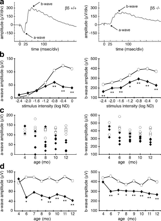
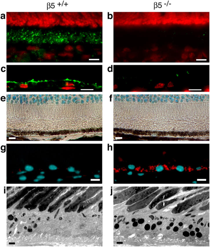
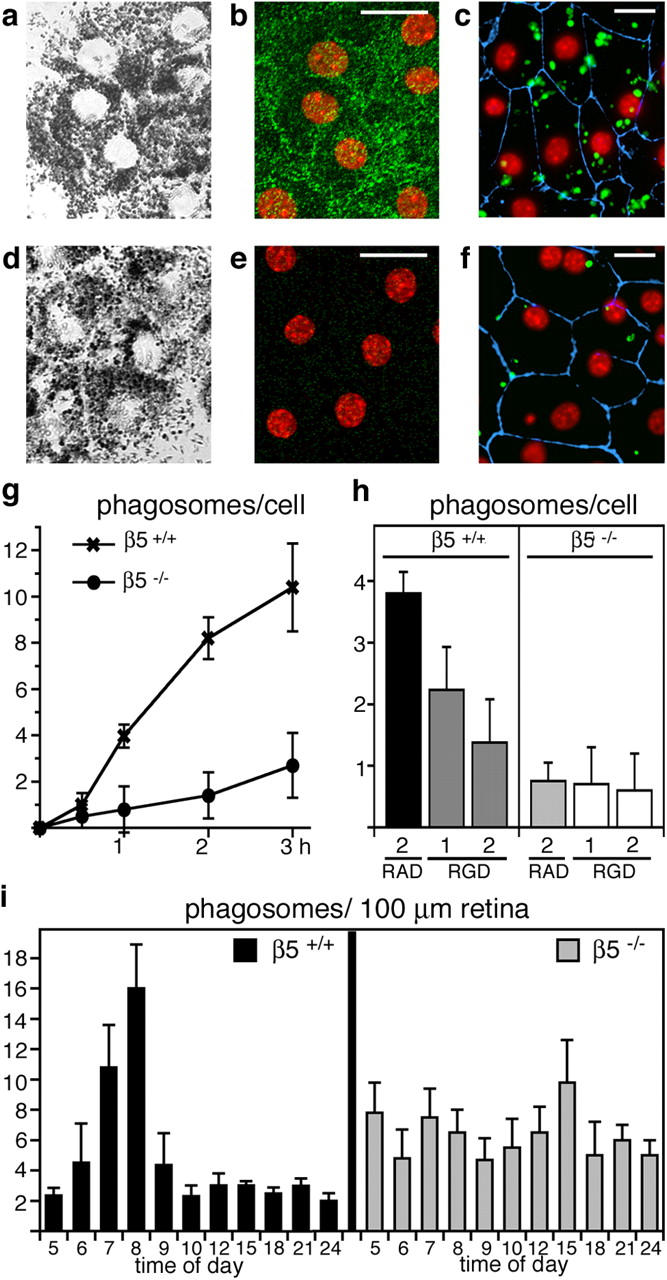
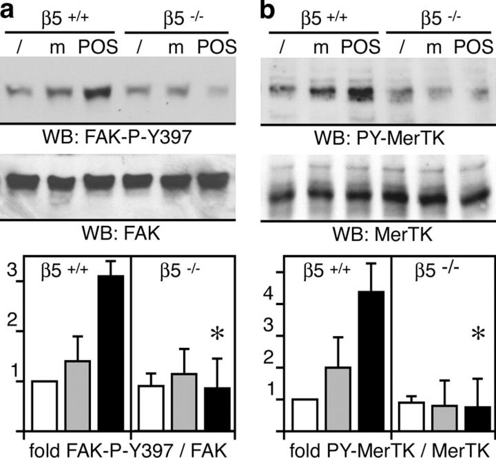
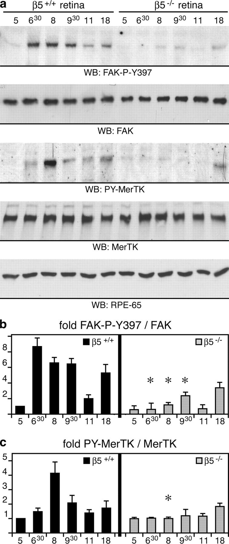
Daily phagocytosis by the retinal pigment epithelium (RPE) of spent photoreceptor outer segment fragments is critical for vision. In the retina, early morning circadian photoreceptor rod shedding precedes synchronized uptake of shed photoreceptor particles by RPE cells. In vitro, RPE cells use the integrin receptor alphavbeta5 for particle binding. Here, we tested RPE phagocytosis and retinal function in beta5 integrin--deficient mice, which specifically lack alphavbeta5 receptors. Retinal photoresponses severely declined with age in beta5-/- mice, whose RPE accumulated autofluorescent storage bodies that are hallmarks of human retinal aging and disease. beta5-/- RPE in culture failed to take up isolated photoreceptor particles. beta5-/- RPE in vivo retained basal uptake levels but lacked the burst of phagocytic activity that followed circadian photoreceptor shedding in wild-type RPE. Rhythmic activation of focal adhesion and Mer tyrosine kinases that mediate wild-type retinal phagocytosis was also completely absent in beta5-/- retina. These results demonstrate an essential role for alphavbeta5 integrin receptors and their downstream signaling pathways in synchronizing retinal phagocytosis. Furthermore, they identify the beta5-/- integrin mouse strain as a new animal model of age-related retinal dysfunction.


Figures

Figure 1.
Figure 1.
β5 integrin–deficient mice lose vision with age. We recorded scotopic ERG responses obtained after a light flash stimulus attenuated with 0.4 log ND (a, c, and d) or attenuated as indicated (b). We compared a-wave (left) and b-wave (right) amplitudes of control (open circles) and β5−/− (closed diamonds) mice. A-wave amplitudes are given as absolute values. (a) Representative ERG responses are diminished in 12-mo-old β5−/− mice (right) compared with wild-type mice (left). Both a- and b-wave peaks (arrows) are affected. (b) Mean scotopic responses to flashes of different intensities are decreased in 12-mo-old β5−/− mice. Asterisks indicate significant differences between wild-type and β5−/− amplitudes at the same flash intensity (n = 5–7, Student's t test, *, P < 0.05; **, P < 0.01). (c) Responses of individual animals (c) and of averages (d) illustrate decline of retinal function in β5−/− mice with age. Asterisks indicate significant differences between wild-type and β5−/− amplitudes at the same age (n = 5–7, Student's t test, *, P < 0.05; **, P < 0.01).
Figure 2.
Figure 2.
β5 integrin–deficient RPE accumulates autofluorescent lipofuscin with age. (a–d) Cryosections of 1-mo-old eyecups of wild-type (a and c) and β5−/− (b and d) mice were labeled with β5 antibody (green). Nuclei are shown in red. (a) In the wild-type mouse, whole retina sections with the neural retina present and β5 integrin was present in the photoreceptor outer segment layer, which also contains apical microvillar processes of RPE ensheathing photoreceptor outer segments (reference 28). (b) As expected, β5 integrin was absent from β5−/− retina. (c and d) To determine whether β5 integrin resided on photoreceptor outer segments or on RPE apical extensions, we prepared sections from eyecups from which the neural retina was peeled off before fixation. Prominent β5 integrin labeling at the apical surface of wild-type but not β5−/− RPE indicated that mouse RPE in vivo expresses apical αvβ5 integrin. (e–f) Methyl green–stained paraffin sections of eyecups from 12-mo-old wild-type (e) and β5−/− (f) mice were examined by light microscopy. Wild-type and β5−/− retina did not differ in appearance of photoreceptor nuclei (cyan at the top of the field), inner and outer segments (clear areas), and RPE (brown pigment and cyan nuclei). (g–h) Cryosections of eyecups from 12-mo-old wild-type (g) and β5−/− (h) mice were examined by wide-field fluorescence microscopy to detect DAPI-stained nuclei (cyan) and autofluorescence (detected in the rhodamine channel, red). β5−/−, but not wild-type, RPE contained numerous autofluorescent inclusion bodies resembling the characteristic age pigment lipofuscin in human RPE (32). (i–j) Ultrathin sections of eyecups from 12-mo-old wild-type (i) and β5−/− (j) mice were examined by transmission electron microscopy. Increased presence of electron dense inclusion bodies in β5−/− RPE compared with wild-type RPE confirmed the results obtained by fluorescence microscopy. Bars: (a–h) 10 μm; (i and j) 1 μm.
Figure 3.
Figure 3.
Lack of αvβ5 integrin impairs RPE phagocytosis of POS. We examined primary RPE in culture from wild-type (a–c) and β5−/− (d–f) mice. Confocal x-y scans of transmitted light (a and d) and of apical αvβ5 integrin (green) and nuclei (red) in the same fields (b and e) were used to compare general cell morphology and to illustrate apical αvβ5 receptors in wild-type, but not in β5−/−, RPE. (c and f) Junction marker ZO-1 (blue) and nuclei (red) appeared similar by wide-field fluorescence microscopy. However, β5−/− RPE in primary culture phagocytosed fewer FITC-POS (green) than wild-type RPE during a 1-h phagocytic challenge. (g) Quantification of in vitro phagocytosis assays showed reduced POS uptake by β5−/− RPE compared with wild-type RPE at all time points. Results represent means ± SD, n = 3, Student's t test, P < 0.01 at 1–3 h. (h) RGD peptides inhibited POS uptake by wild-type but not by β5−/− RPE in a concentration-dependent manner. Bars show 1-h FITC-POS uptake by wild-type and β5−/− RPE in the presence of 2 mM RAD-inactive control peptide, 1 or 2 mM integrin inhibiting RGD peptide as indicated. Since RGD peptides affected cell–substrate adhesion of both wild-type and β5−/− RPE at concentrations above 2 mM, these concentrations were not used for phagocytosis assays. Thus, the 63% inhibition we observed in the presence of 2 mM RGD peptide may not represent maximal possible inhibition of POS uptake by RGD peptides. Results represent means ± SD, n = 3, Student's t test, P < 0.01 for wild-type RPE, RGD versus RAD peptide at 2 mM. (i) Phagosome quantification in electron micrographs of retinal cross sections revealed that β5−/− retina lacked the characteristic burst of phagocytosis that followed the light onset (at 6.00 h) in wild-type retina. Mean (numbers of phagosomes) ± SD is shown. Sections from three eyecups per time point harvested in three separate experiments were examined.
Figure 4.
Figure 4.
β5 integrin–deficient RPE in culture fails to activate phagocytic signaling. (a) Primary wild-type and β5−/− RPE in culture were untreated (lanes /) or received assay medium (lanes m) or POS in assay medium (lanes POS) for 1.5 h. Lysates normalized for RPE protein content were analyzed by immunoblotting with antibodies specific for FAK-P-Y397 and FAK protein. Specific activity of FAK is given as a ratio of FAK-P-Y397 to total FAK and given as fold change compared with untreated wild-type RPE. (n = 3). (b) The same experiment was performed to determine changes in MerTK activity. Specific activity of MerTK is given as a ratio of PY-MerTK to total MerTK and given as fold change compared with untreated wild-type RPE. (a and b) Results are presented as means ± SD. Asterisks indicate significant differences in ratios between wild-type and β5−/− samples of the same treatment (Student's t test, P < 0.001).
Figure 5.
Figure 5.
β5 integrin–deficient retina lacks synchronized activation of FAK and MerTK. (a) Eyecup detergent lysates were prepared from 3-wk-old wild-type and β5−/− mice at different times of the day, as indicated. Expression and phosphorylation profiles of proteins were compared by immunoblotting with primary antibodies as indicated in the panels. (b and c) Specific activities of FAK and MerTK were determined as in Fig. 4. Promptly after light onset, levels of active, phosphorylated FAK and MerTK increased in wild-type eyecups. This increase was absent in β5−/− eyecups. Similar results were obtained comparing expression and activity profiles of FAK and MerTK in wild-type and β5−/− mice at 7 and 12 mo (unpublished data). Note that the rise in FAK phosphorylation preceded the increase of MerTK phosphorylation. This agreed well with our earlier data from in vitro phagocytosis assays showing that MerTK activation upon POS challenge requires FAK activation (reference 29). Ratios of active, phosphorylated protein/total protein are given as fold change compared with ratios in wild-type eyes at 5.00 h, which were set at 1.0 arbitrarily. Results are presented as means ± SD. Asterisks indicate significant differences in ratios (n = 3, Student's t test, P < 0.05) between wild-type and β5−/− samples at the same time points.

Similar articles

Cited by

References

    1. Young, R.W. 1967. The renewal of photoreceptor cell outer segments. J. Cell Biol. 33:61–72. - PMC - PubMed
    1. Young, R.W. 1971. The renewal of rod and cone outer segments in the rhesus monkey. J. Cell Biol. 49:303–318. - PMC - PubMed
    1. LaVail, M.M. 1976. Rod outer segment disk shedding in rat retina: relationship to cyclic lighting. Science. 194:1071–1074. - PubMed
    1. Young, R.W. 1977. The daily rhythm of shedding and degradation of cone outer segment membranes in the lizard retina. J. Ultrastruct. Res. 61:172–185. - PubMed
    1. Young, R.W., and D. Bok. 1969. Participation of the retinal pigment epithelium in the rod outer segment renewal process. J. Cell Biol. 42:392–403. - PMC - PubMed

Publication types

MeSH terms