. 2004 Dec 20;167(6):1099-112.
doi: 10.1083/jcb.200404119. Epub 2004 Dec 13.

Quantitative mass spectrometry reveals a role for the GTPase Rho1p in actin organization on the peroxisome membrane

Affiliations

Quantitative mass spectrometry reveals a role for the GTPase Rho1p in actin organization on the peroxisome membrane

Marcello Marelli et al. J Cell Biol. .

Abstract

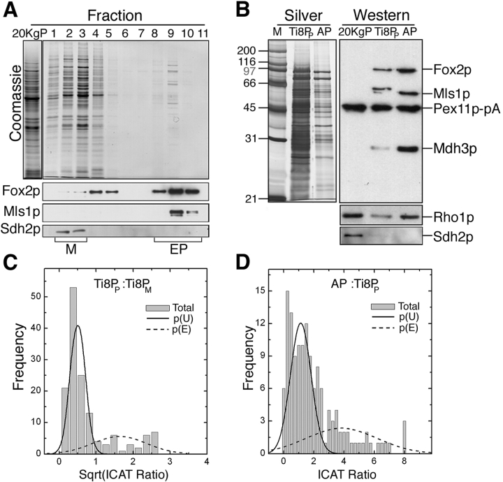
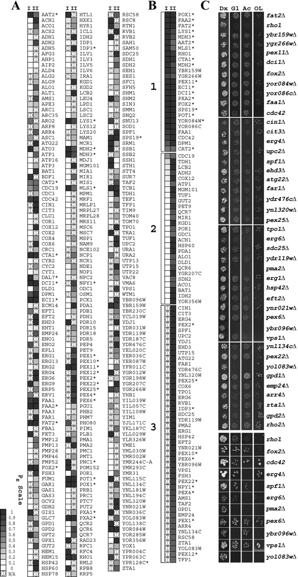
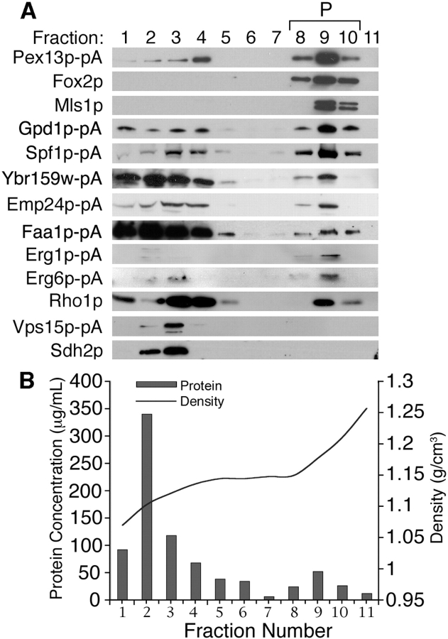
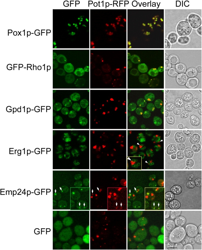
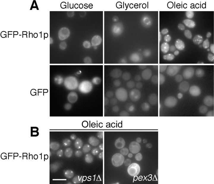
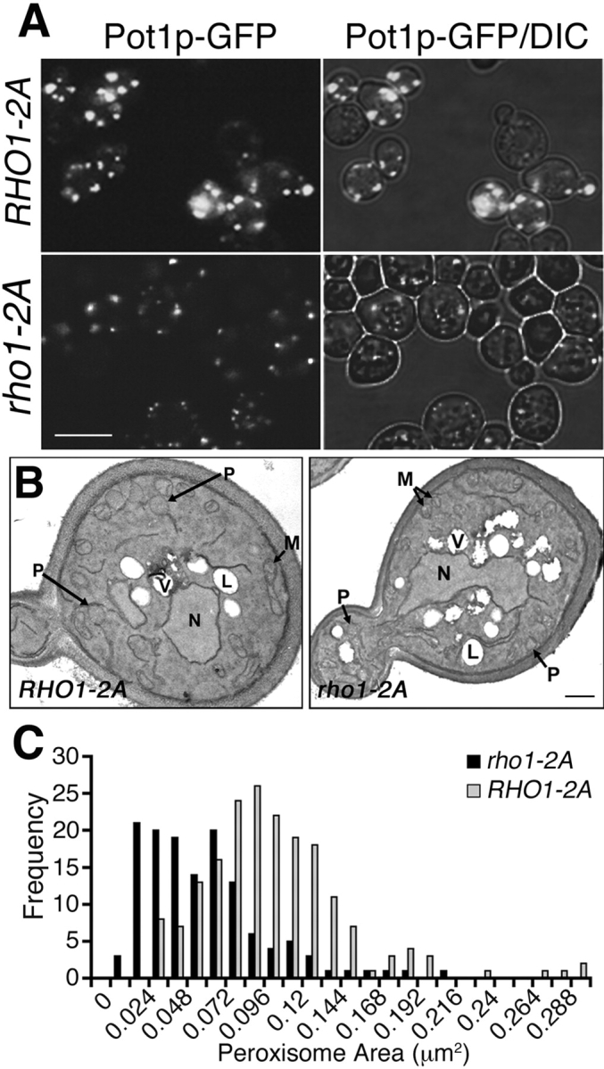
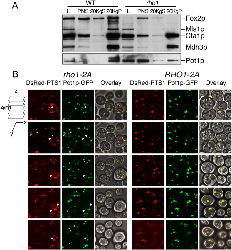
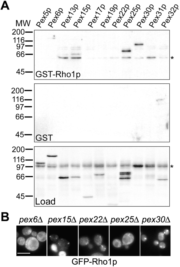
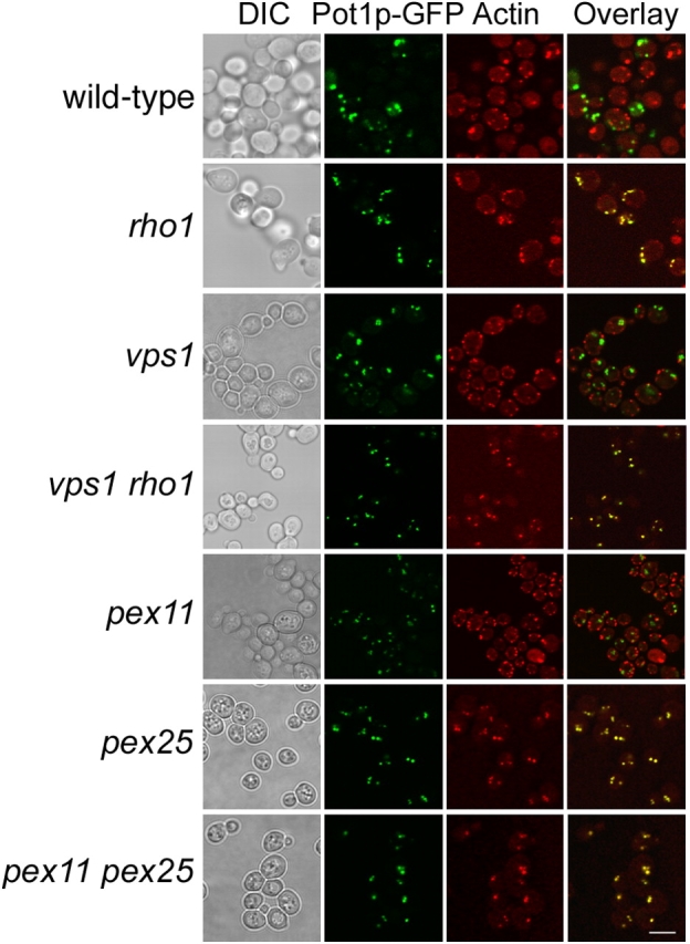
We have combined classical subcellular fractionation with large-scale quantitative mass spectrometry to identify proteins that enrich specifically with peroxisomes of Saccharomyces cerevisiae. In two complementary experiments, isotope-coded affinity tags and tandem mass spectrometry were used to quantify the relative enrichment of proteins during the purification of peroxisomes. Mathematical modeling of the data from 306 quantified proteins led to a prioritized list of 70 candidates whose enrichment scores indicated a high likelihood of them being peroxisomal. Among these proteins, eight novel peroxisome-associated proteins were identified. The top novel peroxisomal candidate was the small GTPase Rho1p. Although Rho1p has been shown to be tethered to membranes of the secretory pathway, we show that it is specifically recruited to peroxisomes upon their induction in a process dependent on its interaction with the peroxisome membrane protein Pex25p. Rho1p regulates the assembly state of actin on the peroxisome membrane, thereby controlling peroxisome membrane dynamics and biogenesis.


Figures

Figure 1.
Figure 1.
Sample preparation and analysis. (A) An organellar 20KgP fraction was subjected to isopycnic density gradient centrifugation and analyzed by SDS-PAGE and Coomassie blue staining (top panel). Fractions enriched for peroxisomes (EP) or mitochondria (M) were identified by Western blotting as shown. Equal amounts of protein derived from each of the hypotonically lysed M and EP fractions were combined and analyzed by ICAT MS/MS. (B) Peroxisomal membranes isolated from a yeast strain synthesizing Pex11p-pA were affinity purified (AP) from a fraction enriched for peroxisomal membranes (Ti8PP). Equal cellular equivalents of each were analyzed by SDS-PAGE and silver staining. Equal amounts of protein from the 20KgP, Ti8PP, and AP fractions were analyzed by Western blotting. Ti8PP and AP fractions were analyzed by ICAT MS/MS. (C and D) Histograms of ICAT ratios (heavy:light) for 192 proteins quantified in ICAT I (C) and 193 proteins quantified in ICAT II (D). The distributions were modeled by two overlapping Gaussian curves using a partially supervised mixture model Expectation-Maximization algorithm. Note that because of the nature of the data in ICAT I (dominance by mitochondrial proteins with low ICAT ratios and relatively few peroxisomal proteins with high ratios), the ICAT ratios in this experiment were transformed to their square root for modeling. For any quantified protein, the probability of being enriched (p(E); dashed line) or not being enriched (p(U); solid line) with peroxisomes was calculated as a function of its ICAT ratio.
Figure 2.
Figure 2.
Prioritization of candidates. (A) 306 candidate proteins identified by ICAT MS/MS are listed alphabetically, and their peroxisome enrichment scores (P E) for ICAT I or ICAT II are represented by shaded squares. See Tables S1 and S2 for details. (B) 52 candidates with PE values > 0.65 in ICAT II, and which were also quantified in ICAT I, were clustered with a Spearman similarity metric into two groups (Groups 1 and 2). Also listed are 46 candidates with high PE values quantified in ICAT II alone (Group 3). Known peroxisomal proteins are indicated with an asterisk. (C) Yeast mutants of selected candidates from Groups 1 and 3 were assayed for their ability to grow on rich medium (YPB) containing glucose (Dx) or an oleic acid/lauric acid mixture (OL), and, as controls, the nonfermentable carbon sources glycerol (Gl) and acetate (Ac) at 25°C. Growth was assayed 2 d (Dx), 4 d (Gl and Ac), and 7 d (OL) after spotting. Slowly growing strains (bottom panel) were also examined after 3 d (Dx), 8 d (Gl and Ac), or 20 d (OL) of growth at 25°C.
Figure 3.
Figure 3.
Rho1p enriches with peroxisomes. (A) Organellar 20KgP fractions from cells expressing different pA chimeras or wild-type cells were separated by isopycnic density gradient centrifugation and analyzed by Western blotting. Fractions enriched for peroxisomes (P; 8–10) were identified by the peroxisomal proteins Pex13p-pA, Fox2p, and Mls1p. Peak mitochondrial and Golgi fractions were identified by Sdh2p and Vps15p-pA, respectively. (B) The protein concentration and density profiles for each gradient fraction are presented.
Figure 4.
Figure 4.
Rho1p, Gpd1p, and Emp24p localize to peroxisomes. Double labeling fluorescence confocal microscopy of yeast cells synthesizing the indicated GFP fusions and containing a plasmid coding for peroxisomal thiolase tagged with RFP (Pot1p-RFP). The GFP chimera of Pox1p (acyl-CoA oxidase) is shown as a control. GFP chimeras of Rho1p and Gpd1p showed punctate signals colocalizing with peroxisomes. The Erg1p-GFP chimera revealed a close association between peroxisomes and lipid bodies (arrowheads; inset is a higher magnification). Emp24p-GFP colocalized with small, Pot1p-RFP–labeled peroxisomes (arrows; insets are higher magnification and longer exposure). Bar, 10 μm.
Figure 5.
Figure 5.
Rho1p associates dynamically with peroxisomes. The distribution of GFP-Rho1p was observed in glucose-, glycerol-, and oleic acid–incubated cells. GFP-Rho1p localized to intracellular membrane structures in glucose- and glycerol-incubated cells. In conditions that induce peroxisomes (oleic acid), GFP-Rho1p localized to distinct punctate structures. (B) In oleic acid–induced vps1Δ cells, which contain few peroxisomes, GFP-Rho1p localized to one or two punctate structures per cell. However, in pex3Δ cells, which are defective in peroxisome biogenesis, GFP-Rho1p failed to accumulate in punctate structures. Bar, 10 μm.
Figure 6.
Figure 6.
rho1 cells have fewer and smaller peroxisomes. (A) RHO1-2A and rho1-2A cells synthesizing Pot1p-GFP were incubated in oleic acid medium for 16 h at the semi-permissive temperature of 27°C and analyzed by confocal microscopy. Bar, 10 μm. (B) rho1-2A and RHO1-2A cells were incubated in oleic acid medium at the permissive temperature of 23°C and processed for EM. N, nucleus; L, lipid body; P, peroxisome; V, vacuole; M, mitochondrion. Bar, 0.5 μm. (C) A histogram of the areas of peroxisomes calculated for 105 randomly chosen cell images of each strain is shown.
Figure 7.
Figure 7.
rho1 cells contain heterotypic peroxisomes. (A) The distribution of peroxisomal enzymes in wild-type and rho1 mutant cells was analyzed by subcellular fractionation. Whole cell lysates (L), postnuclear supernatants (PNS), and 20KgS fractions enriched for cytosol (loaded at one cell equivalent) and 20KgP fractions enriched for peroxisomes and mitochondria (loaded at five cell equivalents) were analyzed by Western blotting using anti-SKL antibodies, which recognizes PTS-1 containing proteins Fox2p, Mls1p, Cta1p, and Mdh3p, and anti-Pot1p antibodies. In rho1 cells, the PTS1-containing proteins Fox2p and Mls1p were not detected in the 20KgP fraction, whereas PTS2-containing Pot1p was partially mislocalized to the 20KgS. (B) rho1-2A and RHO1-2A cells synthesizing the peroxisomal reporters DsRed-PTS1 and Pot1p-GFP were incubated in oleic acid medium at 27°C. A series of optical sections were obtained by confocal microscopy, and the positions of peroxisomes were determined from the signals of the Pot1p-GFP and DsRed-PTS1 reporters. Heterotypic peroxisomes containing Pot1p-GFP or DsRed-PTS1 were numerous in rho-2A cells (arrowheads) but were rarely observed in cells of the complemented strain, RHO1-2A. Bar, 10 μm.
Figure 8.
Figure 8.
Rho1p binds Pex25p and Pex30p. GST-Rho1p and GST were immobilized on glutathione Sepharose and incubated with whole cell lysates derived from strains expressing TAP-tagged peroxins. Whole cell lysates (bottom) and bound fractions (top and middle) were resolved by SDS-PAGE, and TAP chimeras were detected by Western blotting. (top) Proteins bound to GST-Rho1p. Note that Rho1p interacts strongly with Pex25p and Pex30p. (middle) No interactions were detected with GST alone. (bottom) Yeast lysates showing the migration of each chimera. Cross-reacting bands are indicated (asterisks). (B) The distribution of GFP-Rho1p was observed in oleic acid-induced pex6Δ, pex15Δ, pex22Δ, pex25Δ, and pex30Δ cells. Note that GFP-Rho1p is not localized to peroxisomes in pex25Δ or pex6Δ cells. Bar, 10 μm.
Figure 9.
Figure 9.
Actin assembly on peroxisomes is controlled by Rho1p and Pex25p. The subcellular distribution of actin relative to that of peroxisomes was analyzed by double fluorescence confocal microscopy. Yeast deletion mutants expressing the peroxisomal reporter Pot1p-GFP were induced in oleic acid at 30 or 23°C (for rho1 and vps1 rho1) for 16 h, and actin was labeled with phalloidin-RITC. Peroxisomes colocalized with actin patches in rho1 (rho1-2A) and vps1 rho1 (vps1Δ rho1 POT1-G), pex25 (pex25Δ POT1-G), and pex11 pex25 (pex11Δ pex25Δ POT1-G) cells but not in wild-type (POT1-G), vps1 (vps1Δ POT1-G), and pex11 (pex11Δ POT1-G) cells. Bar, 10 μm.

Similar articles

Cited by

References

    1. Aebersold, R., and M. Mann. 2003. Mass spectrometry-based proteomics. Nature. 422:198–207. - PubMed
    1. Anton, M., M. Passreiter, D. Lay, T.P. Thai, K. Gorgas, and W.W. Just. 2000. ARF- and coatomer-mediated peroxisomal vesiculation. Cell Biochem. Biophys. 32 Spring:27–36. - PubMed
    1. Bascom, R.A., H. Chan, and R.A. Rachubinski. 2003. Peroxisome biogenesis occurs in an unsynchronized manner in close association with the endoplasmic reticulum in temperature-sensitive Yarrowia lipolytica Pex3p mutants. Mol. Biol. Cell. 14:939–957. - PMC - PubMed
    1. Baudhuin, P., H. Beaufay, and C. de Duve. 1965. Combined biochemical and morphological study of particulate fractions from rat liver. Analysis of preparations enriched in lysosomes or in particles containing urate oxidase, d-amino acid oxidase, and catalase. J. Cell Biol. 26:219–243. - PMC - PubMed
    1. Birschmann, I., A.K. Stroobants, M. van den Berg, A. Schafer, K. Rosenkranz, W.H. Kunau, and H.F. Tabak. 2003. Pex15p of Saccharomyces cerevisiae provides a molecular basis for recruitment of the AAA peroxin Pex6p to peroxisomal membranes. Mol. Biol. Cell. 14:2226–2236. - PMC - PubMed

Publication types

MeSH terms