. 2004 Dec 20;167(6):1123-35.
doi: 10.1083/jcb.200405148. Epub 2004 Dec 13.

The diabetes-linked transcription factor PAX4 promotes {beta}-cell proliferation and survival in rat and human islets

Affiliations

The diabetes-linked transcription factor PAX4 promotes {beta}-cell proliferation and survival in rat and human islets

Thierry Brun et al. J Cell Biol. .

Abstract

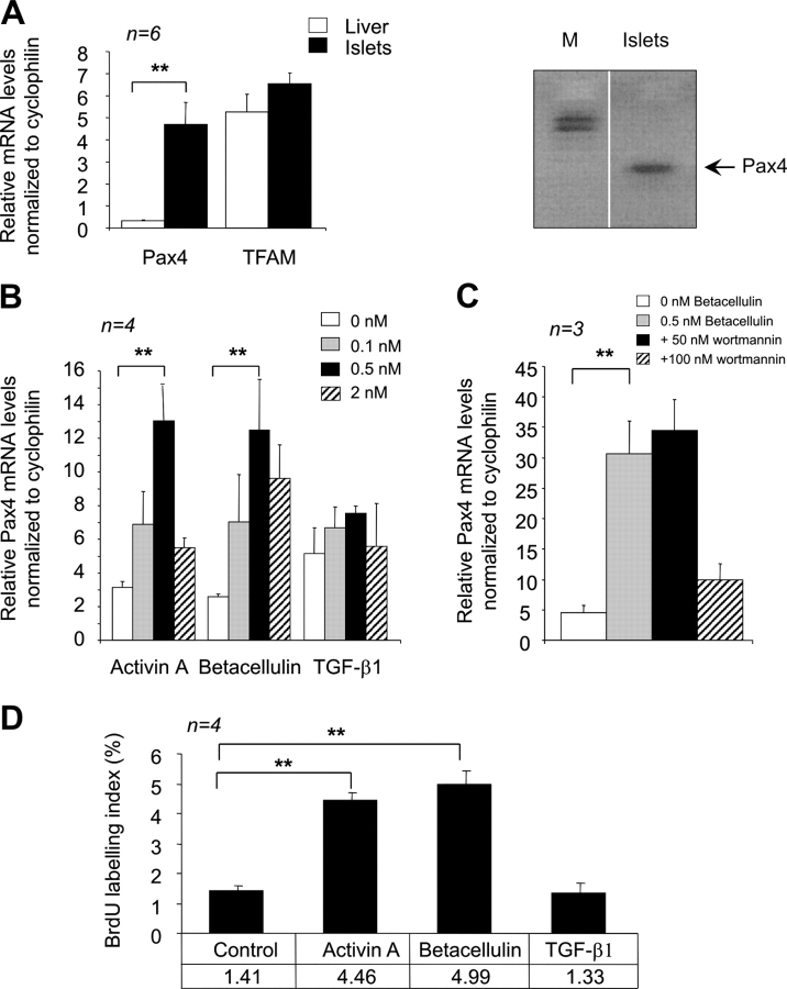
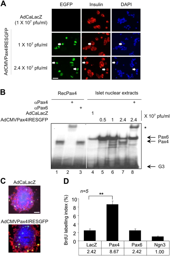
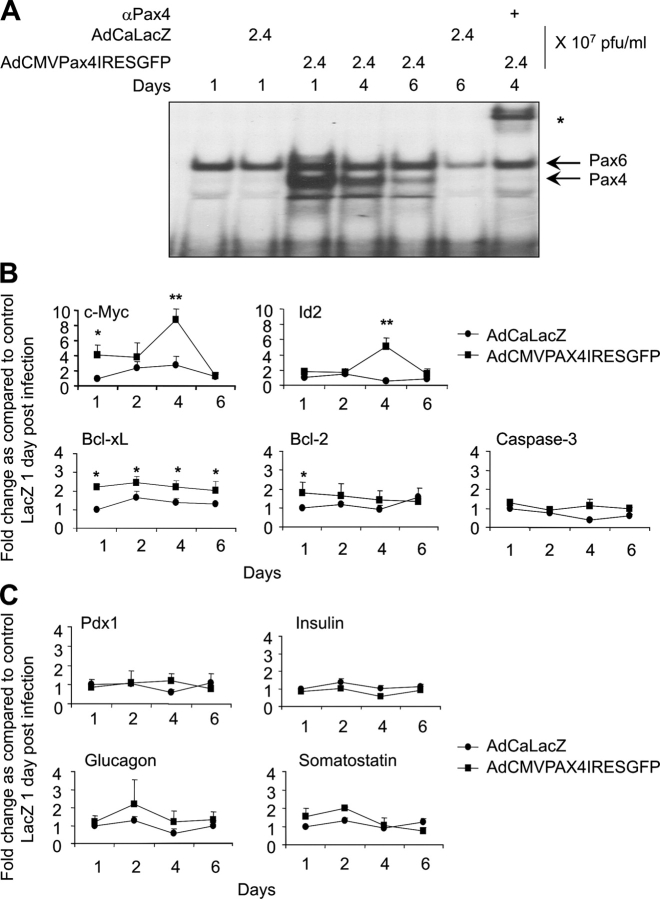
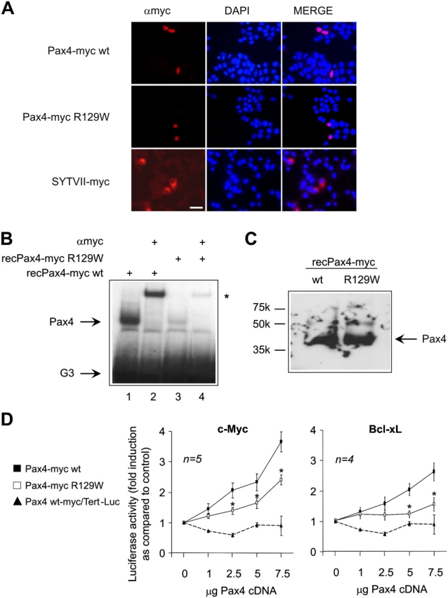
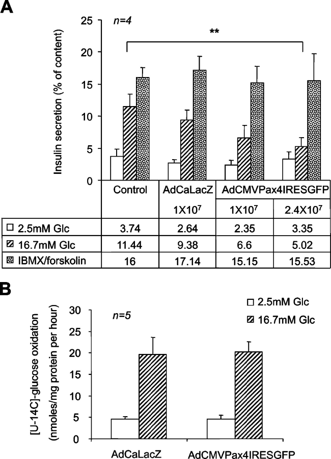
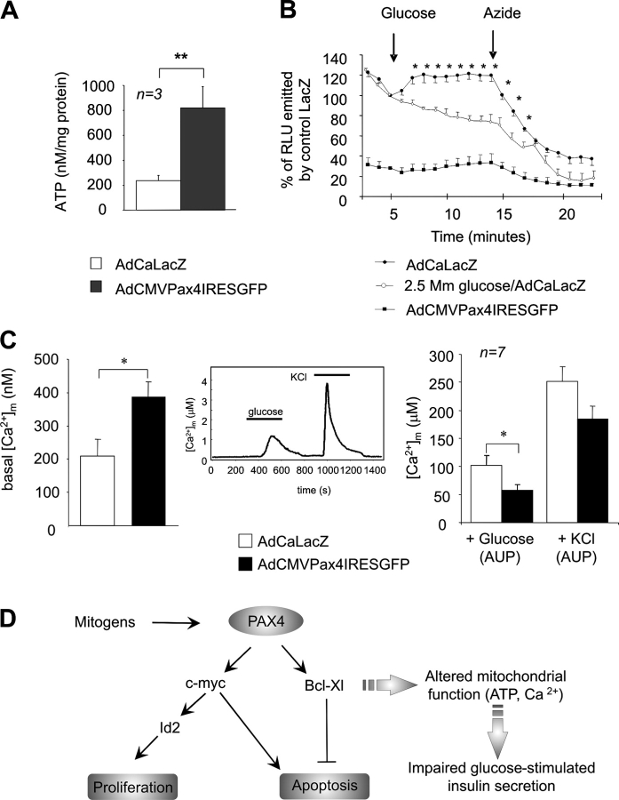
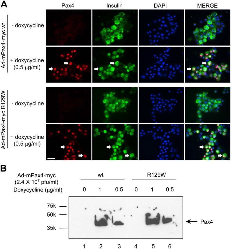
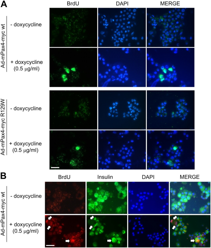
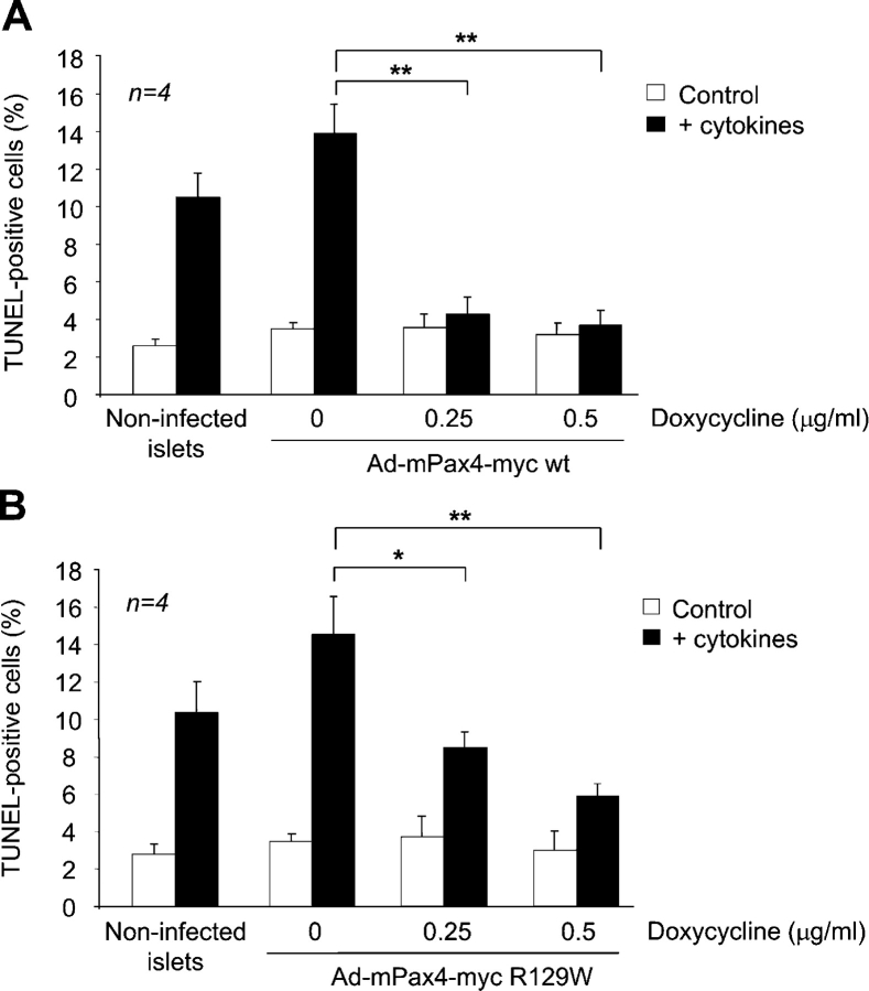
The mechanism by which the beta-cell transcription factor Pax4 influences cell function/mass was studied in rat and human islets of Langerhans. Pax4 transcripts were detected in adult rat islets, and levels were induced by the mitogens activin A and betacellulin. Wortmannin suppressed betacellulin-induced Pax4 expression, implicating the phosphatidylinositol 3-kinase signaling pathway. Adenoviral overexpression of Pax4 caused a 3.5-fold increase in beta-cell proliferation with a concomitant 1.9-, 4-, and 5-fold increase in Bcl-xL (antiapoptotic), c-myc, and Id2 mRNA levels, respectively. Accordingly, Pax4 transactivated the Bcl-xL and c-myc promoters, whereas its diabetes-linked mutant was less efficient. Bcl-xL activity resulted in altered mitochondrial calcium levels and ATP production, explaining impaired glucose-induced insulin secretion in transduced islets. Infection of human islets with an inducible adenoviral Pax4 construct caused proliferation and protection against cytokine-evoked apoptosis, whereas the mutant was less effective. We propose that Pax4 is implicated in beta-cell plasticity through the activation of c-myc and potentially protected from apoptosis through Bcl-xL gene expression.


Figures

Figure 1.
Figure 1.
Activin A and betacellulin increase Pax4 mRNA levels as well as β-cell proliferation in rat islets. (A) Pax4 is expressed in adult rat islets but not in the liver. Quantitative RT-PCR using RNA purified from freshly isolated islets and hepatocytes were performed on Pax4 and TFAM. Data are presented as relative mRNA abundance levels normalized to the transcript cyclophilin and represent the mean ± SEM of six independent experiments performed in triplicates. A representative agarose gel depicting the amplified Pax4 transcript is shown on the right. The fragment was subcloned and confirmed to be Pax4 by sequencing. White line indicates that intervening lanes have been spliced out. (B) PAX4 mRNA levels in islets treated with increasing doses of activin A, betacellulin, or TGF-β1 as indicated. (C) Islets were incubated with 0.5 nM of betacellulin in the absence or presence of 50 and 100 nM of the PI3-kinase inhibitor wortmannin. Pax4 transcript abundance levels were estimated by quantitative RT-PCR. (D) β-Cell proliferation was measured by BrdU incorporation in islets treated with the indicated growth factors at 0.5 nM. Data represent the mean ± SEM of four independent experiments, comprising more than 900 cells per condition. Statistical significance was tested by t test. *, P < 0.05; **, P < 0.01.
Figure 2.
Figure 2.
AdCMVPax4IRESGFP-transduced rat islets express Pax4 and exhibit increased β-cell replication. (A) Immunofluorescent detection of EGFP (green) and insulin (red) as well as DAPI nuclei staining (blue) in dispersed islet cells 48 h after infection with the indicated doses of adenovirus. Pax4 is identified via the reporter cotranslated EGFP in insulin-positive cells (arrows). (B) EMSA using a radiolabeled G3 element of the glucagon gene promoter and full-length mouse Pax4, produced by the coupled TNT system (lanes 1–3), as well as 6 μg of nuclear protein extracts from infected rat islets (lanes 4–8). Infection for 48 h with the indicated amounts of the adenovirus increased Pax4 DNA binding activity to the G3 element in a dose-dependent manner (lanes 5–7). The asterisk delineates the formation of a supershift complex in the presence of anti-Pax4 serum (lanes 2 and 8). White line indicates that intervening lanes have been spliced out. (C) β-Cell proliferation was measured by BrdU incorporation in islets infected either with AdCaLacZ or AdCMVPax4IRESGFP (2.4 × 10⁷ pfu/ml). A representative composite image of an islet immunostained for BrdU (green), insulin (red), and DAPI (blue) is shown. (D) Dispersed β-cells immunostained for both insulin and BrdU were counted under a fluorescent microscope and results are depicted as a percentage of BrdU/insulin-positive cells over the total amount of insulin-positive cells. Data show the mean ± SEM of five independent experiments, each representing more than 1,000 cells per condition. **, P < 0.01. Bars, 50 μM.
Figure 3.
Figure 3.
Time-dependent gene expression profiling of Pax4-overexpressing islets. (A) EMSA using 6 μg of nuclear protein extracts from AdCMVPax4IRESGFP-transduced rat islets cultured in RPMI 1640 medium over a period of 6 d. Pax4 DNA binding activity to the G3 element is maximal 1 d after infection. The asterisk represents the supershifted complex in the presence of anti-Pax4 serum. (B and C) Quantitative RT-PCR analysis performed on RNA isolated from AdCaLacZ (LacZ; •)- and AdCMVPaxIRESGFP (PAX4; ▪)-infected islets (2.4 × 10⁷ pfu/ml, 50% infectibility). Transcript levels were grouped into four categories: proliferative genes comprising c-myc and Id2; apoptotic genes composed of Bcl-xL, Bcl-2, and caspase-3; the transcription factor Pdx-1; and endocrine hormone genes comprising insulin, glucagon, and somatostatin. Expression patterns were measured over a period of 6 d. Each value represents mean ± SEM of three independent experiments. Statistical significance was tested between LacZ- and PAX4-infected islets by unpaired t test. *, P < 0.05; **, P < 0.01.
Figure 4.
Figure 4.
Analysis of the expression and function of Pax4 wt and its mutant R129W. (A) Immunofluorescent detection of the myc-tagged Pax4 or synaptotagmin VII proteins (red) and DAPI nuclei staining (blue) in INS-1E cells 48 h after transfection with the indicated constructs. Pax4 and synaptotagmin VII were detected via the myc epitope in the nuclei and cytoplasm of INS-1E cells, respectively. (B) EMSA using the G3 element and the recombinant proteins Pax4-myc wt (lanes 1 and 2) and Pax4-myc R129W (lanes 3 and 4). An equal amount of protein was applied in each lane (see Fig. 4 C). Pax4 wt bound to the G3 element (lane 1), whereas the binding of the R129W mutant was less efficient (lane 3). The asterisk delineates the formation of a supershift complex due to the addition of anti-myc epitope antibody (lanes 2 and 4). (C) Western blotting of the recombinant proteins Pax4-myc wt and R129W using an anti-myc epitope antibody. (D) Effects of Pax4-myc wt (▪) and its mutant R129W (□) on the human c-myc and murine Bcl-xL promoters. Cotransfection studies using BHK-21 cells were performed with increasing amounts of wt and R129W Pax4. The telomerase promoter construct (▴) was used as a negative control. The pSV-β-galactosidase control vector was used as internal control to normalize for transfection efficiency (∼15%). Data are presented as fold induction of basal luciferase activity and expressed as the mean ± SEM of four to five independent experiments. *, P < 0.05, for comparison between Pax4 wt and R129W for each of the promoter constructs. Bar, 50 μM.
Figure 5.
Figure 5.
Effects of Pax4 overexpression on insulin secretion and glucose oxidation in isolated rat islets. (A) Glucose-induced insulin secretion was inhibited by AdCMVPax4IRESGFP. 2 d after infection, islet hormone secretion was assayed as described in Materials and methods. Data are expressed as the mean ± SEM of four independent experiments. **, P < 0.01. (B) 2 d after infection with 2.4 × 10⁷ pfu/ml of indicated adenoviruses, islet CO2 generation was measured in the presence of 2.5 or 16.7 mM glucose to assess glucose oxidation rate as described in the experimental procedures. Data represent the mean ± SEM of five independent experiments.
Figure 6.
Figure 6.
Total cellular ATP and mitochondrial calcium levels are increased in AdCMVPax4IRESGFP-infected islets. (A) Total cellular ATP levels were measured in islets overexpressing either β-galactosidase or PAX4 (2.4 × 10⁷ pfu/ml, 50% of cell infected) and maintained in 1 mM glucose for 10 min. Results represent the means ± SEM. **, P < 0.01. (B) Cytosolic ATP production in response to 2.5 or 16.5 mM glucose was determined over a period of 20 min using the ATP-sensitive bioluminescence probe luciferase (3.6 × 10⁷ pfu/ml). Glucose and azide were added at indicated times (arrows). Results are the mean ± SEM of at least five experiments performed in duplicates (*, P < 0.05). (C) Mitochondrial calcium was monitored in β-galactosidase or PAX4 overexpressing islets using β-cell–specific/mitochondrial-targeted aequorin as described in Materials and methods. After the establishment of baseline luminescence (30 min; LacZ = 210 ± 49 nM and Pax4 = 387 ± 46 nM, left), islets were superfused for 5 min in basal conditions (0 glucose) before stimulation with glucose (16.7 mM), and then KCl (60 mM) for 5 min intervals each, as shown (middle). The induced increases in [Ca2+]m were evaluated on the basis of the AUP and a presented on the right. Each value represents the mean ± SEM of a minimum of six separate experiments. *, P < 0.05. (D) Proposed model of Pax4-induced β-cell proliferation. Mitogens activate Pax4, which will stimulate c-myc and Bcl-xL gene transcription. c-Myc will promote Id2 gene expression and activate the cell cycle replication program. Bcl-xL increased expression will promote survival by preventing mitochondria from initiating the apoptotic program. However, cells become refractory to glucose-evoked insulin secretion due to altered ATP production and calcium handling.
Figure 7.
Figure 7.
Pax4 and its diabetes-linked mutant are induced by doxycycline in a dose-dependent manner in human islets. (A) Islets were coinfected with either Ad-mPax4-myc wt or R129W as described in Materials and methods. Doxycycline-dependent activation of PAX4 wt and mutant was assessed 48 h later by immunohistochemistry; myc epitope (red), insulin (green), and DAPI (blue). Arrows depict Pax4-expressing β-cells. Pax4 was detected in the nuclei of ∼70% of human islet cells cultured in the presence of doxycycline, whereas no basal induction of Pax4 was observed in the absence of doxycycline. Bar, 50 μM. (B) Western blotting of nuclear extracts derived from infected islet cells cultured in the presence of 0 (lanes 1 and 4), 0.5 (lanes 3 and 6), and 1 μg/ml (lanes 2 and 5) of doxycycline. The same myc anti-serum was used for Western blotting and immunofluorescence.
Figure 8.
Figure 8.
Doxycycline-induced Pax4 stimulates β-cell proliferation in human islets. (A) Proliferation was measured by BrdU incorporation in dispersed islets expressing either Pax4 wt or R129W. Islets were cultured with or without 0.5 μg/ml of doxycycline in the presence of 10 μM BrdU. Immunocytochemical detection of BrdU incorporation (green) and nuclei staining (DAPI in blue) was performed 48 h later. (B) Forced expression of Pax4-induced proliferation in human islet β-cell replication as assessed by costaining of BrdU and insulin (arrows). Bars, 50 μM.
Figure 9.
Figure 9.
Doxycycline-stimulated Pax4 expression protects human islets from cytokine-induced apoptosis. Islets were infected with either Ad-mPax4-myc wt (A) or R129W (B) as described in Materials and methods and cultured for 24 h with the indicated concentrations of doxycycline. Islets were subsequently treated for 24 h with IFN-γ, IL-1β, and TNF-α (2 ng/ml each) to promote apoptosis. Cell death was measured by the TUNEL assay. More than 700 cells were counted for each condition. *, P < 0.05; **, P < 0.01. An ANOVA with Bonferroni/Dunn post hoc analysis between Pax4 wt and R129W revealed a statistical significance of P < 0.00245 at 0.25 μg/ml of doxycycline.

Similar articles

Cited by

References

    1. Baumann Kubetzko, F.B., C. Di Paolo, C. Maag, R. Meier, B.W. Schafer, D.R. Betts, R.A. Stahel, and A. Himmelmann. 2004. The PAX5 oncogene is expressed in N-type neuroblastoma cells and increases tumorigenicity of a S-type cell line. Carcinogenesis. 25:1839–1846. - PubMed
    1. Bonner-Weir, S. 2000. Life and death of the pancreatic beta cells. Trends Endocrinol. Metab. 11:375–378. - PubMed
    1. Buteau, J., S. Foisy, C.J. Rhodes, L. Carpenter, T.J. Biden, and M. Prentki. 2001. Protein kinase Czeta activation mediates glucagon-like peptide-1-induced pancreatic beta-cell proliferation. Diabetes. 50:2237–2243. - PubMed
    1. Buteau, J., S. Foisy, E. Joly, and M. Prentki. 2003. Glucagon-like peptide 1 induces pancreatic beta-cell proliferation via transactivation of the epidermal growth factor receptor. Diabetes. 52:124–132. - PubMed
    1. Campbell, S.C., H. Cragg, L.J. Elrick, W.M. Macfarlane, K.I. Shennan, and K. Docherty. 1999. Inhibitory effect of Pax4 on the human insulin and islet amyloid polypeptide (IAPP) promoters. FEBS Lett. 463:53–57. - PubMed

Publication types

MeSH terms