. 2004 Dec 21;101(51):17825-30.
doi: 10.1073/pnas.0408340102. Epub 2004 Dec 13.

gamma-Tocopherol or combinations of vitamin E forms induce cell death in human prostate cancer cells by interrupting sphingolipid synthesis

Affiliations

gamma-Tocopherol or combinations of vitamin E forms induce cell death in human prostate cancer cells by interrupting sphingolipid synthesis

Qing Jiang et al. Proc Natl Acad Sci U S A. .

Abstract

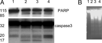
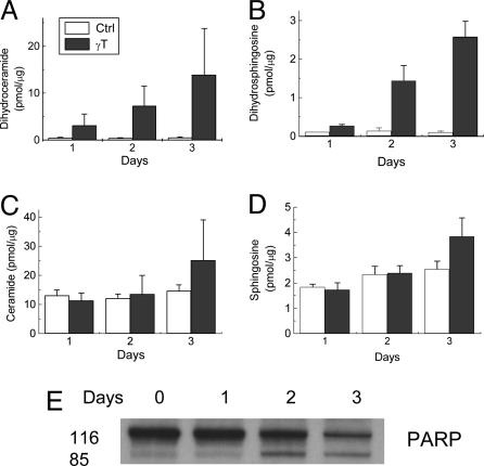
gamma-Tocopherol (gammaT), the predominant form of vitamin E in diets, but not alpha-tocopherol, the major vitamin E form in tissues and supplements, inhibits proliferation of prostate cancer cells (LNCaP and PC-3) and lung cancer cells (A549). In contrast, at similar concentrations, gammaT has no effect on normal prostate epithelial cells. Combinations of some vitamin E forms, such as gammaT and delta-tocopherol, exhibit additive or synergistic inhibitory effects. In this study, gammaT or its combination with delta-tocopherol induced apoptosis in androgen-sensitive prostate LNCaP, but not in androgen-resistant PC-3 cells, by the induction of cytochrome c release, activation of caspase 9 and caspase 3, cleavage of poly-ADP-ribose polymerase (PARP), and involvement of caspase-independent pathways. Myriocin and fumonisin B1, specific inhibitors of key enzymes (serine palmitoyltransferase and dihydroceramide synthase, respectively) in de novo synthesis of sphingolipids, significantly protected cells from gammaT-induced DNA fragmentation, cytochrome c release, PARP cleavage, and the formation of active caspase 3. Compared with vehicle-treated controls, gammaT treatment led to pronounced dihydroceramide and dihydrosphingosine accumulation, which preceded morphological and biochemical manifestations of apoptosis. In contrast, ceramide and shpingosine levels did not increase until day 3, when substantial cell death took place. Our study demonstrates that gammaT and mixed vitamin E forms induce cell death by interrupting the de novo sphingolipid pathway in a prostate cancer cell line. Thus, certain vitamin E forms may be valuable as anticancer agents.


Figures

Fig. 1.
Fig. 1.
Effect of γT on proliferation of PC-3 (A), LNCaP (B), A549 (C), and PrEC (D). In 24-well plates, cells were seeded at a density of 1.8-2.5 × 10⁴ cells per well. After 24-48 h, tocopherols (indicated in μM) and the corresponding amount of DMSO in controls were added. Cell viability (indicated as absorbance in A and B) was evaluated on days 1-4 by using MTT assays. A549 (C) or PrEC (D) cells were treated with vitamin E forms for 4 days, and the viability is expressed as the relative ratio of absorbance between treatments and controls. Data are the averages of two to three independent experiments. * (P < 0.05) and ** (P < 0.01) indicate a significant difference between treated and control cells.
Fig. 2.
Fig. 2.
Effect of AA and LA on γT-induced antiproliferation. After cells were seeded for 24-48 h, tocopherols (50 μM) or DMSO were added with 10 μMAA or LA in PC-3 (A) and LNCaP (B). After a 4-day incubation, cell viability was evaluated by MTT assays and is expressed as the ratio of absorbance between treatments and controls. * (P < 0.05) indicates a significant counteracting effect of AA or LA on γT treatment.
Fig. 3.
Fig. 3.
Effect of combinations of vitamin E forms on cell growth in PC-3 (A) and LNCaP (B). Cells were treated with various forms of vitamin E for 3-4 days. The cell-culture conditions are the same as those described in Fig. 1.
Fig. 4.
Fig. 4.
γT (50 μM) or the combination of γT (25 μM) and δT (10 μM) induced apoptosis in LNCaP but not PC-3. Apoptosis was evaluated in cells after 3-day treatment with tocopherols by means of DNA fragmentation (A) and annexin V-propidium iodide double staining (B). Experimental procedures are described in Materials and Methods.
Fig. 5.
Fig. 5.
γT treatment led to cytochrome c (Cyto C) release, caspase 9 activation, and PARP cleavage. (A) γT caused a time-dependent release of cytochrome c in LNCaP cells but not in PC-3 cells, with no effect on Bax translocation. (B and C) After 72-h treatment, an activation of caspase 9 (B) and PARP cleavage (C) were observed in treated cells.
Fig. 6.
Fig. 6.
Z-VAD-fmk did not protect cells from γT-induced PARP cleavage (A) or DNA fragmentation (B), although it significantly inhibited the formation of active caspase 3. LNCaP cells were treated as follows: lane 1, DMSO (control); lane 2, γT (50 μM); lane 3, γT (25 μM) and δT (10 μM); lane 4, γT (50 μM) and Z-VAD-fmk (30 μM) for 3 days.
Scheme 1.
Scheme 1.
Fig. 7.
Fig. 7.
Fumonisin and myriocin significantly protected cells from γT-induced DNA fragmentation (A), and myriosin prevented cytochrome c release, PARP cleavage, and caspase 3 activation (B). In A, LNCaP cells were treated with DMSO (ctrl), γT (50 μM), fumonisin (40 μM) (c + f), myriocin (6 μM) (c + M), γT (50 μM) with fumonisin (40 μM) (γT + f), or myriocin (6 μM) (γT + M) for 3 days. In B, cells were treated with γT (50 μM) with or without myriosin for 3 days.
Fig. 8.
Fig. 8.
Time course for γT-caused changes in dihydroceramide (A), dihydrosphingosine (B), ceramide (C), and sphingosine (D), as well as PARP cleavage (E). The results of sphingolipids were expressed as the ratio of sphingolipid intermediates (in pmol) to total choline-containing phospholipids (in μg) (see Materials and Methods).

References

    1. Eichholzer, M., Stahelin, H. B., Gey, K. F., Ludin, E. & Bernasconi, F. (1996) Int. J. Cancer 66, 145-150. - PubMed
    1. Toth, B. & Patil, K. (1983) J. Natl. Cancer Inst. 70, 1107-1111. - PubMed
    1. Chung, H., Wu, D., Han, S. N., Gay, R., Goldin, B., Bronson, R. E., Mason, J. B., Smith, D. E. & Meydani, S. N. (2003) J. Nutr. 133, 528-532. - PubMed
    1. Chan, J. M., Stampfer, M. J., Ma, J., Rimm, E. B., Willett, W. C. & Giovannucci, E. L. (1999) Cancer Epidemiol. Biomarkers Prev. 8, 893-899. - PubMed
    1. Moyad, M. A. (2002) Urology 59, 9-19. - PubMed

Publication types

MeSH terms