. 2004 Dec 21;101(51):17795-800.
doi: 10.1073/pnas.0408096101. Epub 2004 Dec 13.

Major reduction of atherosclerosis in fractalkine (CX3CL1)-deficient mice is at the brachiocephalic artery, not the aortic root

Affiliations

Major reduction of atherosclerosis in fractalkine (CX3CL1)-deficient mice is at the brachiocephalic artery, not the aortic root

Daniel Teupser et al. Proc Natl Acad Sci U S A. .

Abstract

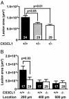
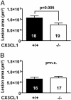
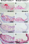
Fractalkine (CX3CL1) is of particular interest in atherogenesis because it can serve as an adhesion molecule and a chemokine. Fractalkine and its receptor CX3CR1 are expressed in atherosclerotic lesions of humans and mice. However, the effect of fractalkine deficiency on atherosclerosis susceptibility is unknown. Fractalkine-deficient mice on the C57BL/6 (B6) background were bred to the atherosclerosis-sensitizing B6.ApoE(-/-) and B6.LDLR(-/-) backgrounds. Compared with controls, aortic-root lesion area was unchanged in fractalkine-deficient male and female B6.ApoE(-/-) mice at 16 weeks of age and males at 12 weeks of age, but it was mildly reduced (30%, P = 0.005) in females at 12 weeks of age. In contrast, lesion area at the brachiocephalic artery (BCA) was reduced dramatically by approximately 85% in fractalkine-deficient females [42,251 +/- 26,136 microm(2) (n = 15) vs. 6,538 +/- 11,320 microm(2);(n = 24), P < 0.0001] and males [36,911 +/- 32,504 microm(2) (n = 24) vs. 6,768 +/- 8,595 microm(2) (n = 14); P = 0.001] at 16 weeks of age. Fractalkine-deficient B6.ApoE(-/-) mice were comparable with controls in body weight, plasma cholesterol, plasma high-density lipoprotein cholesterol and white blood cell counts. On the B6.LDLR(-/-) background, lesion areas were reduced by 35% at the aortic root (P < 0.01) and by 50% at the BCA (P < 0.05) in fractalkine-deficient females at 16 weeks of age. Lesions in fractalkine-deficient mice on the B6.ApoE(-/-) and B6.LDLR(-/-) backgrounds were less complex and contained significantly fewer macrophages than controls. In conclusion, the major reduction of atherosclerosis in fractalkine-deficient mice appears to be at the BCA rather than the aortic root.


Figures

Fig. 1.
Fig. 1.
Atherosclerotic lesion area in CX3CL1-deficient B6.ApoE-/- mice at 16 weeks of age at the aortic root and BCA. Sections at the BCA were quantified 200, 400, and 600 μm proximal to the branching point of the BCA into the subclavian and carotid arteries. (A) Aortic-root lesion area in female B6.ApoE-/- mice. (B) BCA lesion area in female B6.ApoE-/- mice. (C) Aortic-root lesion area in male B6.ApoE-/- mice. (D) BCA lesion area in male B6.ApoE-/- mice. Bars represent means ± SEM, and the numbers of animals in the individual groups are given on the bars.
Fig. 2.
Fig. 2.
Atherosclerotic lesion area in CX3CL1-deficient B6.LDLR-/- mice at 16 weeks of age at the aortic root and BCA. Sections at the BCA were quantified 200, 400, and 600 μm proximal to the branching point of the BCA into the subclavian and carotid arteries. (A) Aortic-root lesion area in female B6.LDLR-/- mice. (B) BCA lesion area in female B6.LDLR-/- mice. Bars represent means ± SEM, and the numbers of animals in the individual groups are given on the bars.
Fig. 3.
Fig. 3.
Atherosclerotic lesion area in CX3CL1-deficient B6.ApoE-/- mice at 12 weeks of age. (A) Aortic-root lesion area in female B6.ApoE-/- mice. (B) Aortic-root lesion area in male B6.ApoE-/- mice. Bars represent means ± SEM, and the numbers of animals in the individual groups are given on the bars. n.s., Not significant.
Fig. 4.
Fig. 4.
CD68-stained area of atherosclerotic lesions at the BCA in CX3CL1-deficient B6.ApoE-/- and B6.LDLR-/- mice at 16 weeks of age. (A) Female B6.ApoE-/- mice. (B) Female B6.LDLR-/- mice.
Fig. 5.
Fig. 5.
Representative serial sections through the BCA of CX3CL1-deficient B6.ApoE-/- mice at 16 weeks of age. Oil red O staining in CX3CL1+/+ (A) and CX3CL1-/- (B) mice. CD68 staining (red) in CX3CL1+/+ (C) and CX3CL1-/- (D) mice. CX3CL1 staining (red) in CX3CL+/+ (E) and CX3CL1-/- (F) mice. α-Actin staining (red) in CX3CL1+/+ (G) and CX3CL1-/- (H) mice.
Fig. 6.
Fig. 6.
Representative serial sections through the BCA of CX3CL1-deficent B6.LDLR-/- mice at 16 weeks of age. Oil red O staining in CX3CL1+/+ (A) and CX3CL1-/- (B) mice. CD68 staining (red) in CX3CL1+/+ (C) and CX3CL1-/- (D) mice. CX3CL1 staining (red; underneath internal elastic lamina marked by arrows) in CX3CL+/+ (E) and CX3CL1-/- (F) mice. α-Actin staining (red) in CX3CL1+/+ (G) and CX3CL1-/- (H) mice.

Similar articles

Cited by

References

    1. Ross, R. (1999) N. Engl. J. Med. 340, 115-126. - PubMed
    1. Glass, C. K. & Witztum, J. L. (2001) Cell 104, 503-516. - PubMed
    1. Smith, J. D., Trogan, E., Ginsberg, M., Grigaux, C., Tian, J. & Miyata, M. (1995) Proc. Natl. Acad. Sci. USA 92, 8264-8268. - PMC - PubMed
    1. Cybulsky, M. I., Iiyama, K., Li, H., Zhu, S., Chen, M., Iiyama, M., Davis, V., Gutierrez-Ramos, J. C., Connelly, P. W. & Milstone, D. S. (2001) J. Clin. Invest. 107, 1255-1262. - PMC - PubMed
    1. Dansky, H. M., Barlow, C. B., Lominska, C., Sikes, J. L., Kao, C., Weinsaft, J., Cybulsky, M. I. & Smith, J. D. (2001) Arterioscler. Thromb. Vasc. Biol. 21, 1662-1667. - PubMed

Publication types

MeSH terms