. 2005 Jan;79(1):87-94.
doi: 10.1128/JVI.79.1.87-94.2005.

Improved enzootic nasal tumor virus pseudotype packaging cell lines reveal virus entry requirements in addition to the primary receptor Hyal2

Affiliations

Improved enzootic nasal tumor virus pseudotype packaging cell lines reveal virus entry requirements in addition to the primary receptor Hyal2

Neal S Van Hoeven et al. J Virol. 2005 Jan.

Abstract

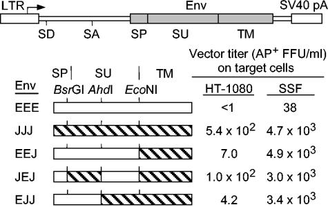
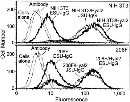
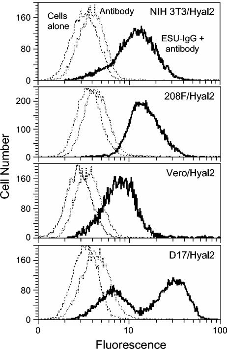
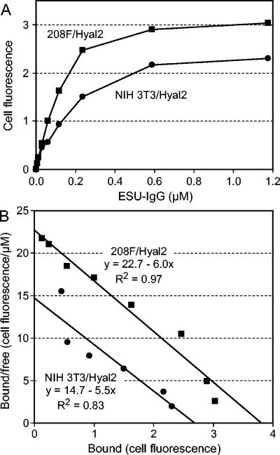
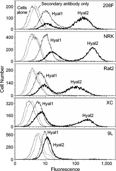
Enzootic nasal tumor virus (ENTV) and jaagsiekte sheep retrovirus (JSRV) are closely related retroviruses that cause epithelial cancers of the respiratory tract in sheep and goats. Both viruses use the glycosylphosphatidylinositol (GPI)-anchored cell surface protein hyaluronidase 2 (Hyal2) as a receptor for cell entry, and entry is mediated by the envelope (Env) proteins encoded by these viruses. Retroviral vectors bearing JSRV Env can transduce cells from a wide range of species, with the exception of rodent cells. Because of the low titer of vectors bearing ENTV Env, it has been difficult to determine the tropism of ENTV vectors, which appeared to transduce cells from sheep and humans only. Here we have developed high-titer ENTV packaging cells and confirm that ENTV has a restricted host range compared to that of JSRV. Most cells that are not transduced by JSRV or ENTV vectors can be made susceptible following expression of human Hyal2 on the cells. However, five rat cell lines from different rat strains and different tissues that were engineered to express human Hyal2 were still only poorly infected by ENTV vectors, even though the ENTV Env protein could bind well to human Hyal2 expressed on four of these cell lines. These results indicate the possibility of a coreceptor requirement for these viruses.


Figures

FIG. 1.
FIG. 1.
Transduction of human and sheep cells by vectors bearing chimeric ENTV/JSRV Env proteins. At the top is the expression cassette used to express the Env proteins. The arrow indicates the transcription start site, and the abbreviations are the following: LTR, retroviral long terminal repeat promoter; SD, splice donor; SA, splice acceptor; SP, endoplasmic reticulum signal peptide; SU, Env surface subunit; TM, Env transmembrane subunit; and SV40 pA, simian virus 40 polyadenylation signal. Hybrid Env proteins (open boxes represent ENTV Env, hatched boxes represent JSRV Env) were made using the indicated restriction enzyme sites, and the relationship of these sites and the SP/SU and SU/TM cleavage sites are shown. LAPSN vectors bearing the indicated Env proteins were made by transient transfection as described in Materials and Methods. SSF and HT-1080 cells, seeded at 105 per well (diameter, 3.5 cm) of 6-well plates, were exposed to the vectors 1 day later and were stained to detect AP+ foci 2 days after vector exposure. Values are averages of two experiments with duplicate determinations in each experiment. Data are from reference .
FIG. 2.
FIG. 2.
Binding levels of ENTV and JSRV Env SU domains to NIH 3T3 and 208F cells expressing human Hyal2 are similar. Dashed lines represent Hyal2-expressing cells without SU-IgG or antibody addition; dotted lines, Hyal2-expressing cells incubated with secondary antibody only; solid heavy lines, parental or Hyal2-expressing cells incubated with ESU-IgG and antibody; and solid thin lines, Hyal2-expressing cells incubated with JSU-IgG and antibody. All profiles were generated in the same experiment with the same reagents. Cells were incubated with ESU-IgG and JSU-IgG at a concentration of 10 μg/ml (0.12 μM).
FIG. 3.
FIG. 3.
Binding of the ENTV Env SU domain to human Hyal2 expressed on cells from different species. The human Hyal2-expressing cell type analyzed is given at the top right of each panel. Dashed lines represent cells without ESU-IgG or antibody addition; solid thin lines, cells incubated with secondary antibody only; solid heavy lines, cells incubated with ESU-IgG and secondary antibody. All profiles were generated in the same experiment with the same reagents. Cells were incubated with ESU-IgG at a concentration of ∼0.5 μg/ml.
FIG. 4.
FIG. 4.
Specific binding of ESU-IgG to human Hyal2 expressed on 208F and NIH 3T3 cells. (A) Parental or human Hyal2-expressing 208F or NIH 3T3 cells were incubated with various concentrations of ESU-IgG, were incubated with secondary antibody, and were analyzed by FACS. At each ESU-IgG concentration, the geometric mean fluorescence of parental cells (208F or NIH 3T3) was subtracted from that of human Hyal2-expressing cells (208F/Hyal2 or NIH 3T3/Hyal2) to obtain the human Hyal2-specific binding values, in arbitrary units, and these values are plotted against the ESU-IgG concentration. All results were generated using the same reagents and the same FACS settings and were repeated once, with similar results. (B) Scatchard analysis of the data from panel A is shown. Best-fit lines, their equations, and the Pearson correlation coefficients are indicated. Kd values (micromolars) are equal to −1/slope.
FIG. 5.
FIG. 5.
The ENTV SU domain binds specifically and at high levels to most rat cells transduced with a vector encoding human Hyal2. The cell type analyzed is given at the top right of each panel. Dashed lines represent Hyal2-expressing cells without SU-IgG or antibody addition; dotted lines, Hyal2-expressing cells incubated with secondary antibody only; solid heavy lines, cells transduced with a vector encoding human Hyal2 and incubated with ESU-IgG and secondary antibody; solid thin lines, cells transduced with a vector encoding human Hyal1 and incubated with ESU-IgG and secondary antibody. All profiles were generated in the same experiment with the same reagents. Cells were incubated with ESU-IgG at a concentration of 10 μg/ml (0.12 μM).

Similar articles

Cited by

References

    1. Alberti, A., C. Murgia, S.-L. Liu, M. Mura, C. Cousens, M. Sharp, A. D. Miller, and M. Palmarini. 2002. Envelope-induced cell transformation by ovine betaretroviruses. J. Virol. 76:5387-5394. - PMC - PubMed
    1. Barker, M., T. Hoshino, O. Gurcay, C. B. Wilson, S. L. Nielsen, R. Downie, and J. Eliason. 1973. Development of an animal brain tumor model and its response to therapy with 1,3-bis(2-chloroethyl)-1-nitrosourea. Cancer Res. 33:976-986. - PubMed
    1. Dirks, C., F.-M. Duh, S. K. Rai, M. I. Lerman, and A. D. Miller. 2002. Mechanism of cell entry and transformation by enzootic nasal tumor virus. J. Virol. 76:2141-2149. - PMC - PubMed
    1. DuBridge, R. B., P. Tang, H. C. Hsia, P. M. Leong, J. H. Miller, and M. P. Calos. 1987. Analysis of mutation in human cells by using an Epstein-Barr virus shuttle system. Mol. Cell. Biol. 7:379-387. - PMC - PubMed
    1. Duc-Nguyen, H., E. N. Rosenblum, and R. F. Zeigel. 1966. Persistent infection of a rat kidney cell line with Rauscher murine leukemia virus. J. Bacteriol. 92:1133-1140. - PMC - PubMed

Publication types

MeSH terms

LinkOut - more resources