. 2005 Jan;79(1):106-15.
doi: 10.1128/JVI.79.1.106-115.2005.

The cytoplasmic tail slows the folding of human immunodeficiency virus type 1 Env from a late prebundle configuration into the six-helix bundle

Affiliations

The cytoplasmic tail slows the folding of human immunodeficiency virus type 1 Env from a late prebundle configuration into the six-helix bundle

Levon G Abrahamyan et al. J Virol. 2005 Jan.

Abstract

Effects of the cytoplasmic tail (CT) of human immunodeficiency virus type 1 Env on the process of membrane fusion were investigated. Full-length Env (wild type [WT]) and Env with its CT truncated (DeltaCT) were expressed on cell surfaces, these cells were fused to target cells, and the inhibition of fusion by peptides that prevent Env from folding into a six-helix bundle conformation was measured. For both X4-tropic and R5-tropic Env proteins, DeltaCT induced faster fusion kinetics than did the WT, and peptides were less effective at inhibiting DeltaCT-induced fusion. We tested the hypothesis that the inhibitory peptides were less effective at inhibiting DeltaCT-induced fusion because DeltaCT folds more quickly into a six-helix bundle. Early and late intermediates of WT- and DeltaCT-induced fusion were captured, and the ability of peptides to block fusion when added at the intermediate stages was quantified. When added at the early intermediate, the peptides were still less effective at inhibiting DeltaCT-induced fusion but they were equally effective at preventing WT- and DeltaCT-induced fusion when added at the late intermediate. We conclude that for both X4-tropic and R5-tropic Env proteins, the CT facilitates conformational changes that allow the trimeric coiled coil of prebundles to become optimally exposed. But once Env does favorably expose its coiled coil to inhibitory peptides, the CT hinders subsequent folding into a six-helix bundle. Because of this facilitation of maximal exposure and hindrance of bundle formation, the coiled coil is optimally exposed for a longer time for WT than for DeltaCT. This accounts for the greater peptide inhibition of WT-induced fusion.


Figures
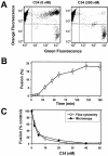
FIG. 1.
FIG. 1.
Flow cytometry analysis of fusion between TF228.1.16 (effector) and HeLaT4+ (target) cells. (A) Effector cells loaded with green cytoplasmic marker were coincubated with target cells loaded with orange marker for 2.5 h at 37°C either in the presence (right) or in the absence (left) of 300 nM C34 peptide. (B) Kinetics of cell-cell fusion were obtained by varying the time E-T cells were coincubated at 37°C, lowering the temperature to halt further fusion, and then lifting cells off the culture dishes with a trypsin-EDTA solution. (C) Dose-response curves for C34 inhibition measured by flow cytometry (open circles) and microscopy (filled circles) assays. A small background signal not related to fusion (about 1.5% of the total number of effector cells) was subtracted from the data in panels B and C. Unless stated otherwise, error bars represent standard errors of the mean for at least three independent duplicate measurements.
FIG. 2.
FIG. 2.
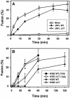
Kinetics of cell-cell fusion induced by the WT and ΔCT. (A) Fusion between 293T cells transiently expressing JRFL WT (filled triangles) or ΔCT (open triangles) and target 3T3.CD4.CCR5 cells was monitored by the flow cytometry assay as described in the legend to Fig. 1. Open squares are the background signal, as observed with mock-transfected 293T cells. (B) The kinetics of fusion induced by HXB2 WT (filled symbols) or ΔCT (open symbols) transiently expressed in 293T and HeLaT4+ cells were measured by fluorescence microscopy. Fusion was induced by coculturing the cells either directly at 37°C (triangles) or after preincubation at 23°C for 2.5 h (circles).
FIG. 3.
FIG. 3.
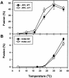
Temperature dependence of fusion promoted by the WT (filled symbols) and ΔCT (open symbols) from the JRFL (A) and HXB2 (B) strains. Cells were coincubated for 2.5 (HXB2) or 2 (JRFL) h at the indicated temperature and then analyzed by flow cytometry. The extent of fusion induced by JRFL Env at 37°C was determined after a 1 h of incubation to reduce the formation of large syncytia. The background signal (<2% of the effector cells after a 37°C incubation) was subtracted from the plotted data. The apparent reduced fusion of JRFL Env at 37°C was caused by leakage of calcein from the effector cells and by syncytium formation, as verified by fluorescence microscopy (data not shown).
FIG. 4.
FIG. 4.
Efficiency of proteolytic cleavage of cell surface-expressed HXB2 WT and ΔCT. The amounts of WT- and ΔCT-bearing plasmids used to transfect a 6-cm-diameter dish of 239T cells are indicated above the lanes. For details, see Materials and Methods.
FIG. 5.
FIG. 5.
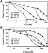
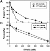
Inhibition of HIV Env-induced cell-cell fusion by the C34 (A) and 5-helix (B) peptides. 293T cells expressing JRFL (circles) or HXB2 (triangles) were coincubated with appropriate target cells in the presence of varied concentrations of HIV fusion inhibitors for 1 or 2.5 h, respectively. WT-induced fusion (open symbols) was more sensitive to inhibitors than was fusion induced by ΔCT (filled symbols). Fusion was quantified by flow cytometry.
FIG. 6.
FIG. 6.
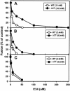
Efficacy of C34 peptide at sequential intermediate stages of fusion induced by HXB2 WT (open circles) and ΔCT (filled circles). Varied concentrations of C34 were added either at the beginning of E-T cell coincubation (A), at the TAS (B), or at the LAS (C). Fusion was then triggered by incubating the cells at 37°C for 2.5 h (A) or for 1 h (B and C). The IC50s obtained by curve fitting (see Materials and Methods) are given in parentheses in the insets. Fusion was quantified by fluorescence microscopy.
FIG. 7.
FIG. 7.
Relative potency of the 5-helix peptide added at the beginning of E-T cell coincubation (A) or at the LAS (B). The IC50s for the HXB2 WT (open circles) and ΔCT (filled circles) are given in parentheses. Details are given in the legend to Fig. 6.
FIG. 8.
FIG. 8.
The time from maximal groove exposure to formation of the 6HB is briefer for ΔCT. C34 inhibits WT-induced fusion more effectively than ΔCT-induced fusion at the TAS, indicating that the grooves of ΔCT are less well exposed than those of the WT at this stage. Inhibition of fusion by C34 is the same for the WT and ΔCT at the LAS. Once grooves are fully exposed, ΔCT more quickly folds into a 6HB, illustrated as shorter transition arrows between the TAS and the LAS and between the LAS and fusion for ΔCT. The progression from left to right denotes a reaction coordinate (rather than time) for fusion.

Similar articles

Cited by

References

    1. Abrahamyan, L. G., R. M. Markosyan, J. P. Moore, F. S. Cohen, and G. B. Melikyan. 2003. Human immunodeficiency virus type 1 Env with an intersubunit disulfide bond engages coreceptors but requires bond reduction after engagement to induce fusion. J. Virol. 77:5829-5836. - PMC - PubMed
    1. Armand-Ugon, M., A. Gutierrez, B. Clotet, and J. A. Este. 2003. HIV-1 resistance to the gp41-dependent fusion inhibitor C-34. Antiviral Res. 59:137-142. - PubMed
    1. Bhattacharya, J., P. J. Peters, and P. R. Clapham. 2004. Human immunodeficiency virus type 1 envelope glycoproteins that lack cytoplasmic domain cysteines: impact on association with membrane lipid rafts and incorporation onto budding virus particles. J. Virol. 78:5500-5506. - PMC - PubMed
    1. Binley, J. M., C. S. Cayanan, C. Wiley, N. Schulke, W. C. Olson, and D. R. Burton. 2003. Redox-triggered infection by disulfide-shackled human immunodeficiency virus type 1 pseudovirions. J. Virol. 77:5678-5684. - PMC - PubMed
    1. Blacklow, S. C., M. Lu, and P. S. Kim. 1995. A trimeric subdomain of the simian immunodeficiency virus envelope glycoprotein. Biochemistry 34:14955-14962. - PubMed

Publication types

LinkOut - more resources