. 2005 Jan;79(1):159-75.
doi: 10.1128/JVI.79.1.159-175.2005.

Systemic priming-boosting immunization with a trivalent plasmid DNA and inactivated murine cytomegalovirus (MCMV) vaccine provides long-term protection against viral replication following systemic or mucosal MCMV challenge

Affiliations

Systemic priming-boosting immunization with a trivalent plasmid DNA and inactivated murine cytomegalovirus (MCMV) vaccine provides long-term protection against viral replication following systemic or mucosal MCMV challenge

Christopher S Morello et al. J Virol. 2005 Jan.

Abstract

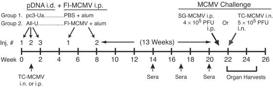
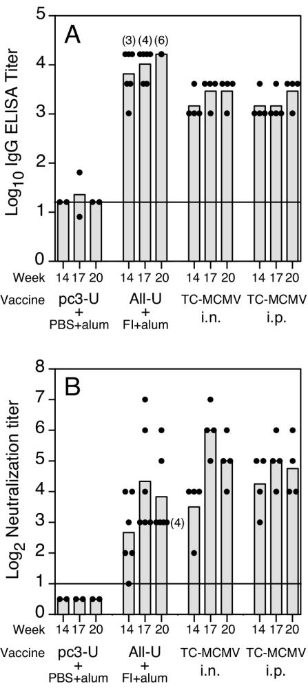
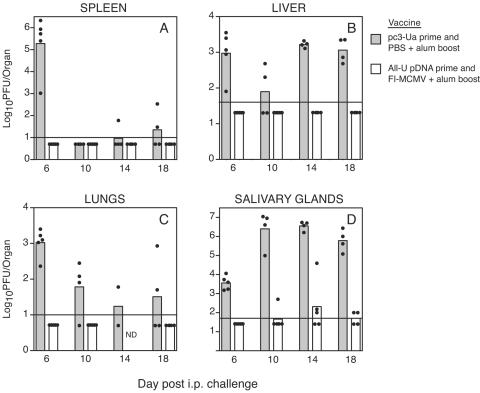
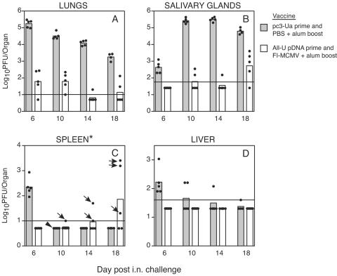
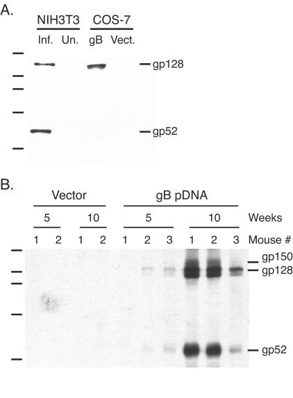
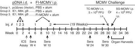
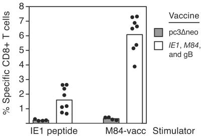
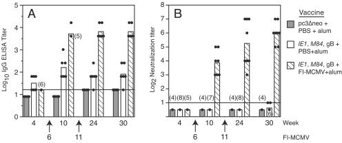
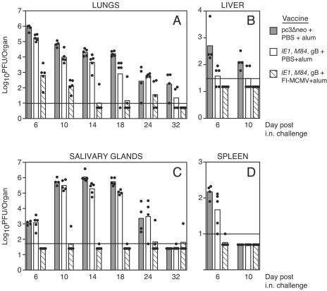
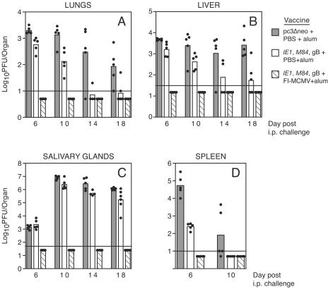
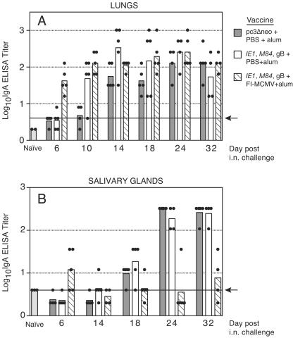
We previously demonstrated that vaccination of BALB/c mice with a pool of 13 plasmid DNAs (pDNAs) expressing murine cytomegalovirus (MCMV) genes followed by formalin-inactivated MCMV (FI-MCMV) resulted in complete protection against viral replication in the spleen and salivary glands following sublethal intraperitoneal (i.p.) challenge. Here, we found that following intranasal (i.n.) challenge, titers of virus in the lungs of the immunized mice were reduced approximately 1,000-fold relative to those for mock-immunized controls. We next sought to extend these results and to determine whether similar protection levels could be achieved by priming with a pool of three pDNAs containing three key plasmids (IE1, M84, and gB). We found that the three-pDNA priming elicited IE1- and M84-p65-specific CD8+ T lymphocytes and, following FI-MCMV boost, high levels of virion-specific immunoglobulin G (IgG) and virus-neutralizing antibodies. When mice were i.n. challenged 4 months after the last boost, titers of virus in the lungs of immunized mice were reduced 1,000- to 2,000-fold from those for controls during the peak of viral replication. Additionally, titers of virus were either at or below the detection limits for the salivary glands, liver, and spleen of the majority of the immunized mice. Following sublethal i.p. challenge, virus was undetectable in all of the above target organs of the immunized mice. Virion-specific IgA in the lungs was consistently detected by day 6 post-i.n. challenge for the immunized mice and by day 14 for controls. These results demonstrate the immunity and high levels of protection of the priming-boosting vaccination against both systemic and mucosal challenge.


Figures

FIG. 1.
FIG. 1.
Immunization groups and schedule for mice primed with All-U pDNA and boosted with FI-MCMV. Two groups of BALB/c mice were i.d. immunized on the weeks shown with 26 μg of either empty vector DNA (pc3-Ua) or the 13 DNA cocktail All-U (2 μg each of m04, M32, M48, M56, M69, M82, M83, M85, M86, M99, e1, U-IE1, and U-M84 DNAs). On the weeks shown, the pc3-Ua-immunized mice were then i.p. boosted twice with PBS plus alum, while the All-U-immunized mice were boosted with 107 PFU equivalents of FI-MCMV plus alum. As controls, two additional mouse groups were immunized on week 1 with 2.5 × 10⁴ PFU of TC-MCMV given either i.n. or i.p. Sera were obtained on the weeks shown for antibody analysis, and mice were then challenged either i.n. or i.p. as shown. Mice were sacrificed postchallenge for virus titer determinations in the spleen, lungs, liver, and salivary glands.
FIG. 2.
FIG. 2.
Virion-specific IgG and neutralizing-antibody responses in the DNA primed/FI-MCMV-boosted or live virus-vaccinated mice. Mice from each group vaccinated with either pc3-Ua plus PBS plus alum (two mice per group), All-U pDNA plus FI+alum (six mice per group), or live TC-MCMV given i.n. or i.p. (four mice each per group) were bled on weeks 14, 17, and 20 of the experiment, and sera were prepared. (A) The levels of virion-specific IgG in serum were measured by ELISA as described in Materials and Methods. Results are represented as the log10 of individual mouse titers (closed circles) and the group mean of the log10 titers (bars). (B) Virus neutralization antibody levels in the sera were measured by in vitro neutralization assay as described in Materials in Methods. Neutralization titers shown are log2 of the highest reciprocal serum dilution that resulted in a ≥50% reduction of the number of input PFU (ca. 50 PFU). Mean log2 and individual log2 titers are shown as in (A). When the ELISA or neutralization titer of a serum was below the detection limit (indicated by horizontal line), the titer was arbitrarily set to the log10 or log2, respectively, of one-half the detection limit for display purposes and mean calculation. Parenthetical numbers indicate the number of sera in a group with identical titers.
FIG. 3.
FIG. 3.
Protection against viral replication in the target organs of All-U plus FI+alum-vaccinated mice following i.p. MCMV challenge. On the days post-i.p. challenge shown, four to five mice per vaccine group (as described in the legend to Fig. 1) were sacrificed, and the (A) spleen, (B) liver, (C) lungs, and (D) salivary glands were aseptically removed, homogenized, and stored for MCMV titer determination as described in Materials and Methods. Bars and closed circles represent the mean log10 of the virus titers for each vaccine group and the log10 viral titers of individual organs, respectively. The horizontal lines show the plaque assay sensitivity limits for each organ as determined by the fraction of the organ homogenate used to infect the NIH 3T3 cells. These limits were chosen as the maximal fraction of homogenate for each organ type that could be used to infect NIH 3T3 cells without the homogenate toxicity inhibiting the plaque formation of 40 PFU of exogenously added MCMV. When virus was undetectable in a given organ, the individual titer of virus for that organ was arbitrarily set to the log10 of one-half the respective detection limit for display purposes and mean calculation. Note that in panel C, the day 14 pc3-Ua-primed and PBS-plus-alum-boosted group had two mice. ND, not determined.
FIG. 4.
FIG. 4.
Protection against viral replication in the target organs of All-U plus FI+alum-vaccinated mice following i.n. MCMV challenge. On the days post-i.n. challenge shown, four to five mice per vaccine group (as described in the legend to Fig. 1) were sacrificed, and the (A) lungs, (B) salivary glands, (C) spleen, and (D) liver were removed, homogenized, and stored for MCMV titer determination as described in Materials and Methods. Data are presented as in Fig. 3. For the spleen samples (C), independent, highly sensitive plaque assays for aliquots of these homogenates yielded different results. Black arrows indicate that the viral levels shown were detected in the first assay, while the second assay yielded no detectable virus. The arrowhead indicates that virus was detectable only in the second assay (log10 titer of 1.70), with five plaques scoring in the assay.
FIG. 5.
FIG. 5.
Expression and immunogenicity of a plasmid encoding gB of MCMV strain K181. (A) Expression of gB in COS-7 cells. NIH 3T3 cells were infected with MCMV for 48 h (Inf.) or left uninfected (Un.). COS-7 cells were transfected with either the vector DNA (Vect.) or the gB-expressing pDNA (gB), and lysates were made 48 h later. Lysate proteins were resolved on an SDS-7.5% PAGE gel and transferred to nitrocellulose. The blot was incubated with the gB-specific monoclonal antibody 2E8.12A, and bound antibody was detected by enhanced chemiluminescence. The migration of the molecular size standard is shown at left, with sizes from the top of 200, 116, 97, 66.5, and 45 kDa. (B) A lysate from NIH 3T3 cells that were infected with MCMV for 48 h was loaded into the preparative lane of an SDS-10% PAGE gel, resolved with the same molecular weight marker as in (A), and transferred to nitrocellulose. Using a Multiscreen apparatus, the blot was simultaneously incubated with sera from individual BALB/c mice that were i.d. immunized with either vector pDNA (Vector) or the gB plasmid (gB pDNA). Sera obtained either 5 or 10 weeks after the first immunization were tested.
FIG. 6.
FIG. 6.
Immunization groups and schedule for mice primed with a three-pDNA cocktail (IE1, M84, and gB) and boosted with FI-MCMV. Three groups of BALB/c mice were i.d. immunized on the weeks shown with either 15 μg of empty vector DNA (pc3Δneo) or a cocktail of 5 μg each of IE1, M84, and gB DNAs. Vector-immunized mice were then i.p. boosted twice with PBS plus alum, while the IE1, M84, and gB pDNA-immunized mice were boosted with either PBS plus alum or 107 PFU equivalents of FI-MCMV plus alum. Eight days following the third DNA injection, four mice per group were sacrificed for measurement of MCMV-specific CD8+ T cells by ICS assay. Sera were obtained on the weeks shown for antibody analysis, and mice were then challenged either i.n. or i.p. as shown. Mice were sacrificed postchallenge for virus titer determinations in the spleen, lungs, liver, and salivary glands as well as IgA measurement in the lungs and salivary glands.
FIG. 7.
FIG. 7.
CD8+-T-cell responses elicited by i.d. immunization with the IE1, M84, and gB pDNA cocktail measured by ICS. Eight days after the last immunization with either vector (pc3Δneo) pDNA or the IE1, M84, and gB pDNA cocktail, four mice from each group were sacrificed to measure the levels of CD8+ T lymphocytes specific for either IE1 or M84-p65 by ICS assay as described in Materials and Methods. Splenocytes from the immunized mice were stimulated with either the immunodominant IE1 epitope peptide (IE1 peptide) or J774 macrophage cells infected for 10 h with an M84-expressing recombinant vaccinia virus (M84-vacc). The levels of specific CD8+ T cells are shown as the percentages of CD8+ T cells that were IFN-γ positive in response to stimulation. Bars represent the group means, and closed circles represent the values for individual mice. Note that four mice from each of the two groups immunized with the IE1, M84, and gB pDNAs (plus or minus subsequent FI-MCMV boost) were combined, since both groups had received only the three-pDNA cocktail at the time of analysis.
FIG. 8.
FIG. 8.
Virion-specific IgG and neutralizing-antibody responses in vaccinated mice. On the weeks of the experiment shown in Fig. 6, four to eight mice per vaccine group were retroorbitally bled and sera were prepared. Arrows and numbers indicate the week numbers when the boosts with either PBS plus alum or FI-MCMV plus alum were given. (A) The levels of virion-specific serum IgG were measured by ELISA as described in Materials and Methods. Titers were calculated and displayed as described in the legend to Fig. 2. (B) Virus neutralization antibody levels in the sera were measured as described in Materials in Methods, and neutralization titers are shown as in Fig. 2.
FIG. 9.
FIG. 9.
Protection against viral replication in the target organs of vaccinated mice following i.n. MCMV challenge. On the days post-i.n. challenge shown, four to five mice per vaccine group (as described in the legend to Fig. 6) were sacrificed, and the (A) lungs, (B) liver, (C) salivary glands, and (D) spleen were aseptically removed, homogenized, and stored for MCMV titer determination on NIH 3T3 cells as described in Materials and Methods. MCMV titers were calculated and are displayed as described in the legend to Fig. 3.
FIG. 10.
FIG. 10.
Protection against viral replication in the target organs of vaccinated mice following i.p. MCMV challenge. On the days post-i.p. challenge shown, four to six mice per vaccine group (as described in the legend to Fig. 5) were sacrificed for the determination of viral titers in the (A) lungs, (B) liver, (C) salivary glands, and (D) spleen as described in the legend to Fig. 3.
FIG. 11.
FIG. 11.
Levels of virion-specific IgA in the lungs and salivary glands of the vaccinated mice following i.n. challenge. (A) Lung and (B) salivary gland homogenates were diluted, and the virion-specific IgA titers were determined by ELISA with plates coated with partially purified MCMV and a mouse α-chain-specific-alkaline phosphatase conjugate as described in Materials and Methods. As controls, the homogenates of the lungs and salivary glands of two naive mice were also tested. The limit of assay sensitivity is shown by the solid horizontal line and arrow. ELISA titers below the detection limit were treated as described in the legend to Fig. 2.

Similar articles

Cited by

References

    1. BenMohamed, L., R. Krishnan, C. Auge, J. F. Primus, and D. J. Diamond. 2002. Intranasal administration of a synthetic lipopeptide without adjuvant induces systemic immune responses. Immunology 106:113-121. - PMC - PubMed
    1. Boeckh, M., W. G. Nichols, G. Papanicolaou, R. Rubin, J. R. Wingard, and J. Zaia. 2003. Cytomegalovirus in hematopoietic stem cell transplant recipients: current status, known challenges, and future strategies. Biol. Blood Marrow Transplant. 9:543-558. - PubMed
    1. Boppana, S. B., K. B. Fowler, W. J. Britt, S. Stagno, and R. F. Pass. 1999. Symptomatic congenital cytomegalovirus infection in infants born to mothers with preexisting immunity to cytomegalovirus. Pediatrics 104:55-60. - PubMed
    1. Boppana, S. B., L. B. Rivera, K. B. Fowler, M. Mach, and W. J. Britt. 2001. Intrauterine transmission of cytomegalovirus to infants of women with preconceptional immunity. N. Engl. J. Med. 344:1366-1371. - PubMed
    1. Britt, W. J. 1996. Vaccines against human cytomegalovirus: time to test. Trends Microbiol. 4:34-38. - PubMed

Publication types

MeSH terms