. 2005 Jan;79(1):184-92.
doi: 10.1128/JVI.79.1.184-192.2005.

Silencing the morphogenesis of rotavirus

Affiliations

Silencing the morphogenesis of rotavirus

Tomas López et al. J Virol. 2005 Jan.

Abstract

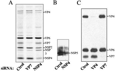
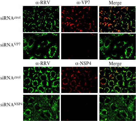
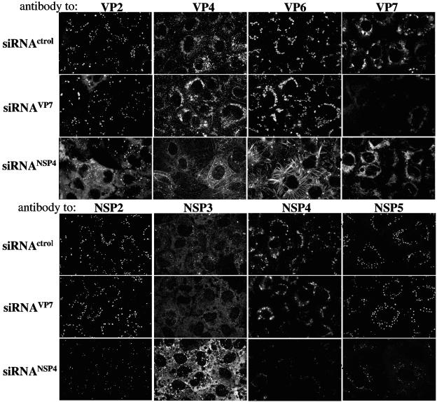
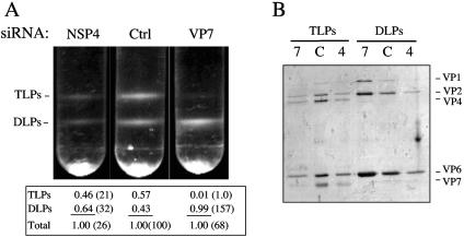
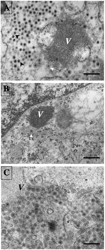
The morphogenesis of rotaviruses follows a unique pathway in which immature double-layered particles (DLPs) assembled in the cytoplasm bud across the membrane of the endoplasmic reticulum (ER), acquiring during this process a transient lipid membrane which is modified with the ER resident viral glycoproteins NSP4 and VP7; these enveloped particles also contain VP4. As the particles move towards the interior of the ER cisternae, the transient lipid membrane and the nonstructural protein NSP4 are lost, while the virus surface proteins VP4 and VP7 rearrange to form the outermost virus protein layer, yielding mature infectious triple-layered particles (TLPs). In this work, we have characterized the role of NSP4 and VP7 in rotavirus morphogenesis by silencing the expression of both glycoproteins through RNA interference. Silencing the expression of either NSP4 or VP7 reduced the yield of viral progeny by 75 to 80%, although the underlying mechanism of this reduction was different in each case. Blocking the synthesis of NSP4 affected the intracellular accumulation and the cellular distribution of several viral proteins, and little or no virus particles (neither DLPs nor TLPs) were assembled. VP7 silencing, in contrast, did not affect the expression or distribution of other viral proteins, but in its absence, enveloped particles accumulated within the lumen of the ER, and no mature infectious virus was produced. Altogether, these results indicate that during a viral infection, NSP4 serves as a receptor for DLPs on the ER membrane and drives the budding of these particles into the ER lumen, while VP7 is required for removing the lipid envelope during the final step of virus morphogenesis.


Figures

FIG. 1.
FIG. 1.
Effect of siRNAVP7 and siRNANSP4 on the synthesis of viral proteins. MA104 cells in 48-well plates were transfected with the indicated siRNA, and after 8 h, the Lipofectamine-siRNA mixture was removed and replaced by MEM. Forty-eight hours posttransfection, the cells were infected with rotavirus RRV at an MOI of 3, and 12 h postinfection, the cells were harvested and processed for SDS-PAGE. (A) Fluorography of cells transfected with the indicated siRNAs in which the cells were labeled with 50 μCi of Easy Tag EXPRESS-35S/ml at 4 hpi and harvested 12 hpi. (B) A lysate of cells transfected with the indicated siRNAs was harvested at 12 hpi and subjected to SDS-PAGE. The proteins were transferred to nitrocellulose membrane and immunostained with a rabbit polyclonal antibody to rotavirus NSP5. (C) Immunoblot analysis of lysates of cells transfected with the indicated siRNAs and harvested at 12 hpi. The proteins were incubated with a rabbit anti-rotavirus polyclonal antiserum. The relative positions of the viral proteins are indicated in each gel. Ctrol, control.
FIG. 2.
FIG. 2.
Effect of siRNAs to NSP4 and VP7 on the expression of the corresponding protein in infected cells. MA104 cells transfected with the indicated siRNAs were infected 48 h posttransfection, and at 8 hpi, the cells were fixed and processed for immunofluorescence. The expression levels of VP7 and NSP4 were monitored by using MAbs 60 (α-VP7) and B4 (α-NSP4), respectively, and the cell infection by RRV was monitored by using a rabbit polyclonal antiserum to rotavirus (α-RRV) followed by anti-mouse Alexa-568 and anti-rabbit Alexa-488 antibodies, respectively.
FIG. 3.
FIG. 3.
Cellular distribution of viral proteins in cells transfected with siRNAVP7 or siRNANSP4. MA104 cells grown on coverslips were transfected with the indicated siRNAs and infected 48 h posttransfection. At 8 hpi, the cells were fixed and processed for immunofluorescence. The distribution of VP2, VP4, VP6, VP7, and NSP4 was monitored by using MAbs 3A8, HS2, 255/60, 60, and B4, respectively. NSP2, NSP3, and NSP5 were stained by using rabbit monospecific antibodies. Anti-mouse Alexa-568 and anti-rabbit Alexa-488 were used as secondary antibodies. siRNActrol, control.
FIG. 4.
FIG. 4.
Virus particles synthesized in the presence of siRNAVP7 or siRNANSP4. (A) Isopycnic CsCl gradients of viral particles assembled in the presence of the indicated siRNAs. The density of each band in the CsCl gradients was determined by scanning digital photographs of the gradients. The numbers below the gradients represent the relative amount of each band with respect to the total amount of viral particles detected in each gradient. The numbers in parentheses indicate the percentage of each band with respect to the amount of the corresponding band found in the control gradient (Ctrl). (B) Gel electrophoresis analysis of the viral particles present in the bands detected in the isopycnic gradients shown in panel A. The same proportion of each collected band was loaded onto the gel, which was silver stained. 4, C, and 7 correspond to the gradients labeled NSP4, Ctrl, and VP7, respectively. The migration of the viral structural proteins in the gels is indicated.
FIG. 5.
FIG. 5.
Morphogenesis of RRV rotavirus particles in MA104 cells transfected with siRNAs. MA104 cells transfected with (A) the siRNA control (siRNActrol), (B) siRNANSP4, or (C) siRNAVP7 were infected 48 h posttransfection, and at 8 hpi, the cells were fixed and prepared for electron microscopy as indicated in Materials and Methods. Dense viroplasmic inclusions (V) are present in the cytoplasm of rotavirus-infected cells, adjacent to the ER. From these structures, DLPs bud into the lumen of the ER, resulting in membrane-enveloped particles (arrows) which later lose the membrane to produce mature triple-layered virions (arrowheads). The pictures shown are representative of at least 20 different virus-infected cells. Magnification, ×15,000. Scale bars, 400 nm.

References

    1. Arias, C. F., M. A. Dector, L. Segovia, T. Lopez, M. Camacho, P. Isa, R. Espinosa, and S. Lopez. 2004. RNA silencing of rotavirus gene expression. Virus Res. 102:43-51. - PMC - PubMed
    1. Arias, C. F., S. Lopez, and R. T. Espejo. 1982. Gene protein products of SA11 simian rotavirus genome. J. Virol. 41:42-50. - PMC - PubMed
    1. Au, K. S., W. K. Chan, J. W. Burns, and M. K. Estes. 1989. Receptor activity of rotavirus nonstructural glycoprotein NS28. J. Virol. 63:4553-4562. - PMC - PubMed
    1. Ball, J. M., P. Tian, C. Q. Zeng, A. P. Morris, and M. K. Estes. 1996. Age-dependent diarrhea induced by a rotaviral nonstructural glycoprotein. Science 272:101-104. - PubMed
    1. Bergmann, C. C., D. Maass, M. S. Poruchynsky, P. H. Atkinson, and A. R. Bellamy. 1989. Topology of the non-structural rotavirus receptor glycoprotein NS28 in the rough endoplasmic reticulum. EMBO J. 8:1695-1703. - PMC - PubMed

Publication types

MeSH terms

LinkOut - more resources