. 2005 Jan;79(1):503-11.
doi: 10.1128/JVI.79.1.503-511.2005.

Severe acute respiratory syndrome coronavirus infection of golden Syrian hamsters

Affiliations

Severe acute respiratory syndrome coronavirus infection of golden Syrian hamsters

Anjeanette Roberts et al. J Virol. 2005 Jan.

Abstract

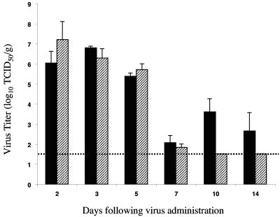
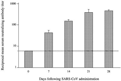
Small animal models are needed in order to evaluate the efficacy of candidate vaccines and antivirals directed against the severe acute respiratory syndrome coronavirus (SARS CoV). We investigated the ability of SARS CoV to infect 5-week-old Golden Syrian hamsters. When administered intranasally, SARS CoV replicates to high titers in the lungs and nasal turbinates. Peak replication in the lower respiratory tract was noted on day 2 postinfection (p.i.) and was cleared by day 7 p.i. Low levels of virus were present in the nasal turbinates of a few hamsters at 14 days p.i. Viral replication in epithelial cells of the respiratory tract was accompanied by cellular necrosis early in infection, followed by an inflammatory response coincident with viral clearance, focal consolidation in pulmonary tissue, and eventual pulmonary tissue repair. Despite high levels of virus replication and associated pathology in the respiratory tract, the hamsters showed no evidence of disease. Neutralizing antibodies were detected in sera at day 7 p.i., and mean titers at day 28 p.i. exceeded 1:400. Hamsters challenged with SARS CoV at day 28 p.i. were completely protected from virus replication and accompanying pathology in the respiratory tract. Comparing these data to the mouse model, SARS CoV replicates to a higher titer and for a longer duration in the respiratory tract of hamsters and is accompanied by significant pathology that is absent in mice. Viremia and extrapulmonary spread of SARS CoV to liver and spleen, which are seen in hamsters, were not detected in mice. The hamster, therefore, is superior to the mouse as a model for the evaluation of antiviral agents and candidate vaccines against SARS CoV replication.


Figures

FIG. 1.
FIG. 1.
Replication of SARS CoV in the upper and lower respiratory tracts of hamsters. Virus titers in NTs (solid bars) and lung homogenates (hatched bars) are the mean values calculated from three hamsters per day. Error bars indicate standard errors. The dotted line designates the lower limit of detection (101.5 TCID50/g of tissue). Hamsters were inoculated with 103TCID50 of SARS CoV on day 0.
FIG. 2.
FIG. 2.
Pathology in NTs and tracheae of hamsters at days 2 and 3 of SARS CoV infection. NTs at day 2 p.i. show the presence of SARS CoV antigens (red) in cells of the epithelial surface lining (arrows in A and B), necrotic debris in the nasal passages (arrowhead in A), and endothelial cells (arrowhead in B). The mononuclear nature of the inflammatory cell infiltrate is seen in B. NTs at day 3 p.i. show mononuclear inflammatory infiltrate in the submucosa and the presence of SARS CoV antigens (red) in cells of the epithelial surface lining and in mucous glands (C). The trachea at day 2 p.i. shows tracheal epithelial cells with loss of the apical portion of the cytoplasm including cilia (arrow in D; compare to arrowhead, where cilia are still present), debris with desquamated epithelial and inflammatory cells in the lumen, and submucosal mononuclear inflammatory infiltrate (D and E). SARS CoV antigens (red) are observed in the epithelial cells of the trachea (E) at 2 days p.i. and are accompanied by mild inflammatory infiltrate. A, B, C, and E, IHC assays; D, hematoxylin-and-eosin stain.
FIG. 3.
FIG. 3.
Pathology in hamster lungs at days 3 and 5 of SARS CoV infection. The lung at 3 days p.i. shows focal pneumonic consolidation around bronchi (asterisk in A). SARS CoV antigens (red) are noted in bronchial epithelium and in consolidated areas of the lung (B), which at higher magnification are seen inside alveolar lining cells (pneumocytes; arrow in C) and possibly in endothelial cells (arrowhead in C). The inflammatory infiltrate at 3 days p.i. is localized to the alveolar septal walls (B and C). The lung at 5 days p.i. shows confluent pneumonic consolidation (D) and decreasing amounts of SARS CoV antigens (red) in both consolidated areas and bronchial epithelium (E). A and D, hematoxylin-and-eosin stain; B, C, and E, IHC assays.
FIG. 4.
FIG. 4.
Pathology in hamster lungs at days 7 and 14 of SARS CoV infection and after challenge. The lung at 7 days p.i. shows a decrease of the inflammatory infiltrate in the alveolar septae and proliferation of reparative cuboidal epithelial cells lining slender air spaces (A). The lung at 14 days p.i. shows normal alveolar air spaces with minimal amounts of thickening of the alveolar septae (B). The lung 3 days after SARS CoV rechallenge shows normal alveolar air spaces and no pneumonic consolidation (C). In contrast, SARS CoV antigens (red) are observed in the bronchial epithelium and in the peribronchial pneumonic consolidations (D) of the lung of a hamster that was mock infected initially and sacrificed 3 days after challenge with SARS CoV, similar to what was seen at day 3 p.i. (Fig. 3A, B, and C). A, B, and C, hematoxylin-and-eosin stain; D, IHC assay.
FIG. 5.
FIG. 5.
Reciprocal SARS CoV-specific serum neutralizing-antibody titers in SARS-infected hamsters on the indicated days following intranasal inoculation. Mean antibody titers from three hamsters per group are shown on a logarithmic scale. Error bars indicate standard errors. The dotted line indicates the lower limit of detection (<1:8).

References

    1. Afzelius, B. A. 1994. Ultrastructure of human nasal epithelium during an episode of coronavirus infection. Virchows Archiv. 424:295-300. - PMC - PubMed
    1. Bukreyev, A., E. W. Lamirande, U. J. Buchholz, L. N. Vogel, W. R. Elkins, M. St. Claire, B. R. Murphy, K. Subbarao, and P. L. Collins. 2004. Mucosal immunisation of African green monkeys (Cercopithecus aethiops) with an attenuated parainfluenza virus expressing the SARS coronavirus spike protein for the prevention of SARS. Lancet 363:2122-2127. - PMC - PubMed
    1. Chong, P. Y., P. Chui, A. E. Ling, T. J. Franks, D. Y. Tai, Y. S. Leo, G. J. Kaw, G. Wansaicheong, K. P. Chan, L. L. Ean Oon, E. S. Teo, K. B. Tan, N. Nakajima, T. Sata, and W. D. Travis. 2004. Analysis of deaths during the severe acute respiratory syndrome (SARS) epidemic in Singapore: challenges in determining a SARS diagnosis. Arch. Pathol. Lab. Med. 128:195-204. - PubMed
    1. Donoghue, M., F. Hsieh, E. Baronas, K. Godbout, M. Gosselin, N. Stagliano, M. Donovan, B. Woolf, K. Robison, R. Jeyaseelan, R. E. Breitbart, and S. Acton. 2000. A novel angiotensin-converting enzyme-related carboxypeptidase (ACE2) converts angiotensin I to angiotensin 1-9. Circulation Res. 87:1-9. - PubMed
    1. Drosten, C., S. Gunther, W. Preiser, S. van der Werf, H. R. Brodt, S. Becker, H. Rabenau, M. Panning, L. Kolesnikova, R. A. Fouchier, A. Berger, A. M. Burguiere, J. Cinatl, M. Eickmann, N. Escriou, K. Grywna, S. Kramme, J. C. Manuguerra, S. Muller, V. Rickerts, M. Sturmer, S. Vieth, H. D. Klenk, A. D. Osterhaus, H. Schmitz, and H. W. Doerr. 2003. Identification of a novel coronavirus in patients with severe acute respiratory syndrome. N. Engl. J. Med. 348:1967-1976. - PubMed

Publication types

LinkOut - more resources