. 2005 Jan;79(1):554-68.
doi: 10.1128/JVI.79.1.554-568.2005.

Evolutionary trace residues in noroviruses: importance in receptor binding, antigenicity, virion assembly, and strain diversity

Affiliations

Evolutionary trace residues in noroviruses: importance in receptor binding, antigenicity, virion assembly, and strain diversity

Sugoto Chakravarty et al. J Virol. 2005 Jan.

Abstract

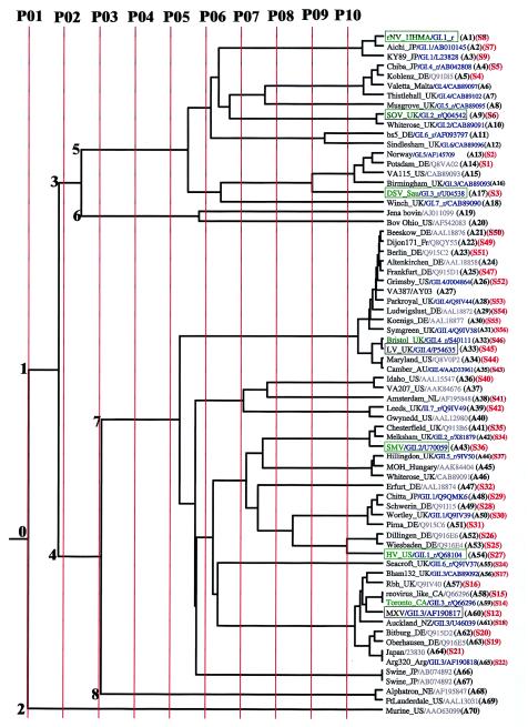
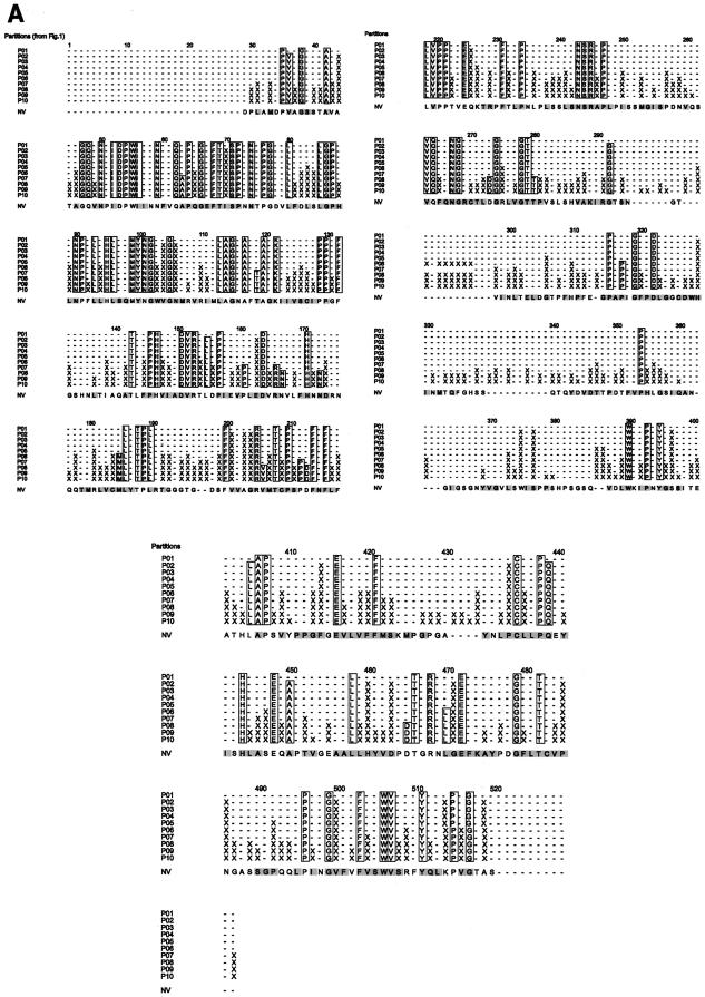
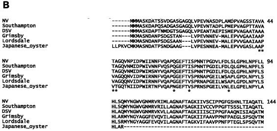
Noroviruses cause major epidemic gastroenteritis in humans. A large number of strains of these single-stranded RNA viruses have been reported. Due to the absence of infectious clones of noroviruses and the high sequence variability in their capsids, it has not been possible to identify functionally important residues in these capsids. Consequently, norovirus strain diversity is not understood on the basis of capsid functions, and the development of therapeutic compounds has been hampered. To determine functionally important residues in noroviruses, we have analyzed a number of norovirus capsid sequences in the context of the Norwalk virus capsid crystal structure by using the evolutionary trace method. This analysis has identified capsid protein residues that uniquely characterize different norovirus strains and provide new insights into capsid assembly and disassembly pathways and the strain diversity of these viruses. Such residues form specific three-dimensional clusters that may be of functional importance in noroviruses. One of these clusters includes residues known to participate in the proteolytic cleavage of these viruses at high pH. Other clusters are formed in capsid regions known to be important in the binding of antibodies to noroviruses, thereby indicating residues that may be important in the antigenicity of these viruses. The highly variable region of the capsid shows a distinct cluster whose residues may participate in norovirus-receptor interactions.


Figures

FIG.1.
FIG.1.
The ET of the norovirus strains (A1 to A70) belonging to the two genogroups for the different partitions. A few randomly chosen duplicate sequences (A58/A59, A64/A65, and A66/A67) as positive controls in the sequence alignments and the tree construction appear correctly on the tree. The partition lines P01 to P10 shown in red indicate the extent of divergence (resolution) of the norovirus sequences. A subset, whose sequence numbers (S1 to S56) are shown in red, has been used for ET trace residue analysis. Sequences of known clusters in genogroups I and II are shown in blue as GI.n or GII.n, respectively, along with the appended “_r” if the sequence is a reference strain for the cluster (10). The EMBL and NCBI database accession numbers are shown following the strain names and cluster numbers if applicable. The strain names reflect the names of the places or regions where the strains were first isolated. The name of the country is shown as a two-letter code (except where the name is obvious) along with the strain name. These codes are as follows: AU, Australia; CA, Canada; DE, Germany; Fr, France; JP, Japan; NL, Netherlands; NZ, New Zealand; Sau, Saudi Arabia; UK, United Kingdom; US, United States. The prototype strains refer to the first strain that was isolated within a given cluster (10). The antigenic strains are shown within boxes, and the prototype strains are shown in green. Antigenic norovirus strains that are also prototypic are shown in green and are enclosed within boxes. The numbers (0 to 8) refer to the different nodes of the tree. Node 0 is the root node that is the parent node of child nodes 1 and 2. Similarly, node 1 is the parent node of child nodes 3 and 4, node 3 is the parent node of child nodes 5 and 6, and node 4 is the parent node of the child nodes 7 and 8.
FIG. 2.
FIG. 2.
(A) Locations of S and P domain trace residues for partitions P01 to P10. The trace residues belonging to a given partition occur in the horizontal row corresponding to the partition. Residues 1 to 218 and residues 219 to 401 are separated from each other by the partition numbers and are shown in the upper part of the figure, while residues 402 to 530 are shown in the lower part of the figure. The ACRs are shown as one-letter amino acid codes within boxes outlined in black, and the evolutionary class-specific residues are shown by the symbol X. The minus (−) signs denote nonconserved neutral residues. Residues are numbered according to the NV residues that are shown at the bottom of each group. (B) ClustalW alignment of partial norovirus sequence from Japanese oyster (NCBI accession no. BAC98461) with five other norovirus strains. Residues are numbered according to NV sequence. The asterisk denotes the class-specific locations that have been compared in the text.
FIG. 2.
FIG. 2.
(A) Locations of S and P domain trace residues for partitions P01 to P10. The trace residues belonging to a given partition occur in the horizontal row corresponding to the partition. Residues 1 to 218 and residues 219 to 401 are separated from each other by the partition numbers and are shown in the upper part of the figure, while residues 402 to 530 are shown in the lower part of the figure. The ACRs are shown as one-letter amino acid codes within boxes outlined in black, and the evolutionary class-specific residues are shown by the symbol X. The minus (−) signs denote nonconserved neutral residues. Residues are numbered according to the NV residues that are shown at the bottom of each group. (B) ClustalW alignment of partial norovirus sequence from Japanese oyster (NCBI accession no. BAC98461) with five other norovirus strains. Residues are numbered according to NV sequence. The asterisk denotes the class-specific locations that have been compared in the text.
FIG.3.
FIG.3.
(A) Interactions of the S domains of the NV subunits across the different icosahedral symmetry axes. The locations of these axes are shown by the corresponding numbers. Subunits A, B, and C are related by a quasi-threefold (shown by a triangle) and C and C2 are related by an icosahedral twofold axis. Subunits A and B5 are related by quasi-twofold axes, and C is related to B5 and B2 by quasi-sixfold symmetry. Subunits A and A5 are related by a strict fivefold axis. The broken lines joining the icosahedral symmetry axes are the icosahedral interfaces indicated by arrows. The line (5-3) joining the fivefold and the threefold axes includes the A-B5 (dimeric), A-A5 (pentameric), and the C-B5 (hexameric) interface regions while the line (3-3) joining the two threefold axes, includes the C-B2 (hexameric) interface region. Similarly, the quasi-trimeric A-B, A-C, and the B-C interface regions lie on the boundaries between the A, B, and the C subunits. (B) NV conserved residues with reference to the secondary structure and icosahedral interfaces. Filled green arrow, β-strand; filled green cylinder, helix. The ACRs are shown in red along with their locations with respect to the icosahedral symmetry axes that are shown, using different symbols: oval, twofold axis; inverted triangle, threefold axis; hexagon, quasi-sixfold axis; pentagon, fivefold axis; and teardrop, quasi-threefold axis. A black border around the symmetry axes symbols indicates a buried residue, while no border around the symbol indicates an exposed residue. Multiple symbols on ACRs indicate the presence of multiple symmetry axes about which these residue(s) interact with other subunits.
FIG. 4.
FIG. 4.
Space-filled representation of the proposed functionally important class-specific residues for the P06 partitions of the phylogenetic tree. The buried atoms of the class-specific residues are shown in blue and the exposed atoms of these residues are cyan. (A) The S domain surface patches 1 to 5 are outlined in black. The red box indicates that the S domain surface patches 2 and 4 may be considered as one single such patch. (B) The different class-specific surface patches CS-2, CS-4, and CS-5 of Table 4 are outlined in red. The surface CS-1 is outlined in black. Another possible class-specific surface consisting of the residues A402 and S447 lies near the CS-1surface. The red outline enclosing the two class-specific patches outlined in black indicates that these patches may be considered as a single patch. The purple outline enclosing the S domain patch number 1 and the P domain CS-1 patch indicates that these patches may be considered as one single patch or a pair of possible neutralizing and nonneutralizing epitope sites. The direction of the icosahedral twofold axis relative to the capsid subunit is also shown. (C) A 180° rotated view of the figure in panel B. This figure indicates the surface patch CS-3 that is not visible in panel B. The surface patch CS-2 is also shown to indicate the relative location of the CS-3 patch.

References

    1. Altschuh, D., A. M. Lesk, A. C. Bloomer, and A. Klug. 1987. Correlation of co-ordinated amino acid substitutions with function in viruses related to tobacco mosaic virus. J. Mol. Biol. 193:693-707. - PubMed
    1. Ando, T., J. S. Noel, and R. L. Fankhauser. 2000. Genetic classification of Norwalk-like viruses. J. Infect. Dis. 181(Suppl. 2):S336-S348. - PubMed
    1. Atchley, W. R., K. R. Wollenberg, W. M. Fitch, W. Terhalle, and A. W. Dress. 2000. Correlations among amino acid sites in bHLH protein domains: an information theoretic analysis. Mol. Biol. Evol. 17:164-178. - PubMed
    1. Bertolotti-Ciarlet, A., L. J. White, R. Chen, B. V. Prasad, and M. K. Estes. 2002. Structural requirements for the assembly of Norwalk virus-like particles. J. Virol. 76:4044-4055. - PMC - PubMed
    1. Blakeney, S. J., A. Cahill, and P. A. Reilly. 2003. Processing of Norwalk virus nonstructural proteins by a 3C-like cysteine proteinase. Virology 308:216-224. - PubMed

Publication types