. 2004 Dec 28;101(52):18200-5.
doi: 10.1073/pnas.0405971102. Epub 2004 Dec 14.

Neurofibromatosis 2 (NF2) tumor suppressor merlin inhibits phosphatidylinositol 3-kinase through binding to PIKE-L

Affiliations

Neurofibromatosis 2 (NF2) tumor suppressor merlin inhibits phosphatidylinositol 3-kinase through binding to PIKE-L

Rong Rong et al. Proc Natl Acad Sci U S A. .

Abstract

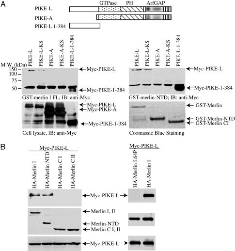
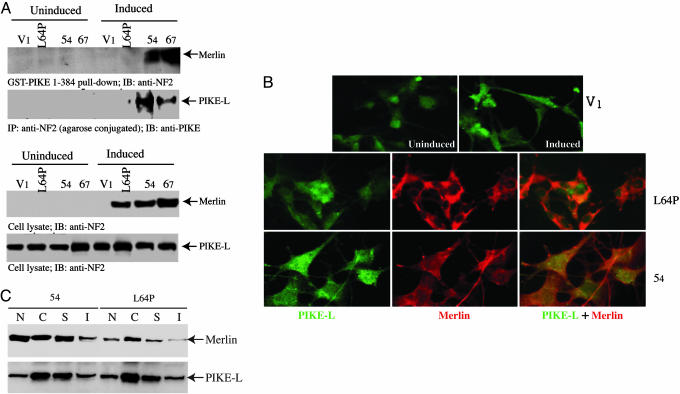
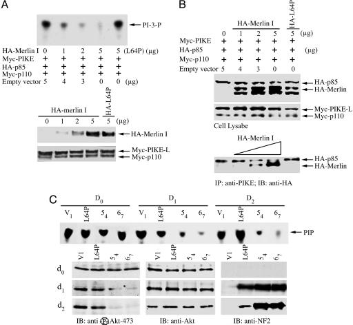
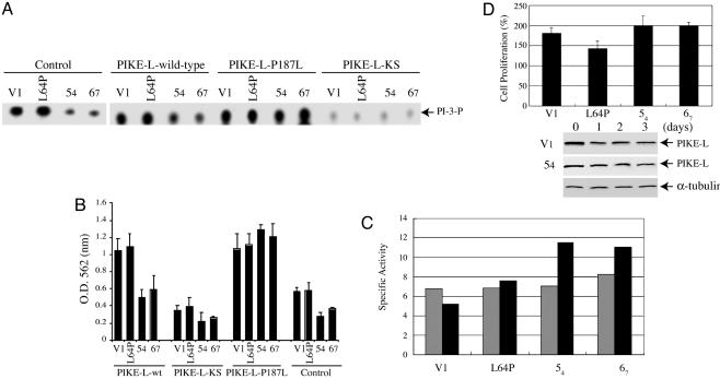
Neurofibromatosis 2 (NF2) is a tumor suppressor, although the molecular mechanism accounting for this effect remains unknown. Here, we show that merlin exerts its activity by inhibiting phosphatidylinositol 3-kinase (PI3-kinase), through binding to PIKE-L. Wild-type merlin, but not patient-derived mutant (L64P), binds PIKE-L and inhibits PI3-kinase activity. This suppression of PI3-kinase activity results from merlin disrupting the binding of PIKE-L to PI3-kinase. In addition, merlin suppression of PI3-kinase activity as well as schwannoma cell growth is abrogated by a single PIKE-L point mutation (P187L) that cannot bind merlin but can still activate PI3-kinase. Knocking down PIKE-L with RNA interference abolishes merlin's tumor-suppressive activity. Our data support the hypothesis that PIKE-L is an important mediator of merlin growth suppression.


Figures

Fig. 1.
Fig. 1.
PIKE-L interacts with merlin. (A) The FERM domain of merlin binds to the N terminus of PIKE-L in vitro. (Top) Illustration of the PIKE constructs used. GST-merlin and GST-FERM domain of merlin bind to PIKE-L but not PIKE-A. (Middle) Purified GST-merlin and GST-merlin-NTD were incubated with HEK293 cell lysates transfected with various myc-tagged PIKE constructs. After 3-h incubation at 4°C, the associated proteins were analyzed by Western blotting with anti-myc antibody. PIKE-L and PIKE-L N terminus (amino acid residues1–384) robustly binds to both merlin and merlin-NTD. The dominant-negative PIKE-L-KS displayed reduced binding activity to merlin. (Left Bottom) Protein expression of transfected constructs was confirmed by myc immunoblotting. (Right Bottom) Levels of GST-merlin recombinant proteins were verified by Coomassie blue staining. (B) PIKE-L interacts with merlin in vivo. Various HA-merlin constructs and myc-PIKE-L were cotransfected into HEK293 cells. PIKE-L was immunoprecipitated with anti-myc antibody, and bound proteins were visualized by Western blot with anti-HA antibody. Both full-length merlin and merlin NTD interacted with PIKE-L (Left Top, lanes 1 and 2). Patient-derived L64P mutant did not bind to PIKE-L (Right Top). Similar levels of all HA-merlin and myc-PIKE constructs were expressed in all experiments (Middle and Bottom).
Fig. 2.
Fig. 2.
PIKE-L interacts with WT merlin in schwannoma cells. (A) Rat RT4-D6P2T schwannoma cells stably transfected with empty vector (V1), merlin mutant L64P, or WT merlin (54 and 67, two clones) were either uninduced or induced with doxycycline for 1 day. One milligram of cell lysate was incubated with purified GST-PIKE-L-1–384. After 3-h incubation at 4°C, the associated proteins were analyzed by Western blotting with anti-NF2. (Top Upper) WT merlin, but not merlin mutant L64P, selectively binds to the N terminus of PIKE-L. (Top Lower) Coimmunoprecipitation with anti-NF2 reveals that PIKE-L specifically associates with WT merlin but not L64P mutant. (Bottom) The induced merlin was verified with anti-NF2 antibody. (B) PIKE-L colocalizes with WT merlin in schwannoma cells. Uninduced and induced schwannoma cells were stained with mouse anti-PIKE antibody and rabbit polyclonal anti-merlin antibody. (Top) PIKE-L resides in both the cytoplasm and the nucleus. (Middle and Bottom) Merlin L64P exclusively distributes in the cytoplasm, whereas WT merlin occurs in both compartments, colocalizing with PIKE-L in 54 cell line. (C) Subcellular fractionation of 54 and L64P cells. The cytosolic (C), membrane soluble (S), membrane insoluble (I), and nuclear (N) fractions were prepared. Immunoblotting analysis was performed with anti-PIKE and anti-merlin antibodies. (Upper) Both WT and mutant merlin display similar distribution patterns in C, S, and I fractions; by contrast, a very faint amount of L64P was observed in the nucleus compared to counterpart in the WT cells. (Lower) PIKE-L reveals identical subcellular distribution.
Fig. 3.
Fig. 3.
merlin blocks the stimulatory effect of PIKE-L on PI3-kinase. (A) TLC was used to assay PI3-kinase activity. HEK293 cells were transfected with the indicated expression constructs. PI3-kinase was immunoprecipitated by anti-p110 antibody and assayed for in vitro lipid kinase activity. (Top) Increasing amount of merlin progressively diminished PI3-kinase activity, but L64P failed to do so. (Middle) Gradually increasing levels of HA-merlin was expressed in transfected cells. (Bottom) Equal amount of Myc-p110 and Myc-PIKE-L was confirmed by anti-myc immunoblotting. (B) merlin competes with PI3-kinase for binding to PIKE-L. (Bottom) Myc-PIKE-L was immunoprecipitated by anti-PIKE antibody, and the coprecipitated HA-p85 and HA-merlin were visualized by Western blot with anti-HA antibody. (Middle) Equal amounts of Myc-p110 and PIKE-L were confirmed with anti-Myc antibody. (Top) Increased expression of merlin was confirmed. (C) Merlin mediates PI3-kinase in schwannoma cells. TLC was used to assay PI3-kinase activity. Rat RT4-D6P2T schwannoma cells stably transfected with empty vector, merlin mutant L64P, or WT merlin (54 or 67) were uninduced or induced with doxycycline for 0, 1, or 2 days. (First blot from top) PI3-kinase activity is abolished by induced WT merlin, but not merlin mutant L64P. Akt and its phosphorylation in the lysate of schwannoma cells were analyzed. (Left, lower three blots) WT, but not patient-derived L64P, merlin blocks Akt phosphorylation. (Center and Right, lower three blots) The expression of Akt and induced merlin was verified.
Fig. 4.
Fig. 4.
merlin suppresses PI3-kinase activity through PIKE-L. (A) TLC was used to assay PI3-kinase activity in rat RT4-D6P2T schwannoma cells, which were infected with control adenovirus or adenovirus expressing WT PIKE-L, PIKE-L-KS, or PIKE-L-P187L. Infection with WT PIKE-L or PIKE-L-P187L resulted in an increase in PI3-kinase activity compared with control infected. This effect was suppressed in the merlin-expressing clones 54 and 67. PI3-kinase activity was unaffected in merlin clones infected with PIKE-L-P187L or merlin mutant L64P infected with either WT or P187L PIKE-L, presumably because of the inability of merlin or PIKE-L to cointeract. As expected, the dominant-negative mutant, PIKE-L-KS, inhibited PI3-kinase. (B) The number of proliferating rat RT4-D6P2T schwannoma cells was measured by MTT assay. Cellular proliferation paralleled PI3-kinase activity. (C) Caspase-3 activity assay. Empty vector (V1), merlin mutant L64P, and WT merlin (54 and 67) cells were induced for 3 days. Caspase-3 activity assay was conducted with 30 μg of protein from day 0 (gray bar) and 3 (black bar) samples. Induction of WT merlin substantially increases apoptosis compared with control or L64P mutant. (D) Knocking down PIKE-L abolishes the tumor-suppressive activity of merlin. The cells was induced and infected with adenovirus expressing sh-PIKE RNA. The expression of PIKE-L in both L64P and 54 cells was decreased substantially upon infection of PIKE RNA interference (blots). Surprisingly, knocking down PIKE-L robustly increases cell proliferation in 54 and 67 cells compared with control and L64P cells (graph). The results were expressed as means ± SEM calculated from five times of determination (P < 0.05).

References

    1. Eldridge, R. (1981) Adv. Neurol. 29, 57–65. - PubMed
    1. Rouleau, G. A., Merel, P., Lutchman, M., Sanson, M., Zucman, J., Marineau, C., Hoang-Xuan, K., Demczuk, S., Desmaze, C., Plougastel, B., et al. (1993) Nature 363, 515–521. - PubMed
    1. Trofatter, J. A., MacCollin, M. M., Rutter, J. L., Murrell, J. R., Duyao, M. P., Parry, D. M., Eldridge, R., Kley, N., Menon, A. G., Pulaski, K., et al. (1993) Cell 72, 791–800. - PubMed
    1. Gutmann, D. H. (1997) Neurobiol. Dis. 3, 247–261. - PubMed
    1. Gonzalez-Agosti, C., Xu, L., Pinney, D., Beauchamp, R., Hobbs, W., Gusella, J. & Ramesh, V. (1996) Oncogene 13, 1239–1247. - PubMed

Publication types

MeSH terms