. 2005 Jan;17(1):103-15.
doi: 10.1105/tpc.104.028357. Epub 2004 Dec 14.

The Rice COLEOPTILE PHOTOTROPISM1 gene encoding an ortholog of Arabidopsis NPH3 is required for phototropism of coleoptiles and lateral translocation of auxin

Affiliations

The Rice COLEOPTILE PHOTOTROPISM1 gene encoding an ortholog of Arabidopsis NPH3 is required for phototropism of coleoptiles and lateral translocation of auxin

Ken Haga et al. Plant Cell. 2005 Jan.

Abstract

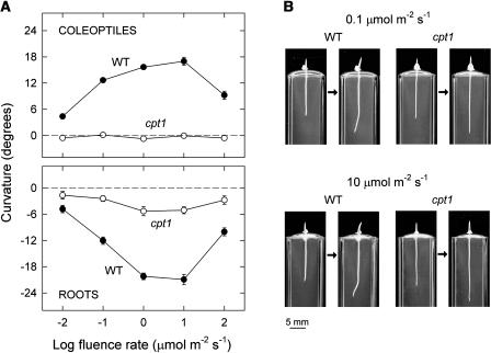
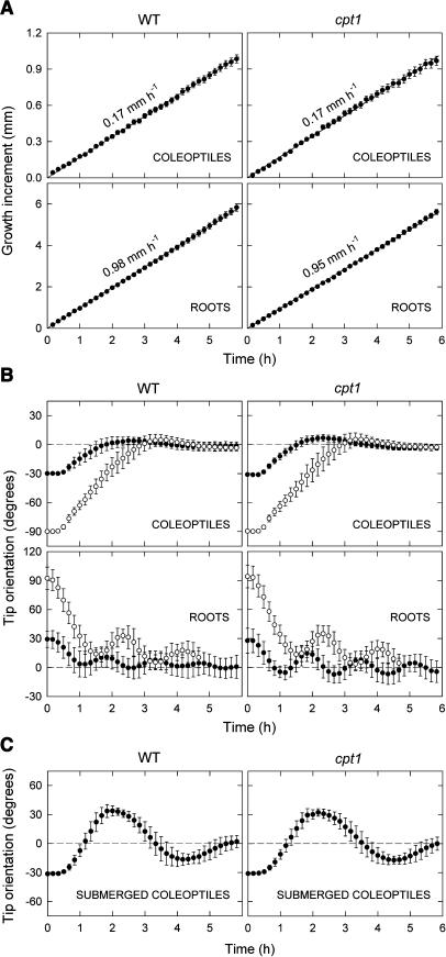
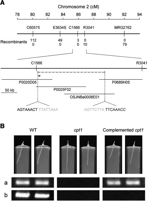
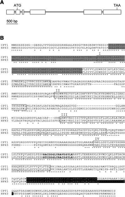
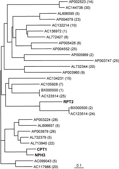
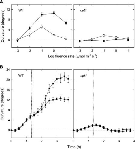
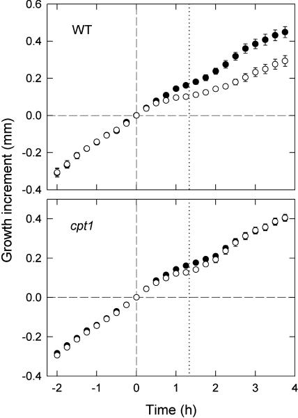
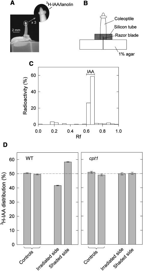
We isolated a mutant, named coleoptile phototropism1 (cpt1), from gamma-ray-mutagenized japonica-type rice (Oryza sativa). This mutant showed no coleoptile phototropism and severely reduced root phototropism after continuous stimulation. A map-based cloning strategy and transgenic complementation test were applied to demonstrate that a NPH3-like gene deleted in the mutant corresponds to CPT1. Phylogenetic analysis of putative CPT1 homologs of rice and related proteins indicated that CPT1 has an orthologous relationship with Arabidopsis thaliana NPH3. These results, along with those for Arabidopsis, demonstrate that NPH3/CPT1 is a key signal transduction component of higher plant phototropism. In an extended study with the cpt1 mutant, it was found that phototropic differential growth is accompanied by a CPT1-independent inhibition of net growth. Kinetic investigation further indicated that a small phototropism occurs in cpt1 coleoptiles. This response, induced only transiently, was thought to be caused by the CPT1-independent growth inhibition. The 3H-indole-3-acetic acid applied to the coleoptile tip was asymmetrically distributed between the two sides of phototropically responding coleoptiles. However, no asymmetry was induced in cpt1 coleoptiles, indicating that lateral translocation of auxin occurs downstream of CPT1. It is concluded that the CPT1-dependent major phototropism of coleoptiles is achieved by lateral auxin translocation and subsequent growth redistribution.


Figures

Figure 1.
Figure 1.
Impaired Phototropisms of cpt1 Coleoptiles and Roots. (A) Phototropic fluence rate-response curves. Seedlings of the cpt1 mutant (open circles) and the wild type Nihonmasari (closed circles), grown for 3 d under red light (2.5 to 3 μmol m−2 s−1), were stimulated continuously with unilateral blue light at the indicated fluence rates. The length of coleoptiles was in the range between 3.5 and 4.5 mm at the onset of blue light. Phototropic curvatures of coleoptiles (top panel) and primary roots (bottom panel) were determined at 6 h of stimulation. The means obtained from 30 to 50 seedlings are shown. The vertical bar indicates ±se. (B) Photographs illustrating impaired phototropisms of cpt1 coleoptiles and roots. Wild-type and cpt1 seedlings were stimulated with unilateral blue light at a fluence rate of 0.1 μmol m−2 s−1 (top panels) or 10 μmol m−2 s−1 (bottom panels). Each pair of photographs was taken just before and after 6-h blue light stimulation. Other conditions were as described for (A).
Figure 2.
Figure 2.
Growth and Gravitropism of cpt1 Seedlings. (A) Growth of coleoptiles and primary roots. Seedlings of the wild type and cpt1 mutant, raised under red light (2.5 μmol m−2 s−1), were monitored for elongation growth of coleoptiles and primary roots. The growth increment (ordinate) indicates the difference in length from the length at time 0. The means ± se from 15 seedlings are shown. Growth rates estimated from the linear regression lines are indicated. The mean initial lengths in millimeters were 3.2 (wild-type coleoptiles), 3.4 (cpt1 coleoptiles), 15.8 (wild-type roots), and 16.3 (cpt1 roots). (B) Gravitropism of coleoptiles and primary roots. Seedlings of the wild type and the cpt1 mutant, prepared as described for Figure 1A, were displaced by 30° (closed circles) or 90° (open circles) and were monitored for the orientation of the coleoptile tip (top panels) and the root tip (bottom panels). The angle of orientation is 0° when it is parallel to the plumb line. In the case of coleoptiles, the orientation to the side of displacement is indicated by negative angles, and the upward gravitropic curvature of coleoptiles is represented by a movement of the tip from a negative angle to 0°. In the case of roots, the orientation to the side of displacement is indicated by positive angles, and the downward gravitropic curvature of roots is represented by a movement of the tip from a positive angle to 0°. The means obtained from 30 (closed circles) or 20 (open circles) seedlings are shown. The vertical bar indicates ±sd. (C) Gravitropism of submerged coleoptiles. Seedlings were submerged in water 6 h before they were displaced by 30°. The length of coleoptiles was ∼3 mm at the time of submergence. The means obtained from 12 seedlings are shown. Other details were as described for (B).
Figure 3.
Figure 3.
Identification of the CPT1 Gene. (A) Map-based cloning. The CPT1 locus was mapped to chromosome 2. C60575 and MRG2762 are STS markers. E3634S, C1566, and R3041 are RFLP markers. Numbers of recombinants between cpt1 and each marker are shown. P0020D05, P0025F02, and P0689H05 are PAC clones, and OSJNBa0008E01 is a BAC clone. The cpt1 mutant was found to have a deletion of ∼209 kb (dashed line) between C1566 and R3041 markers that enclosed the CPT1 locus. Sequences around the two edges of the deletion are indicated (gray letters represent deletion). Within the deletion, a NPH3-like gene (indicated by a horizontal arrow; 4.5 kb) was predicted. cM, centimorgan. (B) Transgenic complementation of the cpt1 mutant. A genomic clone (8.2 kb) containing the NPH3-like gene was introduced into the cpt1 mutant by Agrobacterium-mediated transformation. The photographs show 3-d-old seedlings of the wild type, cpt1 mutant, and complemented cpt1 mutant (T3 generation) grown under irradiation with unilateral white light (3 μmol m−2 s−1; from the right side of the photographs) and overhead red light (3 μmol m−2 s−1). The two complemented cpt1 seedlings originated from independent transformants. Bottom panels show ethidium bromide–stained PCR products obtained for the photographed plants. (a) A 428-bp fragment of the NPH3-like gene was amplified with primers specific to this gene (5′-TTGCAGTGCATAGCCAGTAC-3′ and 5′-TTTCCACGTACTTCTCGTCC-3′). (b) A 502-bp genomic fragment located in the deleted region and at a close distance from the NPH3-like gene was amplified (primers 5′-CGTTGTACGACTGGATGGAC-3′ and 5′-CACTCAACCTTCCGCTTCTC-3′).
Figure 4.
Figure 4.
Structure of CPT1 and Amino Acid Sequence of CPT1. (A) Structure of the genomic CPT1 gene. Exons (boxes) and introns (lines) were determined by a comparison of the genomic and cDNA sequences. The positions of the start and stop codons are indicated. (B) The deduced amino acid sequence of CPT1, shown in comparison with the sequence of NPH3. Asterisks denote residues identical in the two sequences. Gray and black shadings indicate BTB/POZ and coiled-coil domains, respectively. The four regions, I to IV, conserved in the NPH3 family of Arabidopsis and the corresponding regions of CPT1 are boxed. The Tyr phosphorylation site conserved in the NPH3 family of Arabidopsis and the corresponding site of CPT1 are indicated in bold letters.
Figure 5.
Figure 5.
Phylogenetic Tree of Rice CPT1 Homologs and the Two Related Arabidopsis Homologs NPH3 and RPT2. The tree was obtained by the neighbor-joining method using 1000 bootstrap replicates. Homologs of CPT1 were identified in the japonica rice genomic database. Each predicted homolog is indicated by the accession number of a BAC or PAC clone and the number of the predicted gene in the clone (given in parentheses). The accession numbers of CPT1, NPH3, and RPT2 are AB186127, AF180390, and AF181683, respectively. The scale represents 0.1 substitutions per site.
Figure 6.
Figure 6.
Kinetic Investigation of Phototropic Curvature in Wild-Type and cpt1 Coleoptiles. (A) Phototropic fluence rate-response curves. Wild-type and cpt1 seedlings, prepared as described for Figure 1A, were stimulated with unilateral blue light for 80 min at indicated fluence rates. The curvature was determined immediately (open circles) and 80 min (closed circles) after the end of blue light irradiation. (B) Time courses of phototropism. Wild-type and cpt1 seedlings were stimulated with unilateral blue light for 80 min (until the time indicated by the vertical dotted line; circles) or continuously (squares) at 0.3 μmol m−2 s−1. The curvature of coleoptiles was monitored after the onset of blue light. The means ± se from 10 (circles) or four (squares) seedlings are shown.
Figure 7.
Figure 7.
Phototropic Differential Growth in Wild-Type and cpt1 Coleoptiles. Coleoptiles were stimulated with unilateral blue light for 80 min (from time 0 to the time indicated by the vertical dotted line) at 0.3 μmol m−2 s−1. The lengths on irradiated (open circles) and shaded (closed circles) sides of the coleoptile were monitored before and after the onset of blue light. The growth increment (ordinate) indicates the difference in coleoptile length from the length at time 0. The means ± se from 10 seedlings are shown.
Figure 8.
Figure 8.
Phototropic Lateral Translocation of IAA in Wild-Type and cpt1 Coleoptiles. The experiment was conducted using wild-type and cpt1 seedlings prepared as described for Figure 1A. (A) Photographs illustrating the method of 3H-IAA application. Lanolin containing 3H-IAA was applied to the tip region in the form of a ring (∼0.3 mm wide, located ∼0.3 mm below the tip). Two hours after 3H-IAA application, the seedling was irradiated with unilateral blue light as described for Figure 6B. The photograph indicates the side view of a coleoptile to which 3H-IAA was applied. Unilateral blue light was given in the direction perpendicular to this view. (B) A diagram illustrating the method of recovering diffusible substances from the basal cut surface of 3H-IAA–treated coleoptiles. Immediately after 80-min blue light irradiation, the coleoptile was excised and placed on a pair of agar blocks. A razor blade that separated the two agar blocks also divided the base (∼0.5 mm) of the excised coleoptile into irradiated and shaded halves. A ring of silicon tube served as a guide for placing the coleoptile to the right position. After 40-min incubation, the coleoptile was removed from the assembly, and the radioactivity recovered in the agar blocks was determined. (C) Identification of the recovered 3H-IAA. The diffusible substances collected as described above (but without blue light irradiation) were separated by thin layer chromatography. The radioactivity in each zone is shown as a percent of the total radioactivity recovered. The position of authentic IAA is shown. Rf, rate of flow. (D) Distribution of the recovered radioactivity in irradiated and shaded halves of wild-type and cpt1 coleoptiles. The amounts of radioactivity obtained from irradiated and shaded halves were expressed as percentages of the sum from the two halves. Controls were obtained similarly from two halves without blue light irradiation. The means ± se from three (wild type) or four (cpt1) independent experiments (a set of samples per experiment, 12 coleoptiles per sample) are shown.

Similar articles

Cited by

References

    1. Baba, T., et al. (2000). Construction and characterization of rice genomic libraries, PAC library of japonica variety Nipponbare, and BAC library of indica variety Kasalath. Bull. Natl. Inst. Agrobiol. Resour. 14, 41–49.
    1. Biswas, K.K., and Iino, M. (2002). A mutant of rice impaired specifically in the phototropism of coleoptiles. J. Plant Res. 115 (suppl.), 71. - PubMed
    1. Biswas, K.K., Neumann, R., Haga, K., Yatoh, O., and Iino, M. (2003). Photomorphogenesis of rice seedlings: A mutant impaired in phytochrome-mediated inhibition of coleoptile growth. Plant Cell Physiol. 44, 242–254. - PubMed
    1. Blakeslee, J.J., Bandyopadhyay, A., Peer, W.A., Makam, S.N., and Murphy, A.S. (2004). Relocalization of the PIN1 auxin efflux facilitator plays a role in phototropic responses. Plant Physiol. 134, 28–31. - PMC - PubMed
    1. Briggs, W.R., et al. (2001). The phototropin family of photoreceptors. Plant Cell 13, 993–997. - PMC - PubMed

Publication types

MeSH terms

Associated data

LinkOut - more resources