. 2005 Jan 1;19(1):138-51.
doi: 10.1101/gad.1246005. Epub 2004 Dec 14.

Targeted deletion of numb and numblike in sensory neurons reveals their essential functions in axon arborization

Affiliations

Targeted deletion of numb and numblike in sensory neurons reveals their essential functions in axon arborization

Eric J Huang et al. Genes Dev. .

Abstract

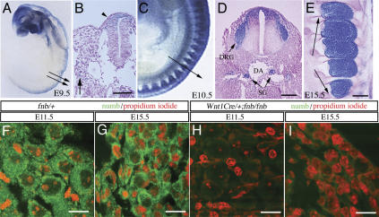
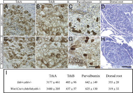
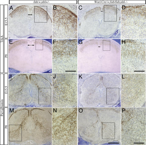
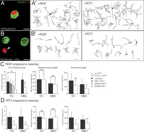
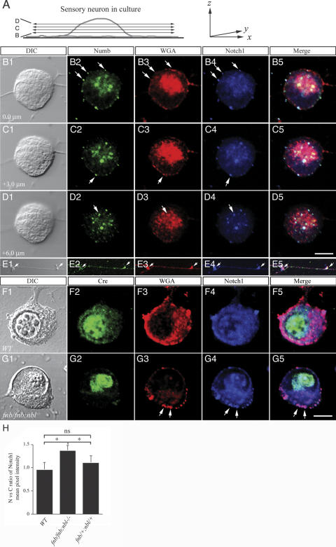
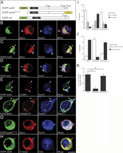
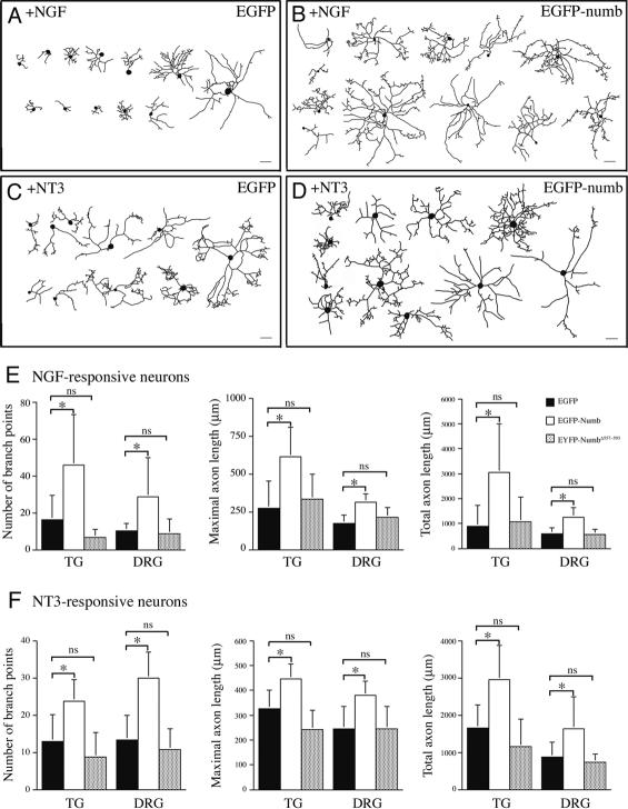
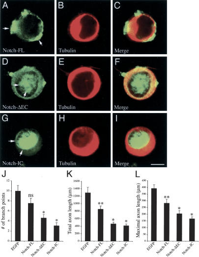
Mouse Numb homologs antagonize Notch1 signaling pathways through largely unknown mechanisms. Here we demonstrate that conditional mouse mutants with deletion of numb and numblike in developing sensory ganglia show a severe reduction in axonal arborization in afferent fibers, but no deficit in neurogenesis. Consistent with these results, expression of Cre recombinase in sensory neurons from numb conditional mutants results in reduced endocytosis, a significant increase in nuclear Notch1, and severe reductions in axon branch points and total axon length. Conversely, overexpression of Numb, but not mutant Numb lacking alpha-adaptin-interacting domain, leads to accumulation of Notch1 in markedly enlarged endocytic-lysosomal vesicles, reduced nuclear Notch1, and dramatic increases in axonal length and branch points. Taken together, our data provide evidence for previously unidentified functions of Numb and Numblike in sensory axon arborization by regulating Notch1 via the endocytic-lysosomal pathways.


Figures

Figure 1.
Figure 1.
Conditional deletion of numb in the developing sensory ganglia. (AE) The recombination pattern of Wnt1Cre in trunk neural crest and developing sensory ganglia was examined by crossing Wnt1Cre transgenic mice with ROSA26 mice. The regions where recombination occurred showed intense lacZ staining. (A,B) Wnt1Cre recombination pattern at E9.5 was revealed in whole-mount staining (A) and tissue sections at the trunk level (indicated by arrows in A). (B,C) lacZ+ cell in the dorsal spinal cord (arrowhead in B) and in scattered cells in the migratory paths of trunk neural crest (arrow). A few lacZ+ cells are identified around dorsal aorta (DA) at the presumptive site for future sympathetic ganglion (arrow in B). (C) Whole-mount lacZ staining in E10.5 embryo showed intense lacZ+ cells in the coalesced DRG. (D) LacZ+ cells were also present in the sympathetic ganglia (SG) adjacent to the DA. (E) At E15.5, essentially every cell within the dorsal root ganglion is positive for lacZ. (FI) No detectable amount of Numb protein was identified in the sensory ganglia of Wnt1Cre/+;fnb/fnb at E11.5 (H) and E15.5 (I). Bars: B,D,E, 50 μm; FI, 20 μm.
Figure 2.
Figure 2.
Neuronal differentiation in the sensory ganglia is not affected in Wnt1Cre/+;fnb/fnb;nbl–/– mice. Differentiation of TrkA, TrkB, and parvalbumin-positive neurons in the DRG of control (AC) and Wnt1Cre/+;fnb/fnb;nbl–/– (EG) mice at P0. (I) The total numbers of TrkA+ neurons were determined at the level of L1, TrkB+ neurons were determined at L2, and parvalbumin+ neurons were determined at the level of L3. (D,H,I) The number of sensory afferent axons in the dorsal roots of control (D) and conditional mutant (H) mice was not significantly different at the thoracic level. At least three animals are used for counting in each group. Numbers represent mean ± SEM.
Figure 3.
Figure 3.
Defects in nociceptive and proprioceptive innervation in the spinal cord of Wnt1Cre/+;fnb/fnb;nbl–/– mice. Nociceptive sensory afferent fibers, highlighted by the TrkA antibody, are examined at E15.5 and P0. (AD) At E15.5, exuberant ingrowth of TrkA+ fibers is identified in the developing dorsal horns of control spinal cord (A,B). Some of the TrkA+ fibers extend into the intermediate layers of the dorsal horn. In contrast, the intensity of TrkA+ fibers is much reduced in the spinal cord of Wnt1Cre/+;fnb/fnb;nbl–/– mice. Note the reduction in the size of dorsal funiculus (DF, double arrows) in mutant spinal cords. Comparisons are made at the similar of levels of the spinal cord. (EH) By P0, TrkA+ fibers form a compact layer of innervation at the dorsal horn of control mice, whereas the staining intensity of TrkA+ fibers continues to be reduced in the mutants. Compare the staining intensity in panels F and H. Proprioceptive fibers are labeled using the antibody that recognizes parvalbumin. (IL) As early as E15.5, parvalbumin+ fibers form compact bundles in the dorsal roots (arrows) and dorsal funiculus (arrowheads) and project into the ventral horn of the spinal cord (I,J). In contrast, only sparse number of parvalbumin+ fibers are identified in the dorsal funiculus of the mutant spinal cord (K,L). The control spinal cord at P0 shows intense parvalbumin+ fibers in the Ia afferent projection to the ventral spinal cord. In mutant spinal cord; however, the number of parvalbumin+ fibers remains significantly reduced and only spare parvalbumin+ fibers are identified in the ventral horn. Bars: F,H, 25 μm; O, 200 μm; P, 50 μm.
Figure 4.
Figure 4.
Loss of Numb and Nbl results in reduced axonal length and arborization in sensory neurons in culture. (A) Expression of Cre recombinase in control sensory neurons did not affect the level of endogenous Numb protein. (B) In contrast, Cre recombinase effectively abolished the expression of Numb in fnb/fnb;nbl–/– neurons (arrow), whereas the level of endogenous Numb protein remained unchanged in neurons without Cre. (A′,B′) Camera lucida drawings of axons in control neurons grown in the presence of NGF or NT3 (A′) and fnb/fnb;nbl–/– (B′) neurons upon expression of Cre recombinase. Bars: AB, 10 μm. (C) For NGF-responsive neuron, cultured from either trigeminal ganglion (TG) or dorsal root ganglia (DRG), loss of Numb and Nbl reduced axon branch points and total axon length, without affecting the maximal axon length. Compared with wild-type neurons, the number of axon branch points and branch index were significantly reduced in fnb/fnb;nbl–/– neurons, but not in fnb/+;nbl–/– or fnb/fnb;nbl+/– neurons. *p < 0.01; **0.01 < p < 0.05. The maximal axon length, however, was not significantly different between wild-type and fnb/fnb; nbl–/– neurons. (D) Loss of Numb and Nbl also had similar effects on the axon branch points and total axon length in NT3-responsive neurons from both TG and DRG. *p < 0.01; *0.01 < p < 0.05. Paired Student's t-test was performed for each statistical analyses, except the axon branch point panel of C, where one way ANOVA was used followed by a Tukey multiple comparison test.
Figure 5.
Figure 5.
Loss of Numb and Nbl leads to a reduction in endocytic vesicles. (A) Schematic diagram depicting the confocal planes obtained from a sensory neuron in culture, which are presented in BD. (BE) Colocalization of Numb endosomal marker WGA and Notch1 ICD in the cytoplasm and axons of sensory neurons. Numb was present in endocytic vesicles and colocalized with Notch1 ICD in the wild-type sensory neurons (arrows in panels BD). Interestingly, the colocalization of Numb and Notch1 ICD was also present in varicosities along sensory axons (E). (F,G) Deletion of numb, using HSV-iCre, in the nbl null background resulted in a marked reduction of WGA-positive endocytic vesicles in the cytoplasm (G3), without affecting the presence of endocytic vesicles in the cell surface. The presence of intracytoplasmic Notch1 vesicles was reduced (G4). Most of the Notch1 protein was present on the cell surface (arrows in G4). DIC images in B1G1 were presented to define the boundary of the nuclear membrane (Q,V). (H) Quantitative analyses of Notch immunofluorescent intensity in nucleus and cytoplasm. Ratio of nucleus-to-cytoplasm Notch was determined using Leica Confocal Software (for details, see Materials and Methods). Whereas expression of Cre recombinase did not alter the nucleus-to-cytoplasm ratio of Notch in either wild-type or fnb/+;nbl/+ neurons, it resulted in a modest increase of such ratio in fnb/fnb;nbl/nbl neurons. Student's t-test, *p < 0.05; ns indicates not significant. Bars: D5, 7 μm; G5, 5 μm.
Figure 6.
Figure 6.
Overexpression of Numb, but not Numb mutant lacking α-adaptin-interacting domain, leads to accumulation of markedly enlarged lysosomal vesicles and a reduction of Notch1 in the nucleus. (A) A Schematic diagram of constructs of wild-type Numb and Nbl, and mutant Numb in which the tripeptide sequences interacting with α-adaptin were deleted. (B) Expression of EGFP in sensory neurons had no effects in the distribution of endocytic vesicles and Notch1. Endocytic vesicles containing Notch1 were present in the cell surface (arrows) as well as in the cytoplasm. Significant amount of Notch1 was detected in the nucleus. (CF) Expression of EGFP-numb led to accumulation of markedly enlarged vesicles in the cytoplasm. Most of these enlarged vesicles showed the properties of endosome (C,D, arrows indicate WGA and transferrin biotin-positivity in these vesicles, respectively) and lysosome (E,F, arrow shows the presence of LAMP1 and cathepsin D). Neurons expressing EGFP-numb showed a very low level of Notch1 in the cytoplasm and nucleus (* in CF). Many of these neurons also showed accumulation of Notch1 in the enlarged vesicles (arrows in CF). (G) Deletion of the α-adaptin-interacting tripeptide sequences in the C terminus of Numb abolished the effects of Numb to induce lysosome formation. (H) The Numb-induced lysosomal vesicles showed accumulation of transferrin-biotin (arrows) without affecting the presence of TrkA vesicles. (I) Quantitative analyses of the percentage of neurons that contained more than one vesicle (<1 μm diameter). Expression of EGFP-Numb, but not Numb mutant without the α-adaptin-interaction domain, induced many markedly enlarged vesicles in the cytoplasm. (J) Quantitative representation of the percentage of neurons that showed a predominant Notch1 staining in cytoplasm or in both nucleus and cytoplasm. (K) The nuclear to cytoplasmic ratios of Notch1 were presented by measuring the mean pixel intensity using Leica Confocal Software. Paired Student's t-tests were used to compare the difference of Notch1 signal intensity between neurons expressing EGFP, EGFP-Numb, or EYFP-NumbΔ557–593. Bar: H, 7 μm.
Figure 7.
Figure 7.
Overexpression of Numb promotes branching and lengthening of sensory axons. (AD) Depiction of representative images of neurons expressing EGFP (A) or EGFP-numb (B) grown in NGF or in the presence of NT3 (C,D). Neurons were infected with HSV expressing EGFP or EGFP-numb, allowed to grow for 48 h, and processed for image analyses. (E) Quantification of changes in axon branch points, maximal axon length, and total axon length in sensory neurons expressing EGFP, EGFP-numb, or EYFP-NumbΔ557–593. Expression of EGFP-numb, but not EYFP-NumbΔ557–593, drastically increased the number of collateral branches (left), maximal axon length (middle), and total axon length (right). (F) Expression of EGFP-numb also had a similar effects on the axon branching and length in NT3-responsive neurons. Bars: AD, 100 μm. Paired Student's t-tests were used to for statistical analyses. *p < 0.01; ns indicates not significant.
Figure 8.
Figure 8.
Constitutively active Notch negatively regulates sensory axon arborization. (AI) Constructs of full length (Notch-FL) and two constitutively active forms of Notch, Notch-ΔEC and Notch-IC, are tagged with EGFP and expressed in cultured sensory neurons using HSV. (AC) When expressed in sensory neurons, Notch-FL shows a prominent presence on the cell membrane and in the cytoplasm, but not in the nucleus (arrows). In contrast, Notch-ΔEC shows a predominant distribution in the nucleus, as well as on the cell membrane and in the cytoplasm (DF, arrows), whereas Notch-IC shows a almost exclusive distribution in the nucleus (GI, arrows). Bar: I, 5 μm. (JL) Expression of Notch-FL in sensory neurons has no effect on the number of axon branch points (J), but induces a slight reduction in the total and maximal axon length (K,L, **0.05 < p < 0.01, n = 25 for each group, one-way ANOVA). In contrast, both Notch-ΔEC and Notch-IC induce significant reduction in axon branch points, total axon length and maximal axon length in sensory neurons. (*p < 0.01).

Similar articles

Cited by

References

    1. Anderson D.J. 1999. Lineages and transcription factors in the specification of vertebrate primary sensory neurons. Curr. Opin. Neurobiol. 9: 517–524. - PubMed
    1. Arber S., Ladle, D.R., Lin, J.H., Frank, E., and Jessell, T.M. 2000. ETS gene Er81 controls the formation of functional connections between group Ia sensory afferents and motor neurons. Cell 101: 485–498. - PubMed
    1. Berdnik D., Torok, T., Gonzalez-Gaitan, M., and Knoblich, J.A. 2002. The endocytic protein α-adaptin is required for numbmediated asymmetric cell division in Drosophila. Dev. Cell 3: 221–231. - PubMed
    1. Berezovska O., McLean, P., Knowles, R., Frosh, M., Lu, F.M., Lux, S.E., and Hyman, B.T. 1999. Notch1 inhibits neurite outgrowth in postmitotic primary neurons. Neuroscience 93: 433–439. - PubMed
    1. Berezovska O., Jack, P., McLean, C., Aster, J.C., Hicks, C., Xia, W., Wolfe, M.S., Kimberly, W.T., Weinmaster, G., Selkoe, D.J., et al. 2000. Aspartate mutations in presenilin and γ-secretase inhibitors both impair notch1 proteolysis and nuclear translocation with relative preservation of notch1 signaling. J. Neurochem. 75: 583–593. - PubMed

Publication types